General histology of organisms - Part 1 (Fish)
Received: 11-Sep-2017 Accepted Date: Sep 15, 2017; Published: 20-Sep-2017
Citation: Genten F. General histology of organisms - Part 1 (Fish) J Histol Histopathol Res 2017;1(1):1-2.
This open-access article is distributed under the terms of the Creative Commons Attribution Non-Commercial License (CC BY-NC) (http://creativecommons.org/licenses/by-nc/4.0/), which permits reuse, distribution and reproduction of the article, provided that the original work is properly cited and the reuse is restricted to noncommercial purposes. For commercial reuse, contact reprints@pulsus.com
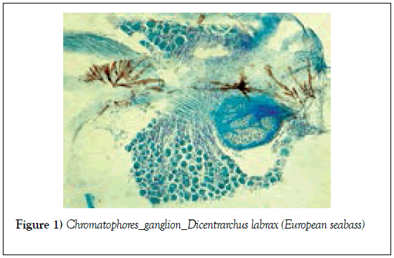
Parasagittal section of the medial trunk in this fish. This picture displays spinal ganglion cells (at the bottom), neurites (in the middle) and beautiful stellate chromatophores (brown) (Figure 1).
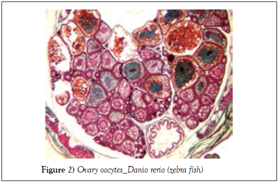
This low magnification view shows various oocyte developmental stages: previtellogenic (small) and vitellogenic (larger, with yolk globules) oocytes. On the bottom, one can see a transverse section through the posterior intestine (unstained lumen) (Figure 2).
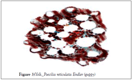
Isolated yolk granules (various colours and sizes) and large lipid droplets (unstained) coming from an embryo’s yolk sac content (pregnant female - guppys are ovoviviparous fish). The yolk sac is an extraembryonic membrane which encloses the yolk and plays major roles in nutrition and respiration (Figure 3).
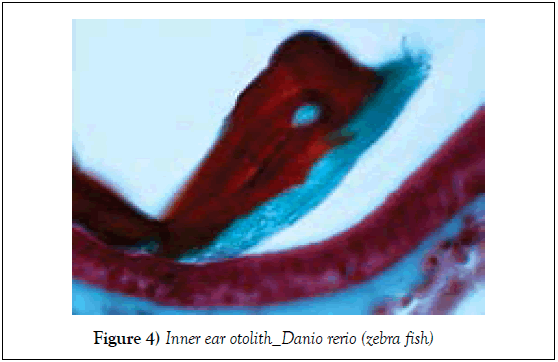
An otolith (red and blue) linked to the sensory macula (dark pink - at the bottom) by a thin otolithic membrane connecting the two structures. Otoliths are free, compact, non-bony structures composed of calcium carbonate deposited in a protein matrix. The present one is a large otolith resembling a dolphin or a sperm whale (removing the blue) (Figure 4).
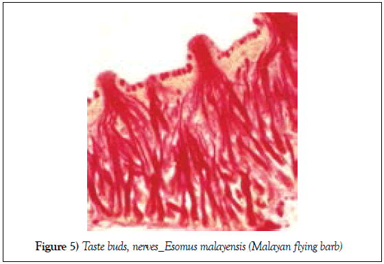
Taste buds innervation. This photomicrograph shows three taste buds and their rich innervation (nerve fibres in bright red). The surface of the epithelium (pale orange) is rich in mucus-secreting cells (about twenty, in bright red too). Taste buds transmit information to the brain via the nerves VII (facial nerve), IX (glossopharyngeal nerve) and X (vagal nerve) (Figure 5).
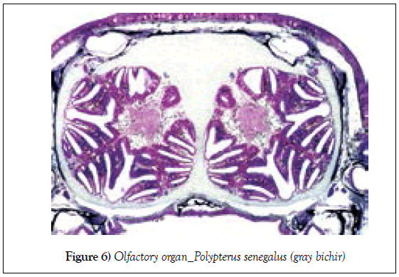
Transverse section through the paired olfactory organs showing the olfactory chambers lined with olfactory lamellae (dark purple). This lamellar arrangement greatly increases the surface area in contact with water and thus the olfaction (Figure 6).
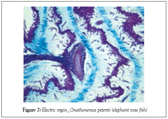
Electric organ of the elephant-nose fish. The electrocytes (“zigzag structures” - in purple) are the morphological and functional elementary units of the electric organ which generate electric signals. They are modified skeletal muscle fibers, are surrounded by two gelatinous layers (pale blue) and separated from each other by collagenous septa (sky blue, darker). At the centre, a large electric nerve (funnel shaped) is demonstrated. Mormyrids have a brain / body weight ratio much higher than in any other fish. This is due to the amazing dimensions of their cerebellum (Figure 7).
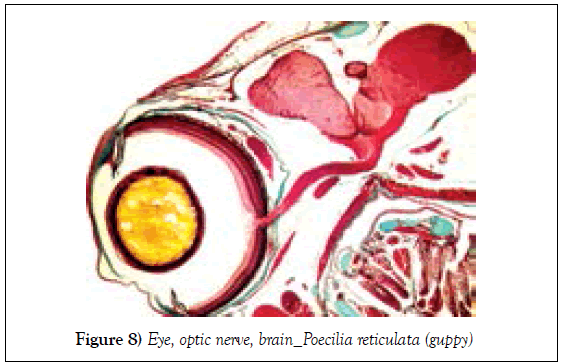
This general view emphasizes the connection between the eye (left – lens in yellow) and the brain (right – pink) via the optic nerve (Figure 8).
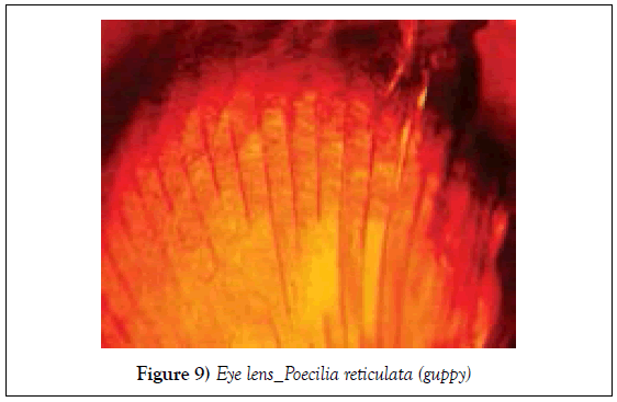
The picture illustrates fully parallel differentiated lens fibers having lost, inter alia, their nuclei. Crystalline protein is the major protein found in the very elongated lens cells, the latter being separated by very little extracellular matrix (Figure 9).
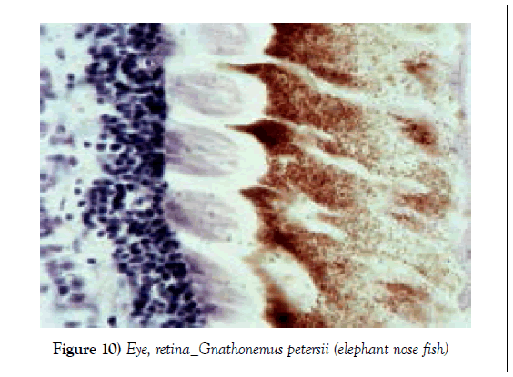
Posterior part of the retina. The pigment epithelium (on the right – melanin in brown), the photoreceptor cell processes (inner and outer segments - light purple at the centre) and the cell bodies of the cone and rod cells (outer nuclear layer - on the left, in dark purple) are clearly visible. Bony fish often have several twin cones (Figure 10) [1].


