General histology of organisms - Part 2 (Fish)
Received: 11-Sep-2017 Accepted Date: Sep 15, 2017; Published: 20-Sep-2017
Citation: Genten F. General histology of organisms - Part 2 (Fish) J Histol Histopathol Res 2017;1(1):3-4.
This open-access article is distributed under the terms of the Creative Commons Attribution Non-Commercial License (CC BY-NC) (http://creativecommons.org/licenses/by-nc/4.0/), which permits reuse, distribution and reproduction of the article, provided that the original work is properly cited and the reuse is restricted to noncommercial purposes. For commercial reuse, contact reprints@pulsus.com
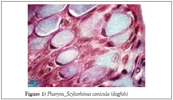
Pharyngeal epithelium. In fishes the most common unicellular glands are mucus-secreting cells. The mucoid substance (in pale blue) distends the apical portion of these large cells. The highly condensed nucleus is moved to the basal cytoplasmic region. The other nuclei are those of the ordinary epithelial cells (Figure 1).
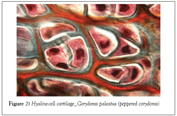
The chondrocytes are closely packed and their cytoplasm (pink), (dark) nuclei as well as lacunae (unstained) are well visible. The matrix is stained grey and orange. This typically type of cartilage is seen in different parts of the skeleton or, like here, in the barbels of catfishes (Siluriforms) (Figure 2).
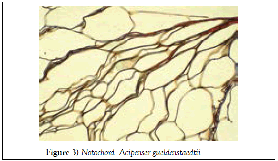
Notochord cells showing their typical appearance. The cells are filled with a gelatinous substance that gives, together with the sheath, the notochord its flexibility and elasticity. These thick-walled cells lie closely pressed together and the tissue resembles that of plants (Figure 3).
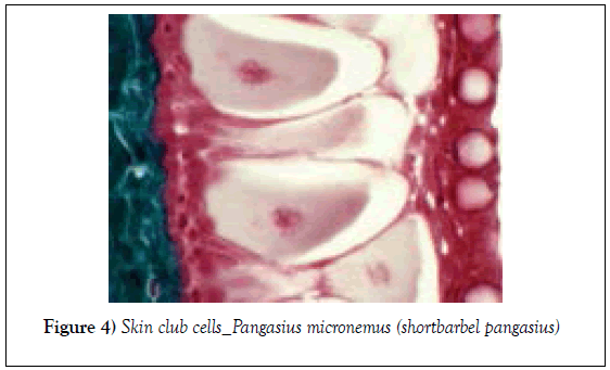
Skin of Pangasius. The Ostariophysi (mostly Siluriformes, Cypriniformes and Characiformes) are characterized by a highly complex physiological feature, in all other groups of teleosts: the fright reaction elicited by alarm substance produced by special epidermal cells: the club cells. These large cells (at the center) have a centrally located nucleus and an eosinophilic cytoplasm (pale pink) As they do not reach the surface, only injury of the skin can release the content of these cells into the water leading to the fright reaction of congeners. On the right, one can see superficial mucous cells and in green, on the left, the dermis (Figure 4).
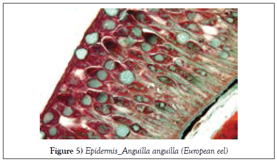
Eel skin with negligible scale cover. Large and numerous unicellular gland cells (green mucous cells and serous cells with a secretory vacuole) are visible. The protein secreting serous cells take up the largest part of the epithelium (Figure 5).
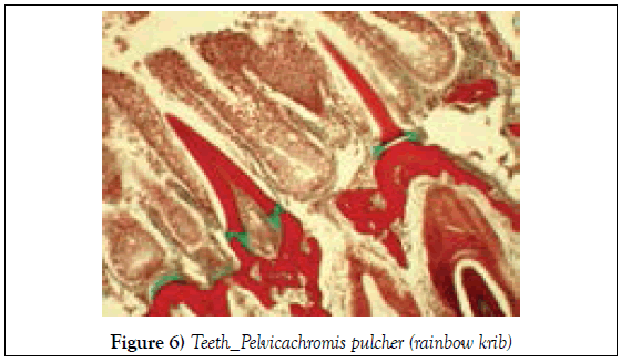
In this carnivorous fish, typical teeth have a pointed apex attached to the maxillary bone through a few elastic ligaments (in green). On the left, buccal stratified epithelium (brown-pink) and in the lower right corner, a growing tooth (Figure 6).
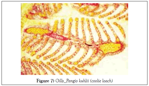
Transverse section through a gill filament (primary lamella). The well vascularized gill filaments are supported by cartilaginous tissue (magenta) and bear numerous flattened parallel secondary lamellae which constitute the respiratory surface. Erythrocytes (bright yellow) are present everywhere as well in the afferent and efferent arterial vessels as in the capillaries between them (Figure 7).
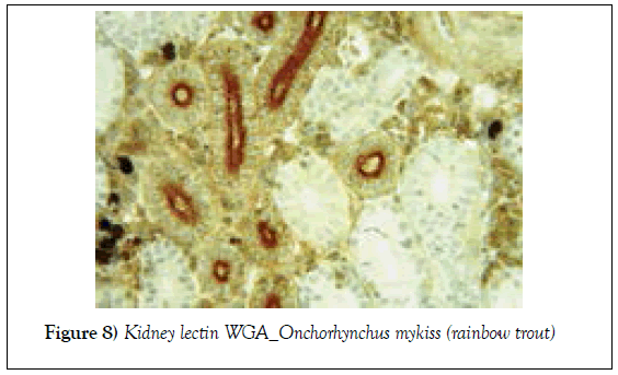
Lectins can bind to specific carbohydrates. This picture reveals WGA-lectin histochemical localization of complex sugar moieties (N-Acetylglucosamin et sialic acid - stained in brown) in the trout kidney. Lectin reactive signal was detected on the brush border (dark brown) of the proximal tubules but not in the distal tubules (unstained) which lack microvilli. In black, melanomacrophage centers (Figure 8).
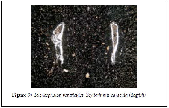
Transverse section of the cerebral hemispheres showing the clearly separated first and second cerebrum ventricles inside which the paired anterior tela choroidea is found. Thanks to the silver impregnation, nervous elements stained black and many capillaries display orange erythrocytes (Figure 9).
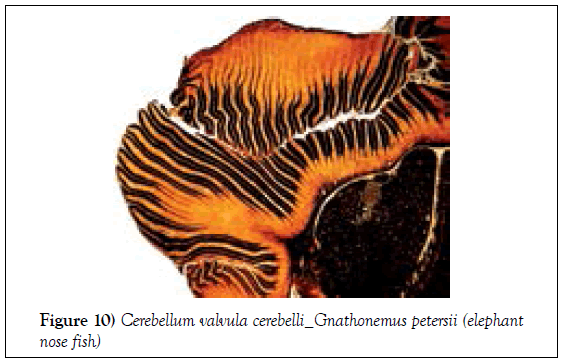
Section through the hypertrophied lateral lobes of the valvula cerebelli. The valvula consists of a series of deeply convoluted folds oriented towards the outer (posterior lobe) or the inner (anterior lobe) side. The axons are stained black with silver. This very remarkable hypertrophy of the mormyrid cerebellum involves an enormous capacity for processing electroreceptive information. Fish with a large valvula cerebelli often have a better developed lateral line system (Figure 10) [1].


