Hepatomesenteric trunk and gastro-splenico-phrenic trunk
Satheesha Nayak B.*
Melaka Manipal Medical College (Manipal Campus), International Centre for Health Sciences, Madhav Nagar, Manipal, Udupi District, Karnataka State, India
- *Corresponding Author:
- Satheesha Nayak B., MSc, PhD
Associate Professor of Anatomy, Melaka Manipal Medical College (Manipal Campus), International Centre for Health Sciences, Madhav Nagar, Manipal, Udupi District, Karnataka State, 576 104, India
Tel: +91 820 2922519
Fax: +91 820 2571905
E-mail: nayaksathish@yahoo.com
Date of Received: June 4th, 2008
Date of Accepted: June 11th, 2008
Published Online: June 17th, 2008
© IJAV. 2008; 1: 2–3.
[ft_below_content] =>Keywords
celiac trunk, hepatic artery, superior mesenteric artery, variations, hepatomesenteric trunk, gastro-splenico-phrenic trunk
Introduction
The celiac trunk is a ventral branch of abdominal aorta. It usually arises from the aorta at the level of twelfth thoracic vertebra and after a short course, divides into left gastric, common hepatic and splenic arteries. The common hepatic artery, after its origin from the celiac trunk, runs downwards and to the right till it reaches the first part of duodenum. At the upper border of the first part of duodenum, it divides into hepatic artery proper and gastroduodenal arteries. Common hepatic artery usually gives a right gastric branch before its termination. The hepatic artery proper ascends in the right free margin of lesser omentum lying anterior to the portal vein and on the left side of the bile duct. It divides into right and left hepatic arteries at or near the porta hepatis.
The inferior phrenic arteries are usually two in number and they arise from the abdominal aorta just below the aortic opening of the diaphragm. They supply the diaphragm and suprarenal glands.
The superior mesenteric artery is the second ventral branch of the abdominal aorta. It supplies the derivatives of the midgut. It runs anterior to the third part of the duodenum and enters the mesentery of the small intestine. Its normal branches include inferior pancreaticoduodenal, jejunal, ileal, iliocolic, right colic and middle colic arteries.
We saw the origin of common hepatic artery from the hepatomesenteric trunk and the presence of inferior phrenic trunk which originated from the gastro-splenico-phrenic trunk.
Case Report
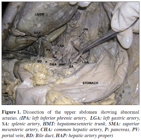
During the regular dissection classes for the first year medical undergraduate students, we observed a few variations in the branches of the celiac trunk and superior mesenteric arteries. The variations were found in a female cadaver aged approximately 55 years. The celiac trunk was smaller than its normal size and gave three branches namely; inferior phrenic trunk, left gastric artery and splenic artery (Figure 1). The left gastric and splenic arteries were of equal size. Their course and distribution were normal. The inferior phrenic trunk divided into right and left inferior phrenic arteries.
Figure 1: Dissection of the upper abdomen showing abnormal arteries. (IPA: left inferior phrenic artery, LGA: left gastric artery, SA: splenic artery, HMT: hepatomesenteric trunk, SMA: superior mesenteric artery, CHA: common hepatic artery, P: pancreas, PV: portal vein, BD: Bile duct, HAP: hepatic artery proper)
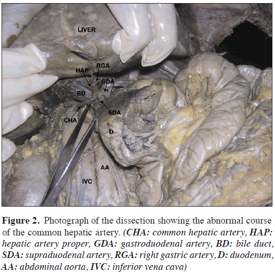
A hepatomesenteric trunk took its origin from aorta, just below the level of origin of celiac trunk. This origin was 2.5 cm above the superior border of the pancreas. At the level of the upper border of the pancreas (Figure 1) the trunk divided into common hepatic and superior mesenteric arteries. The common hepatic artery coursed to the right, behind the portal vein and bile duct and then coursed anterior and to the left by making a loop around the right side of the bile duct (Figure 2). Thus the common hepatic artery was related to the posterior aspect of portal vein and posterior, right and anterior aspects of the bile duct. The artery then divided into three branches namely; hepatic artery proper, gastroduodenal artery and supraduodenal artery (Figure 2). The right gastric artery was a branch of hepatic artery proper. The course and branching pattern of superior mesenteric artery was normal.
Figure 2: Photograph of the dissection showing the abnormal course of the common hepatic artery. (CHA: common hepatic artery, HAP: hepatic artery proper, GDA: gastroduodenal artery, BD: bile duct, SDA: supraduodenal artery, RGA: right gastric artery, D: duodenum, AA: abdominal aorta, IVC: inferior vena cava)
Discussion
Variations of the branching pattern and distribution of the common hepatic artery are common. Variations in its origin and course are relatively rare and are of surgical and radiological importance. One of the rare origins of the hepatic artery is from the superior mesenteric artery. The superior mesenteric artery and the common hepatic artery arising from abdominal aorta as a common trunk named “hepatomesenteric trunk”. In such cases the celiac trunk is reduced in size and is called “gastrosplenic trunk”. The gastrosplenic trunk gives left gastric artery and splenic artery. The existence of the hepatomesenteric trunk has been reported [1,2,3,4]. The common hepatic artery usually passes in front of the portal vein when it originates as a branch of hepatomesenteric trunk. Peschaud et al., [5] have reported a case of common hepatic artery crossing the portal vein from the front. The knowledge of the common hepatic artery arising from superior mesenteric artery is important for surgeons performing pancreaticoduodenectomy. The common hepatic artery is liable to get damaged in such surgical interventions. In the current case, the common hepatic artery passed behind the portal vein and bile duct and looped around the bile duct on its right side. This fact may be functionally important. The bile duct may be compressed by the loop of the artery. Such abnormal course of the artery might confuse the radiologist doing endovascular procedures on the artery.
The inferior mesenteric artery might very rarely arise from the superior mesenteric artery in addition to the common hepatic artery. Clinically, cases like this highlight the importance of knowing the mesenteric artery anatomy and the possibility of its numerous variations in surgical procedures such as right hemicolectomy, resection of the transverse colon, left hemicolectomy, sigmoidectomy, and en bloc resection of the head of the pancreas and the superior mesenteric vessels.
The celiac artery is also known for variations. All the three of its branches might come from the abdominal aorta as independent branches or it might give rise to additional branches such as the superior mesenteric artery and inferior phrenic arteries. One such case of a celiaco-mesenterico-phrenic trunk has been reported by Nayak [6]. In the current case, the two inferior phrenic arteries took their origin from a common inferior phrenic trunk, which was a branch of the celiac trunk. This type of celiac trunk can be named “gastro-splenico-phrenic trunk”. The knowledge of this type of variation is important for the surgeons performing kidney transplants and suprarenal surgeries.
References
- Nakano H, Kikuchi K, Seta S, Katayama M, Horikoshi K, Yamamura T, Otsubo T. A patient undergoing pancreaticoduodenectomy in whom involved common hepatic artery anomalously arising from the superior mesenteric artery was removed and reconstructed. Hepatogastroenterology. 2005; 52: 1883—1885.
- Osawa T, Feng XY, Sasaki N, Nagato S, Matsumoto Y, Onodera M, Nara E, Fujimura A, Nozaka Y. Rare case of the inferior mesenteric artery and the common hepatic artery arising from the superior mesenteric artery. Clin. Anat. 2004; 17: 518—521.
- Nakamura Y, Miyaki T, Hayashi S, Iimura A, Itoh M. Three cases of the gastrosplenic and the hepatomesenteric trunks. Okajimas Folia Anat. Jpn. 2003; 80: 71—76.
- Kahraman G, Marur T, Tanyeli E, Yildirim M. Hepatomesenteric trunk. Surg. Radiol. Anat. 2001; 23: 433—435.
- Peschaud F, El-Hajjam M, Malafosse R, Goere D, Benoist S, Penna C, Nordlinger B. A common hepatic artery passing in front of the portal vein. Surg. Radiol. Anat. 2006; 28: 202—205.
- Nayak S. Common celiaco-mesenterico-phrenic trunk and renal vascular variations. Saudi Med. J. 2006; 27: 1894—1896.
Satheesha Nayak B.*
Melaka Manipal Medical College (Manipal Campus), International Centre for Health Sciences, Madhav Nagar, Manipal, Udupi District, Karnataka State, India
- *Corresponding Author:
- Satheesha Nayak B., MSc, PhD
Associate Professor of Anatomy, Melaka Manipal Medical College (Manipal Campus), International Centre for Health Sciences, Madhav Nagar, Manipal, Udupi District, Karnataka State, 576 104, India
Tel: +91 820 2922519
Fax: +91 820 2571905
E-mail: nayaksathish@yahoo.com
Date of Received: June 4th, 2008
Date of Accepted: June 11th, 2008
Published Online: June 17th, 2008
© IJAV. 2008; 1: 2–3.
Abstract
A thorough knowledge of variation in the branching pattern of celiac trunk and superior mesenteric artery is important for surgeons, radiologists and other medical specialties. We observed some variations in the branching pattern of celiac trunk and superior mesenteric artery during dissection classes for first year medical students. The celiac trunk (gastro-splenico-phrenic trunk) divided into inferior phrenic trunk, left gastric and splenic arteries. The inferior phrenic trunk divided into right and left inferior phrenic arteries. The common hepatic artery took its origin from a hepatomesenteric trunk and passed behind the portal vein and bile duct. It hooked around the bile duct and then divided into three branches; hepatic artery proper, gastroduodenal artery and supraduodenal artery
-Keywords
celiac trunk, hepatic artery, superior mesenteric artery, variations, hepatomesenteric trunk, gastro-splenico-phrenic trunk
Introduction
The celiac trunk is a ventral branch of abdominal aorta. It usually arises from the aorta at the level of twelfth thoracic vertebra and after a short course, divides into left gastric, common hepatic and splenic arteries. The common hepatic artery, after its origin from the celiac trunk, runs downwards and to the right till it reaches the first part of duodenum. At the upper border of the first part of duodenum, it divides into hepatic artery proper and gastroduodenal arteries. Common hepatic artery usually gives a right gastric branch before its termination. The hepatic artery proper ascends in the right free margin of lesser omentum lying anterior to the portal vein and on the left side of the bile duct. It divides into right and left hepatic arteries at or near the porta hepatis.
The inferior phrenic arteries are usually two in number and they arise from the abdominal aorta just below the aortic opening of the diaphragm. They supply the diaphragm and suprarenal glands.
The superior mesenteric artery is the second ventral branch of the abdominal aorta. It supplies the derivatives of the midgut. It runs anterior to the third part of the duodenum and enters the mesentery of the small intestine. Its normal branches include inferior pancreaticoduodenal, jejunal, ileal, iliocolic, right colic and middle colic arteries.
We saw the origin of common hepatic artery from the hepatomesenteric trunk and the presence of inferior phrenic trunk which originated from the gastro-splenico-phrenic trunk.
Case Report
During the regular dissection classes for the first year medical undergraduate students, we observed a few variations in the branches of the celiac trunk and superior mesenteric arteries. The variations were found in a female cadaver aged approximately 55 years. The celiac trunk was smaller than its normal size and gave three branches namely; inferior phrenic trunk, left gastric artery and splenic artery (Figure 1). The left gastric and splenic arteries were of equal size. Their course and distribution were normal. The inferior phrenic trunk divided into right and left inferior phrenic arteries.
Figure 1: Dissection of the upper abdomen showing abnormal arteries. (IPA: left inferior phrenic artery, LGA: left gastric artery, SA: splenic artery, HMT: hepatomesenteric trunk, SMA: superior mesenteric artery, CHA: common hepatic artery, P: pancreas, PV: portal vein, BD: Bile duct, HAP: hepatic artery proper)
A hepatomesenteric trunk took its origin from aorta, just below the level of origin of celiac trunk. This origin was 2.5 cm above the superior border of the pancreas. At the level of the upper border of the pancreas (Figure 1) the trunk divided into common hepatic and superior mesenteric arteries. The common hepatic artery coursed to the right, behind the portal vein and bile duct and then coursed anterior and to the left by making a loop around the right side of the bile duct (Figure 2). Thus the common hepatic artery was related to the posterior aspect of portal vein and posterior, right and anterior aspects of the bile duct. The artery then divided into three branches namely; hepatic artery proper, gastroduodenal artery and supraduodenal artery (Figure 2). The right gastric artery was a branch of hepatic artery proper. The course and branching pattern of superior mesenteric artery was normal.
Figure 2: Photograph of the dissection showing the abnormal course of the common hepatic artery. (CHA: common hepatic artery, HAP: hepatic artery proper, GDA: gastroduodenal artery, BD: bile duct, SDA: supraduodenal artery, RGA: right gastric artery, D: duodenum, AA: abdominal aorta, IVC: inferior vena cava)
Discussion
Variations of the branching pattern and distribution of the common hepatic artery are common. Variations in its origin and course are relatively rare and are of surgical and radiological importance. One of the rare origins of the hepatic artery is from the superior mesenteric artery. The superior mesenteric artery and the common hepatic artery arising from abdominal aorta as a common trunk named “hepatomesenteric trunk”. In such cases the celiac trunk is reduced in size and is called “gastrosplenic trunk”. The gastrosplenic trunk gives left gastric artery and splenic artery. The existence of the hepatomesenteric trunk has been reported [1,2,3,4]. The common hepatic artery usually passes in front of the portal vein when it originates as a branch of hepatomesenteric trunk. Peschaud et al., [5] have reported a case of common hepatic artery crossing the portal vein from the front. The knowledge of the common hepatic artery arising from superior mesenteric artery is important for surgeons performing pancreaticoduodenectomy. The common hepatic artery is liable to get damaged in such surgical interventions. In the current case, the common hepatic artery passed behind the portal vein and bile duct and looped around the bile duct on its right side. This fact may be functionally important. The bile duct may be compressed by the loop of the artery. Such abnormal course of the artery might confuse the radiologist doing endovascular procedures on the artery.
The inferior mesenteric artery might very rarely arise from the superior mesenteric artery in addition to the common hepatic artery. Clinically, cases like this highlight the importance of knowing the mesenteric artery anatomy and the possibility of its numerous variations in surgical procedures such as right hemicolectomy, resection of the transverse colon, left hemicolectomy, sigmoidectomy, and en bloc resection of the head of the pancreas and the superior mesenteric vessels.
The celiac artery is also known for variations. All the three of its branches might come from the abdominal aorta as independent branches or it might give rise to additional branches such as the superior mesenteric artery and inferior phrenic arteries. One such case of a celiaco-mesenterico-phrenic trunk has been reported by Nayak [6]. In the current case, the two inferior phrenic arteries took their origin from a common inferior phrenic trunk, which was a branch of the celiac trunk. This type of celiac trunk can be named “gastro-splenico-phrenic trunk”. The knowledge of this type of variation is important for the surgeons performing kidney transplants and suprarenal surgeries.
References
- Nakano H, Kikuchi K, Seta S, Katayama M, Horikoshi K, Yamamura T, Otsubo T. A patient undergoing pancreaticoduodenectomy in whom involved common hepatic artery anomalously arising from the superior mesenteric artery was removed and reconstructed. Hepatogastroenterology. 2005; 52: 1883—1885.
- Osawa T, Feng XY, Sasaki N, Nagato S, Matsumoto Y, Onodera M, Nara E, Fujimura A, Nozaka Y. Rare case of the inferior mesenteric artery and the common hepatic artery arising from the superior mesenteric artery. Clin. Anat. 2004; 17: 518—521.
- Nakamura Y, Miyaki T, Hayashi S, Iimura A, Itoh M. Three cases of the gastrosplenic and the hepatomesenteric trunks. Okajimas Folia Anat. Jpn. 2003; 80: 71—76.
- Kahraman G, Marur T, Tanyeli E, Yildirim M. Hepatomesenteric trunk. Surg. Radiol. Anat. 2001; 23: 433—435.
- Peschaud F, El-Hajjam M, Malafosse R, Goere D, Benoist S, Penna C, Nordlinger B. A common hepatic artery passing in front of the portal vein. Surg. Radiol. Anat. 2006; 28: 202—205.
- Nayak S. Common celiaco-mesenterico-phrenic trunk and renal vascular variations. Saudi Med. J. 2006; 27: 1894—1896.


