Hepatosplenic and left hepatogastric trunks as variant arteries of foregut - A case report
Santosh K. Sangari* and Estomih P. Mtui
Program in Anatomy and Body Visualization, Department of Radiology, Weill Cornell Medicine, New York; NY, USA
- *Corresponding Author:
- Santosh K. Sangari, MBBS, MS
Program in Anatomy and Body Visualization, Department of Radiology, Weill Cornell Medicine, 1300 York Avenue New York, NY 10065 USA
Tel: +1 (212) 746-6171
E-mail: sks2005@med.cornell.edu
Date of Received: April 15th, 2015
Date of Accepted: January 2nd, 2017
Published Online: January 18th, 2017
© Int J Anat Var (IJAV). 2016; 9: 50–52.
[ft_below_content] =>Keywords
hepatosplenic trunk, left hepatogastric trunk, replaced left hepatic artery, left inferior phrenic artery
Introduction
The celiac trunk is the artery of the foregut and normally arises from the abdominal aorta at the level of T12 vertebra. It is about 1-2 cm long and divides into common hepatic; left gastric and splenic arteries. The right and left inferior phrenic arteries normally are lateral branches of abdominal aorta immediately below the aortic hiatus in the diaphragm [1]. There are many variations of the celiac trunk reported in the literature [2,3]. The knowledge of variations in the arterial supply of the gut is necessary for interventional procedures and have important surgical implications. The present study shows new interesting variant foregut arteries; which have not been reported earlier. This would help surgeon to plan treatment and requires interventional evaluation before management.
Case Report
The observations were made in 80-year-old male cadaver during regular educational dissection at Weill Cornell Medical College; NY.
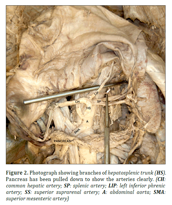
In the present case; instead of the celiac trunk arising from abdominal aorta; there were two arteries for the foregut – hepatosplenic trunk and left hepatogastric trunk; both directly arising from abdominal aorta (Figure 1). The hepatosplenic trunk was a branch of abdominal aorta below the aortic hiatus and divided into common hepatic artery and splenic artery. The common hepatic artery immediately after its origin gave off left inferior phrenic artery; which was seen supplying the diaphragm and in addition also supplied the left suprarenal gland by superior suprarenal arteries (Figure 2).
Figure 1. Hepatosplenic (HS) and left hepatogastric (LHG) trunks arising from abdominal aorta (A) independently. Pancreas has been pulled down to show the arteries clearly. (CH: common hepatic artery; RH: right hepatic artery; GDA: gastroduodenal artery; LH: replaced left hepatic artery; LG: left gastric artery; E: esophageal branches; CBD: common bile duct; PV: portal vein; SMA: superior mesenteric artery)
The common hepatic artery was seen running to the right along the upper border the pancreas and terminated by dividing into gastroduodenal artery and the right hepatic artery (Figure 1). The cystic artery was a branch of right hepatic artery as usual.
The splenic artery was not seen to be tortuous and near its origin divided into two branches; which entered the hilum of spleen to supply it (Figure 2).
The left hepatogastric trunk was a direct branch of abdominal aorta immediately below the origin of hepatosplenic trunk. The left hepatogastric trunk divided into replaced left hepatic artery and left gastric artery. The esophageal branches were seen arising instead from the replaced left hepatic artery (Figure 1).
Discussion
During embryogenesis; the ventral splanchnic branches are paired vessels of the dorsal aortae distributed to the capillary plexus in the wall of the yolk sac. After the fusion of the dorsal aortae; they merge to form unpaired trunks; connected by longitudinal anastomosis. Numerous of these ventral splanchnic branches disappear and only three trunks persist; i.e. celiac trunk for foregut; superior mesenteric artery for midgut and inferior mesenteric artery for hindgut. Many lateral splanchnic branches from aorta supply structures developing from intermediate mesoderm and gives rise to suprarenal; phrenic; renal and gonadal arteries. The retention or disappearance of these anastomotic arteries result in variations of celiac trunk; superior mesenteric artery or inferior mesenteric artery [1,2]. The classic trifurcation of celiac trunk into left gastric; common hepatic and splenic arteries has been reported in 86% of cases. Rarely it may have more than three branches or be absent. The celiac trunk may have two branches; usually splenic and common hepatic arteries; rarely left gastric and splenic arteries. The variations in the branching pattern of celiac trunk has been classified into five types; in which right and left inferior phrenic arteries also branch off from the celiac trunk [3].
In the present case; the celiac trunk was replaced by hepatosplenic and left hepatogastric trunks. The hepatosplenic trunk divided into common hepatic and splenic arteries. The common hepatic artery immediately after origin gave left inferior phrenic artery. The left inferior phrenic artery in addition to supplying the diaphragm also supplied the left suprarenal gland by giving superior suprarenal arteries. This variation has to be kept in mind by the surgeon during surgical/interventional procedures involving common hepatic artery; so that the arterial supply to the diaphragm and the suprarenal gland is not compromised.
Normally the left gastric artery is a branch of celiac trunk. The origin of left gastric artery directly from abdominal aorta has been seen in 4% by MDCT angiography [4]. In the present case; the left hepatogastric trunk is a direct branch from the abdominal aorta; immediately below the origin of hepatosplenic trunk. The left hepatogastric trunk divided into the replaced left hepatic artery and left gastric artery. The esophageal arteries are normally branches of left gastric artery; which supply the lower end of esophagus. Instead in the present case; esophageal arteries branched off from the replaced left hepatic artery. In literature; the left hepatic artery arising from the left gastric branch of celiac trunk has been classified as Type II hepatic arterial variant [3,5,6,7]. For the hepatic arterial infusion chemotherapy/hepatic artery infusion pump placement; the catheters are typically placed in the gastroduodenal artery surgically and in proper hepatic artery percutaneously; so that they infuse both the hepatic arteries [8,9,10]. In the present case; the right hepatic artery is a branch of common hepatic artery and left hepatic artery is arising from left hepatogastric trunk. Since the two hepatic arteries are arising from different sources; it would need more than one catheter for infusion chemotherapy. Also; the ligation of left gastric artery; if required during surgical procedures would lead to the necrosis of the left lobe of liver if attention is not paid to the left hepatic artery arising from the left hepatogastric trunk. In conclusion; a preoperative visceral angiography is a key for many hepatobiliary surgeries and for interventional procedures for hepatic chemotherapy.
References
- Standring S, ed. Gray’s Anatomy. The anatomical basis of clinical practice. 40th Ed.; Churchill Livingstone Elsevier. 2008; 205–209; 1116–1120.
- Tandler J. Über die variatäten der Arteria coeliaca und deren Entwicklung. Anat Hefte. 1904; 25: 473–499.
- Bergman RA, Afifi AK Miyauchi R. Five variations in the celiac trunk. Illustrated Encyclopedia of human anatomic variation: Opus II: cardiovascular system. (accessed January 2015)
- Ognjanovic N, Jeremic D, Zivanovic-Macuzic I, Sazdanovic M, Sazdanovic P. Tanaskovic I, Jovanovic J, Popovic R, Vojinovic R, Milosevic B, Milosavljevic M, Stojadinovic D, Tosevski J, Vulovic M. MDCT angiography of anatomical variation of the coeliac trunk and superior mesenteric artery. Arch Biol Sci. 2014; 66(1): 233–240.
- Gumus H, Bukte Y, Ozdemir E, Senturk S, Tekbas G, Onder H, Ekici F, Bilici A. Variations of the celiac trunk and hepatic arteries: a study with 64-detector computed tomographic angiography. Eur Rev Med Pharmacol Sci. 2013; 17: 1636–1641.
- Covey AM, Brody LA, Maluccio MA, Getrajdman GI, Brown KT. Variant hepatic arterial anatomy revisited: Digital subtraction angiography performed in 600 patients. Radiol. 2002; 224: 542–547.
- De Cecco CN, Ferrari R, Rengo M, Paolantonio P, Vecchietti F, Laghi A. Anatomical variations of the hepatic arteries in 250 patients studied with 64-row CT angiography. Eur Radiol. 2009; 19: 2765–2770.
- Blumgart L, Fong Y. Surgery of the liver and biliary tract. Saunders, New York. 2000; 1625–1628.
- Zanon C, Grosso M, Clara R, Chiappino I, Mancini A, Mussa A. Percutaneous implantation of arterial port-a-cath via trans-subclavian access. Anticancer Res. 1999; 19: 5667–5671.
- Ikebe M, Itasaka H, Adachi E, Shirabe K, Maekawa S, Mutoh Y, Yoshida K, Takenaka K. New method of catheter-port system implantation in hepatic arterial infusion chemotherapy. Am J Surg. 2003; 186(1): 63–66.
Santosh K. Sangari* and Estomih P. Mtui
Program in Anatomy and Body Visualization, Department of Radiology, Weill Cornell Medicine, New York; NY, USA
- *Corresponding Author:
- Santosh K. Sangari, MBBS, MS
Program in Anatomy and Body Visualization, Department of Radiology, Weill Cornell Medicine, 1300 York Avenue New York, NY 10065 USA
Tel: +1 (212) 746-6171
E-mail: sks2005@med.cornell.edu
Date of Received: April 15th, 2015
Date of Accepted: January 2nd, 2017
Published Online: January 18th, 2017
© Int J Anat Var (IJAV). 2016; 9: 50–52.
Abstract
The present case reports 80-year-old male cadaver with hepatosplenic and left hepatogastric trunks supplying foregut. The hepatosplenic trunk was a branch of abdominal aorta; which divided into common hepatic and splenic arteries. The common hepatic artery gave origin to the left inferior phrenic artery before dividing into gastroduodenal artery and right hepatic artery. The left inferior phrenic artery also supplied the left suprarenal gland by superior suprarenal arteries. The left hepatogastric trunk was a direct branch of abdominal aorta immediately below the hepatosplenic trunk and divided into replaced left hepatic artery and left gastric artery. The esophageal arteries; which supplied the lower end of esophagus were branches of replaced left hepatic artery.
-Keywords
hepatosplenic trunk, left hepatogastric trunk, replaced left hepatic artery, left inferior phrenic artery
Introduction
The celiac trunk is the artery of the foregut and normally arises from the abdominal aorta at the level of T12 vertebra. It is about 1-2 cm long and divides into common hepatic; left gastric and splenic arteries. The right and left inferior phrenic arteries normally are lateral branches of abdominal aorta immediately below the aortic hiatus in the diaphragm [1]. There are many variations of the celiac trunk reported in the literature [2,3]. The knowledge of variations in the arterial supply of the gut is necessary for interventional procedures and have important surgical implications. The present study shows new interesting variant foregut arteries; which have not been reported earlier. This would help surgeon to plan treatment and requires interventional evaluation before management.
Case Report
The observations were made in 80-year-old male cadaver during regular educational dissection at Weill Cornell Medical College; NY.
In the present case; instead of the celiac trunk arising from abdominal aorta; there were two arteries for the foregut – hepatosplenic trunk and left hepatogastric trunk; both directly arising from abdominal aorta (Figure 1). The hepatosplenic trunk was a branch of abdominal aorta below the aortic hiatus and divided into common hepatic artery and splenic artery. The common hepatic artery immediately after its origin gave off left inferior phrenic artery; which was seen supplying the diaphragm and in addition also supplied the left suprarenal gland by superior suprarenal arteries (Figure 2).
Figure 1. Hepatosplenic (HS) and left hepatogastric (LHG) trunks arising from abdominal aorta (A) independently. Pancreas has been pulled down to show the arteries clearly. (CH: common hepatic artery; RH: right hepatic artery; GDA: gastroduodenal artery; LH: replaced left hepatic artery; LG: left gastric artery; E: esophageal branches; CBD: common bile duct; PV: portal vein; SMA: superior mesenteric artery)
The common hepatic artery was seen running to the right along the upper border the pancreas and terminated by dividing into gastroduodenal artery and the right hepatic artery (Figure 1). The cystic artery was a branch of right hepatic artery as usual.
The splenic artery was not seen to be tortuous and near its origin divided into two branches; which entered the hilum of spleen to supply it (Figure 2).
The left hepatogastric trunk was a direct branch of abdominal aorta immediately below the origin of hepatosplenic trunk. The left hepatogastric trunk divided into replaced left hepatic artery and left gastric artery. The esophageal branches were seen arising instead from the replaced left hepatic artery (Figure 1).
Discussion
During embryogenesis; the ventral splanchnic branches are paired vessels of the dorsal aortae distributed to the capillary plexus in the wall of the yolk sac. After the fusion of the dorsal aortae; they merge to form unpaired trunks; connected by longitudinal anastomosis. Numerous of these ventral splanchnic branches disappear and only three trunks persist; i.e. celiac trunk for foregut; superior mesenteric artery for midgut and inferior mesenteric artery for hindgut. Many lateral splanchnic branches from aorta supply structures developing from intermediate mesoderm and gives rise to suprarenal; phrenic; renal and gonadal arteries. The retention or disappearance of these anastomotic arteries result in variations of celiac trunk; superior mesenteric artery or inferior mesenteric artery [1,2]. The classic trifurcation of celiac trunk into left gastric; common hepatic and splenic arteries has been reported in 86% of cases. Rarely it may have more than three branches or be absent. The celiac trunk may have two branches; usually splenic and common hepatic arteries; rarely left gastric and splenic arteries. The variations in the branching pattern of celiac trunk has been classified into five types; in which right and left inferior phrenic arteries also branch off from the celiac trunk [3].
In the present case; the celiac trunk was replaced by hepatosplenic and left hepatogastric trunks. The hepatosplenic trunk divided into common hepatic and splenic arteries. The common hepatic artery immediately after origin gave left inferior phrenic artery. The left inferior phrenic artery in addition to supplying the diaphragm also supplied the left suprarenal gland by giving superior suprarenal arteries. This variation has to be kept in mind by the surgeon during surgical/interventional procedures involving common hepatic artery; so that the arterial supply to the diaphragm and the suprarenal gland is not compromised.
Normally the left gastric artery is a branch of celiac trunk. The origin of left gastric artery directly from abdominal aorta has been seen in 4% by MDCT angiography [4]. In the present case; the left hepatogastric trunk is a direct branch from the abdominal aorta; immediately below the origin of hepatosplenic trunk. The left hepatogastric trunk divided into the replaced left hepatic artery and left gastric artery. The esophageal arteries are normally branches of left gastric artery; which supply the lower end of esophagus. Instead in the present case; esophageal arteries branched off from the replaced left hepatic artery. In literature; the left hepatic artery arising from the left gastric branch of celiac trunk has been classified as Type II hepatic arterial variant [3,5,6,7]. For the hepatic arterial infusion chemotherapy/hepatic artery infusion pump placement; the catheters are typically placed in the gastroduodenal artery surgically and in proper hepatic artery percutaneously; so that they infuse both the hepatic arteries [8,9,10]. In the present case; the right hepatic artery is a branch of common hepatic artery and left hepatic artery is arising from left hepatogastric trunk. Since the two hepatic arteries are arising from different sources; it would need more than one catheter for infusion chemotherapy. Also; the ligation of left gastric artery; if required during surgical procedures would lead to the necrosis of the left lobe of liver if attention is not paid to the left hepatic artery arising from the left hepatogastric trunk. In conclusion; a preoperative visceral angiography is a key for many hepatobiliary surgeries and for interventional procedures for hepatic chemotherapy.
References
- Standring S, ed. Gray’s Anatomy. The anatomical basis of clinical practice. 40th Ed.; Churchill Livingstone Elsevier. 2008; 205–209; 1116–1120.
- Tandler J. Über die variatäten der Arteria coeliaca und deren Entwicklung. Anat Hefte. 1904; 25: 473–499.
- Bergman RA, Afifi AK Miyauchi R. Five variations in the celiac trunk. Illustrated Encyclopedia of human anatomic variation: Opus II: cardiovascular system. (accessed January 2015)
- Ognjanovic N, Jeremic D, Zivanovic-Macuzic I, Sazdanovic M, Sazdanovic P. Tanaskovic I, Jovanovic J, Popovic R, Vojinovic R, Milosevic B, Milosavljevic M, Stojadinovic D, Tosevski J, Vulovic M. MDCT angiography of anatomical variation of the coeliac trunk and superior mesenteric artery. Arch Biol Sci. 2014; 66(1): 233–240.
- Gumus H, Bukte Y, Ozdemir E, Senturk S, Tekbas G, Onder H, Ekici F, Bilici A. Variations of the celiac trunk and hepatic arteries: a study with 64-detector computed tomographic angiography. Eur Rev Med Pharmacol Sci. 2013; 17: 1636–1641.
- Covey AM, Brody LA, Maluccio MA, Getrajdman GI, Brown KT. Variant hepatic arterial anatomy revisited: Digital subtraction angiography performed in 600 patients. Radiol. 2002; 224: 542–547.
- De Cecco CN, Ferrari R, Rengo M, Paolantonio P, Vecchietti F, Laghi A. Anatomical variations of the hepatic arteries in 250 patients studied with 64-row CT angiography. Eur Radiol. 2009; 19: 2765–2770.
- Blumgart L, Fong Y. Surgery of the liver and biliary tract. Saunders, New York. 2000; 1625–1628.
- Zanon C, Grosso M, Clara R, Chiappino I, Mancini A, Mussa A. Percutaneous implantation of arterial port-a-cath via trans-subclavian access. Anticancer Res. 1999; 19: 5667–5671.
- Ikebe M, Itasaka H, Adachi E, Shirabe K, Maekawa S, Mutoh Y, Yoshida K, Takenaka K. New method of catheter-port system implantation in hepatic arterial infusion chemotherapy. Am J Surg. 2003; 186(1): 63–66.


