High division of the popliteal artery – a case report
Reena Singla1*, Usha Chabbra2 and Subhash Kaushal1
1Department of Anatomy, Maharishi Markandeshwar Institute of Medical Sciences & Research, Mullana, Ambala, Haryana, India.
2Department of Anatomy, Government Medical College, Patiala, Punjab, India.
- *Corresponding Author:
- Reena Singla
Assistant Professor, Department of Anatomy, Maharishi Markandeshwar Institute of Med. Sci. & Research, Mullana – Ambala, Haryana, India
Tel: +91 (989) 6212241
E-mail: singla.reena@ymail.com
Date of Received: September 4th, 2011
Date of Accepted: August 11th, 2012
Published Online: December 15th, 2012
© Int J Anat Var (IJAV). 2012; 5: 104–106.
[ft_below_content] =>Keywords
popliteal artery, popliteus muscle, anterior tibial artery, posterior tibial artery, peroneal artery
Introduction
The popliteal artery is a common recipient site for above or below knee bypass grafts. It is also frequently affected by penetrating and blunt trauma involving the lower extremity. Exposure of this artery is therefore, often required in both emergent and elective vascular procedures [1]. Knowledge of the anatomical variations in the branching of the popliteal artery is important because damage to its branches can be limb- or life threatening [2].
Popliteal artery is the continuation of the femoral artery beyond the tendinous opening in the adductor magnus muscle. Usually, it runs downwards in the popliteal fossa with a lateral inclination superficial to popliteus muscle and terminates by dividing into an anterior tibial and a posterior tibial artery at the lower border of popliteus muscle [3].
The popliteal artery lies on the anterior wall of the popliteal fossa. It gives several branches during its course, i.e., muscular branches pass to the lower parts of the hamstring muscles and to the upper parts of the muscles of the calf. These are large and give rise to cutaneous twigs, one of which accompanies the sural nerve. Two superior (medial and lateral), two inferior (medial and lateral) and middle genicular arteries lie on the on the anterior wall of the popliteal fossa [4].
Case Report
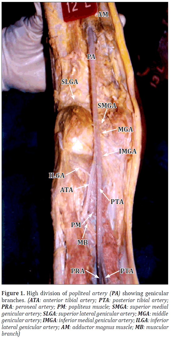
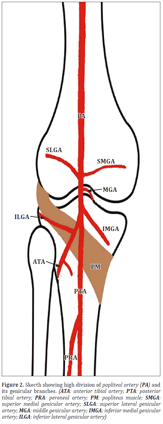
During cadaveric study on the popliteal artery, high division of the popliteal artery was observed on the left side of one of the embalmed male cadaver. Approximate age of cadaver was between 30-35 years. Popliteal artery divided at the level of the upper border of popliteus muscle into anterior tibial and posterior tibial arteries. Inferior lateral genicular artery which is usually a branch of popliteal artery was found to be the branch of anterior tibial artery as shown in Figures 1 and 2. However, on the right side popliteal artery bifurcated at the lower border of popliteus muscle into anterior tibial and posterior tibial arteries and genicular branches, followed the usual pattern.
Figure 1: High division of popliteal artery (PA) showing genicular branches. (ATA: anterior tibial artery; PTA: posterior tibial artery; PRA: peroneal artery; PM: popliteus muscle; SMGA: superior medial genicular artery; SLGA: superior lateral genicular artery; MGA: middle genicular artery; IMGA: inferior medial genicular artery; ILGA: inferior lateral genicular artery; AM: adductor magnus muscle; MB: muscular branch)
Figure 2: Skecth showing high division of popliteal artery (PA) and its genicular branches. (ATA: anterior tibial artery; PTA: posterior tibial artery; PRA: peroneal artery; PM: popliteus muscle; SMGA: superior medial genicular artery; SLGA: superior lateral genicular artery; MGA: middle genicular artery; IMGA: inferior medial genicular artery; ILGA: inferior lateral genicular artery)
Discussion
Variations in the branching pattern of the popliteal artery revolve around the high division of that trunk and the resulting differences in the arrangement of the terminal branches,posterior tibial,anterior tibial and peroneal arteries. Keen reported high division of popliteal artery in 14 out of 280 limbs [5].
Colborn et al. reported popliteal bifurcation above the lower border of popliteus muscle in 3 out of 42 cadavers. In each case, the point of bifurcation was symmetrical in both limbs, and the peroneal artery arose from the anterior tibial artery. In one case, the popliteal artery coursed deep to the popliteus muscle [1].
Somayaji et al. dissected 250 limbs. In 25 specimens, high division of popliteal artery was seen. In 19 out of 25 specimens, popliteal artery divided at the upper border of popliteus muscle into an anterior tibial and posterior tibial arteries. In 6 specimens, popliteal artery divided at upper border of popliteus muscle into anterior tibial artery and peroneal artery, where the posterior tibial artery was absent [3].
These variants can be explained due to the combination of persistent primitive arterial segments, abnormal fusions, or segmental hypoplasia or absence, as embryonic vascular development determines the anatomic variability. Thus embryonic vessels may either persist or degenerate (degeneration of these vessels is normal), or abnormal fusions may occur. Understanding the embryology and variant anatomy has significant clinical implications regarding transluminal angioplasty, embolectomy, vascular grafting, direct surgical repair or the diagnosis of the arterial injury [6].
Conclusion
Variations of the branches of popliteal artery are of paramount importance not only in clinical practice but also in theoretical considerations. Knowledge of variation of popliteal artery bifurcation point and branching pattern is mandatory for vascular surgeons to avoid complications during various surgical approaches and the choice of suitable graft sites in lower extremity. Awareness of these variations will also be beneficial to angiographers for evaluation of arteriograms.
Acknowledgement
We are thankful and grateful to our Department Head and Vice Principal Dr. Patnaik VV Gopichand and Dr. Nidhi Puri for their advice and support.
References
- Colborn GL, Lumsden AB, Taylor BS, Skandalakis JE. The surgical anatomy of the popliteal artery. Am Surg.1994; 60: 238–246.
- Tindall AJ, Shetty AA, James KD, Middleton A, Fernando KW. Prevalence and surgical significance of a high-origin anterior tibial artery. J Orthop Surg (Hong Kong). 2006; 14: 13–16.
- Somayaji SN, Nayak S, Bairy KL. Variations in the branching pattern of the popliteal artery. J Anat Soc India. 1996; 45: 23–26.
- Romanes GJ. Cunningham’s Manual of Practical Anatomy. Vol. 1. 15th Ed., Oxford, Oxford Medical Publications. 2003; 164–165.
- Keen JA. A study of the arterial variations in the limbs with special reference to symmetry of vascular patterns. Am J Anat. 1961; 108: 245–261.
- Mauro MA, Jaques PF, Moore M. The popliteal artery and its branches: Embryologic basis of normal and variant anatomy. Am J Roentgenol.1988; 150: 435–437.
Reena Singla1*, Usha Chabbra2 and Subhash Kaushal1
1Department of Anatomy, Maharishi Markandeshwar Institute of Medical Sciences & Research, Mullana, Ambala, Haryana, India.
2Department of Anatomy, Government Medical College, Patiala, Punjab, India.
- *Corresponding Author:
- Reena Singla
Assistant Professor, Department of Anatomy, Maharishi Markandeshwar Institute of Med. Sci. & Research, Mullana – Ambala, Haryana, India
Tel: +91 (989) 6212241
E-mail: singla.reena@ymail.com
Date of Received: September 4th, 2011
Date of Accepted: August 11th, 2012
Published Online: December 15th, 2012
© Int J Anat Var (IJAV). 2012; 5: 104–106.
Abstract
Cadaveric study on the popliteal artery was conducted in the Department of Anatomy, Government Medical College, Patiala. Common variation of high division of popliteal artery was observed on the left side of one of the embalmed male cadaver. This artery divided at the level of upper border of popliteus muscle into an anterior tibial and posterior tibial artery. Inferior lateral genicular artery, which is usually a branch of popliteal artery, was found to be the branch of anterior tibial artery. However, arterial branching pattern and point of bifurcation on the right side were as usual.
Knowledge of these variations will be beneficial to angiographers for evaluation of arteriograms and vascular surgeons for various surgical approaches in the lower extremity.
Keywords
popliteal artery, popliteus muscle, anterior tibial artery, posterior tibial artery, peroneal artery
Introduction
The popliteal artery is a common recipient site for above or below knee bypass grafts. It is also frequently affected by penetrating and blunt trauma involving the lower extremity. Exposure of this artery is therefore, often required in both emergent and elective vascular procedures [1]. Knowledge of the anatomical variations in the branching of the popliteal artery is important because damage to its branches can be limb- or life threatening [2].
Popliteal artery is the continuation of the femoral artery beyond the tendinous opening in the adductor magnus muscle. Usually, it runs downwards in the popliteal fossa with a lateral inclination superficial to popliteus muscle and terminates by dividing into an anterior tibial and a posterior tibial artery at the lower border of popliteus muscle [3].
The popliteal artery lies on the anterior wall of the popliteal fossa. It gives several branches during its course, i.e., muscular branches pass to the lower parts of the hamstring muscles and to the upper parts of the muscles of the calf. These are large and give rise to cutaneous twigs, one of which accompanies the sural nerve. Two superior (medial and lateral), two inferior (medial and lateral) and middle genicular arteries lie on the on the anterior wall of the popliteal fossa [4].
Case Report
During cadaveric study on the popliteal artery, high division of the popliteal artery was observed on the left side of one of the embalmed male cadaver. Approximate age of cadaver was between 30-35 years. Popliteal artery divided at the level of the upper border of popliteus muscle into anterior tibial and posterior tibial arteries. Inferior lateral genicular artery which is usually a branch of popliteal artery was found to be the branch of anterior tibial artery as shown in Figures 1 and 2. However, on the right side popliteal artery bifurcated at the lower border of popliteus muscle into anterior tibial and posterior tibial arteries and genicular branches, followed the usual pattern.
Figure 1: High division of popliteal artery (PA) showing genicular branches. (ATA: anterior tibial artery; PTA: posterior tibial artery; PRA: peroneal artery; PM: popliteus muscle; SMGA: superior medial genicular artery; SLGA: superior lateral genicular artery; MGA: middle genicular artery; IMGA: inferior medial genicular artery; ILGA: inferior lateral genicular artery; AM: adductor magnus muscle; MB: muscular branch)
Figure 2: Skecth showing high division of popliteal artery (PA) and its genicular branches. (ATA: anterior tibial artery; PTA: posterior tibial artery; PRA: peroneal artery; PM: popliteus muscle; SMGA: superior medial genicular artery; SLGA: superior lateral genicular artery; MGA: middle genicular artery; IMGA: inferior medial genicular artery; ILGA: inferior lateral genicular artery)
Discussion
Variations in the branching pattern of the popliteal artery revolve around the high division of that trunk and the resulting differences in the arrangement of the terminal branches,posterior tibial,anterior tibial and peroneal arteries. Keen reported high division of popliteal artery in 14 out of 280 limbs [5].
Colborn et al. reported popliteal bifurcation above the lower border of popliteus muscle in 3 out of 42 cadavers. In each case, the point of bifurcation was symmetrical in both limbs, and the peroneal artery arose from the anterior tibial artery. In one case, the popliteal artery coursed deep to the popliteus muscle [1].
Somayaji et al. dissected 250 limbs. In 25 specimens, high division of popliteal artery was seen. In 19 out of 25 specimens, popliteal artery divided at the upper border of popliteus muscle into an anterior tibial and posterior tibial arteries. In 6 specimens, popliteal artery divided at upper border of popliteus muscle into anterior tibial artery and peroneal artery, where the posterior tibial artery was absent [3].
These variants can be explained due to the combination of persistent primitive arterial segments, abnormal fusions, or segmental hypoplasia or absence, as embryonic vascular development determines the anatomic variability. Thus embryonic vessels may either persist or degenerate (degeneration of these vessels is normal), or abnormal fusions may occur. Understanding the embryology and variant anatomy has significant clinical implications regarding transluminal angioplasty, embolectomy, vascular grafting, direct surgical repair or the diagnosis of the arterial injury [6].
Conclusion
Variations of the branches of popliteal artery are of paramount importance not only in clinical practice but also in theoretical considerations. Knowledge of variation of popliteal artery bifurcation point and branching pattern is mandatory for vascular surgeons to avoid complications during various surgical approaches and the choice of suitable graft sites in lower extremity. Awareness of these variations will also be beneficial to angiographers for evaluation of arteriograms.
Acknowledgement
We are thankful and grateful to our Department Head and Vice Principal Dr. Patnaik VV Gopichand and Dr. Nidhi Puri for their advice and support.
References
- Colborn GL, Lumsden AB, Taylor BS, Skandalakis JE. The surgical anatomy of the popliteal artery. Am Surg.1994; 60: 238–246.
- Tindall AJ, Shetty AA, James KD, Middleton A, Fernando KW. Prevalence and surgical significance of a high-origin anterior tibial artery. J Orthop Surg (Hong Kong). 2006; 14: 13–16.
- Somayaji SN, Nayak S, Bairy KL. Variations in the branching pattern of the popliteal artery. J Anat Soc India. 1996; 45: 23–26.
- Romanes GJ. Cunningham’s Manual of Practical Anatomy. Vol. 1. 15th Ed., Oxford, Oxford Medical Publications. 2003; 164–165.
- Keen JA. A study of the arterial variations in the limbs with special reference to symmetry of vascular patterns. Am J Anat. 1961; 108: 245–261.
- Mauro MA, Jaques PF, Moore M. The popliteal artery and its branches: Embryologic basis of normal and variant anatomy. Am J Roentgenol.1988; 150: 435–437.


