How to use magnification ratio on a macro lens for dental documentation
2 Department of Conservative and Endodontics Dentistry, Nair Hospital Dental College, Mumbai, India, Email: dentist123@gmail.com
Received: 04-May-2018 Accepted Date: May 05, 2018; Published: 14-May-2018
Citation: Davda M, Pawar AM. How to use magnification ratio on a macro lens for dental documentation. Dentist Case Rep 2018;2(2):40-1.
This open-access article is distributed under the terms of the Creative Commons Attribution Non-Commercial License (CC BY-NC) (http://creativecommons.org/licenses/by-nc/4.0/), which permits reuse, distribution and reproduction of the article, provided that the original work is properly cited and the reuse is restricted to noncommercial purposes. For commercial reuse, contact reprints@pulsus.com
Editorial
Technically magnification ratio is a relation between the object size and the sensor size. When 1 mm of the sensor equals 1 mm of the subject, magnification is said to be 1:1. Similarly when 1 mm of the sensor equals 2 mm of the object, magnification is said to be 1:2.
In dentistry the magnification ratio holds significant importance for 2 reasons:
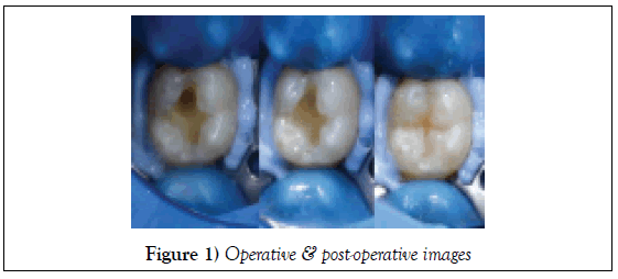
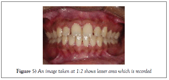
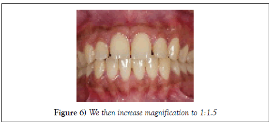
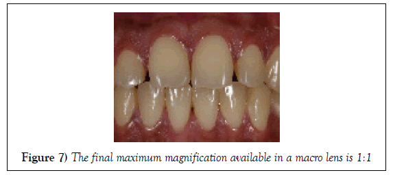
1) It sets a standard distance from which an image has to be made and hence the composition remains standardized. e.g. If pre-operative image was made at a magnification of 1:1.5 then even the operative & post-operative images have to be made at the same magnification. We set the magnification to 1:1.5 on the same 100 mm macro lens and then move physically ahead and backward to focus. When the object appears sharp, image is to be recorded. It is interesting to note that the lens will focus at the same subject – camera distance like that of the pre-operative image at 1:1.5 magnifications (Figures 1-8).
This is the specialty of a macro lens and it cannot be reproduced by other lenses like zoom lenses (18–05 lens, 55–250 lens, 70-300 lens etc. or normal prime lenses like 50 mm, 100 mm, etc.
2) It is a method of communication with our colleagues.
Magnification ratio and its applications
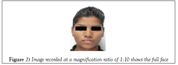
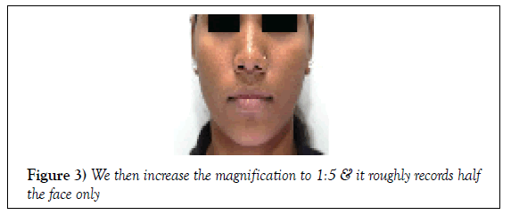
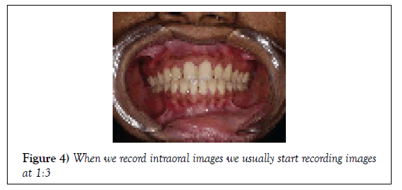
When we increase the magnification we come closer to the subject as follows: (Note that all images are unprocessed or uncropped images & they are not a part of any protocol) Organizations/institutes/journals/companies have their own photography protocols which have fixed number of views that are needed to be documented by their followers. e.g. AACD has 12 view protocols, UCLA has 16 image protocols.
All these images have been allotted a magnification ratio and the dentist is expected to document the image at that particular magnification only.
Important Note
Magnifications of different cameras might look slightly different from one another even when the same lens is used. This depends upon the sensor size of the camera. In a full frame camera like the one used in this article more number of teeth might be visible at a magnification of 1:1 compared to a crop sensor camera in which only 2–3 teeth might be seen. The reader is expected to refer to their camera model catalogue to find out the sensor size of their own camera.


