Human skull with complete metopic suture and multiple sutural bones at lambdoid suture – a case report
Poonam Verma, Sema* and Anupama Mahajan
Department of Anatomy, Sri Guru Ram Das Institute of Medical Sciences and Research, Vallah, Amritsar, India.
- *Corresponding Author:
- Dr. Seema
Professor ,Department of Anatomy, Sri Guru Ram Das Institute of Medical Sciences and Research, Vallah, Amritsar, India.
Tel: +91 991 4754354
E-mail: drseema16@gmail.com
Date of Received: March 2nd, 2012
Date of Accepted: October 21st, 2012
Published Online: May 6th, 2014
© Int J Anat Var (IJAV). 2014; 7: 7–9.
[ft_below_content] =>Keywords
lambdoid suture,skull,variation,wormian bone
Introduction
Frontal bone of the skull develops in two halves during the fetal life separated by the metopic suture. The suture normally disappears soon after the birth [1]. The persistent complete metopic suture extending from the nasion to bregma is called metopism [2]. Lambda represent the meeting point of the sagittal and lambdoid suture. It represents the site of posterior median fontanel in the fetal life. It obliterates by the age of 2 to 3 months. The ‘sutural bones’ or ‘Wormian bones’ are small, irregular bones found at the sutures and fontanels of the skull. Whenever the wormian bones are present they are either two or three but they are present in large numbers in case of hydrocephalic skulls [1]. The mechanism of formation of the wormian bones is not clearly known. Some say they are due to external influences [3]. Other say they are due to normal developmental processes and are genetically determined [4]. They are commonly found in relation to the frontal and occipital bones. It is important to know about these bones because they can mislead in the diagnosis of fracture of skull bones in medicolegal cases.
Case Report
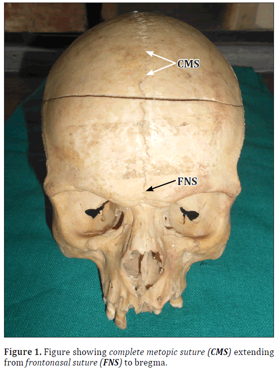
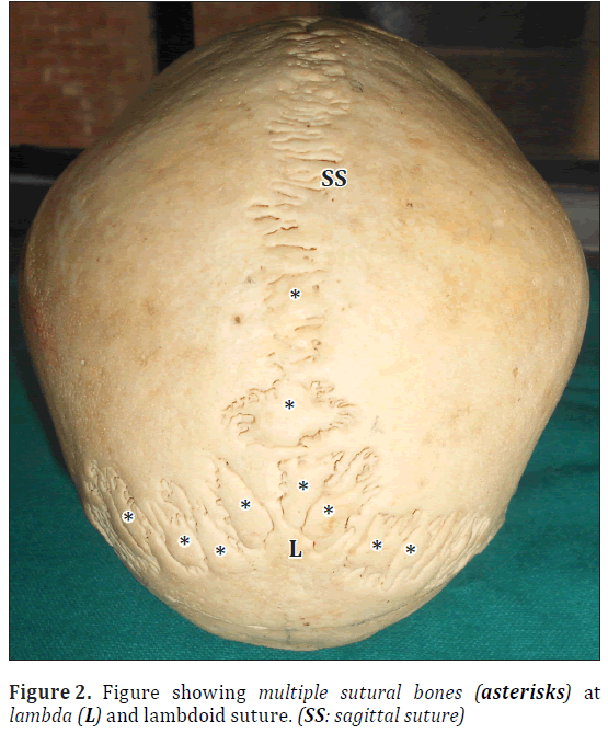
A dry adult human skull with complete metopic suture and a series of sutural bones at the lambda and in the lambdoid suture was found in the Department of Anatomy, Sri Guru Ram Das Institute of Medical Sciences and Research, Amritsar (Figure 1). The frontal bone was doubled because of complete metopic suture. It was a prominent dentate suture extending from the frontonasal suture to the bregma (Figure 2). There were no other notable variations in the skull.
Discussion
The frontal bone is one of the unpaired bones of the skull which ossifies in the fibrous membrane at 8th week of intrauterine life from two primary centers [1]. Metopism is found in 5.1% of Asians and 8.7% in European Caucasians. Sutural bones are very commonly found in the skull. Nearly 40% of skulls contain sutural bones in the vicinity of the lambdoid suture. The next most common sutural bone is the epipteric bone found near the anterolateral fontanel [5]. Presence of sutural bones is almost invariably associated with abnormal development of the central nervous system and may serve as a useful marker for the early identification and treatment of the affected infant or child [6]. Jeanty et al. have reported the presence of wormian bones in four fetuses [7]. But in these cases there were no associated anomalies. Tewari et al. studied 1500 skulls for the presence of sutural bones. They have found the preinterparietal bone in 6 (0.4%) cases [8]. El-Najjar and Dawson are of the opinion that the occurrence of the wormian bones is controlled by the genetic factor [9]. Significant sutural bones as against normal developmental variants were considered to be those more than 10 in number, measuring greater than 6 mm and arranged in a general mosaic pattern. They were found in all the cases of osteogenesis imperfecta but not in the normal skulls [10]. Comparison of cranial capacity in skulls with and without sutural bones showed no significant difference and this is interpreted as indicating that sutural bones are not formed secondary to stress [11]. It has no morphological importance but it certainly has a morphogenetic bearing [12]. The association of the persistent metopic suture and wormian bone at the bregma was reported by Nayak [13]. Also present specimen showed the complete metopic suture with multiple wormian bones at lambda and along the lambdoid suture.
Conclusion
The sutural bones are important from clinical point of view. The presence of series of sutural bones like this may lead to problems in posterior approach to the cranial cavity. These bones might lead to confusions in reading the radiographs in the case of head injuries. The sutural bones may be mistaken for multiple fractures.
References
- Williams PL, Bannister LH, Berry MM, Collins P, Dyson M, Dussek JE, Ferguson MWJ, eds. Gray’s Anatomy. 38th Ed., Baltimore, Churchil and Livingstone. 1995; 595.
- Bryce TH, ed. Quain’s Elements of Anatomy. Vol. 4., 11th Ed., London, Longmans Green. 1915; 177.
- Finkel DJ. Wormian bones – a study of environmental stress. Am J Physical Anthropol. 1971; 35: 278.
- Pal GP, Bhagwat SS, Routal RV. A study of sutural bones in Gujarati (Indian) crania. Anthropol Anz. 1986; 44: 67–76.
- Bergman RA, Afifi AK, Miyauchi R. Compendium of Human Anatomical Variation. Baltimore, Urban and Schwarzenberg. 1988: 197–205.
- Pryles CV, Khan AJ. Wormian bones. A marker of CNS abnormality? Am J Dis Child. 1979; 133: 380–382.
- Jeanty P, Silva SR, Turner C. Prenatal diagnosis of wormian bones. J Ultrasound Med. 2000; 19: 863–869.
- Tewari PS, Malhotra VK, Agarwal SK, Tewari SP. Preinterparietal bone in man. Anat Anz. 1982; 152: 337–339.
- El-Najjar M, Dawson GL. The effect of artificial cranial deformation on the incidence of Wormian bones in the lambdoidal suture. Am J Phys Anthropol. 1977; 46: 155–160.
- Cremin B, Goodman H, Spranger J and Beighton P. Wormian bones in osteogenesis imperfecta and other disorders. Skeletal Radiol. 1982; 8: 35–38.
- Malhotra VK, Tewari PS, Pandey SN, Tewari SP. Interparietal bone. Acta Anat (Basel).1978; 101: 94–96.
- Astley R. Metaphyseal fractures in osteogenesis imperfecta. Br J Radiol. 1979; 52: 441–443.
- Nayak S. Presence of Wormian bone at bregma and paired frontal bone in an Indian skull. Neuroanatomy. 2006; 5: 42–43.
Poonam Verma, Sema* and Anupama Mahajan
Department of Anatomy, Sri Guru Ram Das Institute of Medical Sciences and Research, Vallah, Amritsar, India.
- *Corresponding Author:
- Dr. Seema
Professor ,Department of Anatomy, Sri Guru Ram Das Institute of Medical Sciences and Research, Vallah, Amritsar, India.
Tel: +91 991 4754354
E-mail: drseema16@gmail.com
Date of Received: March 2nd, 2012
Date of Accepted: October 21st, 2012
Published Online: May 6th, 2014
© Int J Anat Var (IJAV). 2014; 7: 7–9.
Abstract
The sutural bones are unnamed bones commonly found at the level of lambda and lambdoid suture in a human skull. They vary from person to person in number and shape. An adult human skull with complete metopic suture lying between two frontal bones was found. The same skull showed a large number of sutural bones at the lambda and along the lambdoid suture. It is important to know about such variation because they can mislead the diagnosis of fracture of skull bones. Knowledge of this variation is very important for forensic experts, anthropologists, radiologists, orthopedists and neurosurgeons.
-Keywords
lambdoid suture,skull,variation,wormian bone
Introduction
Frontal bone of the skull develops in two halves during the fetal life separated by the metopic suture. The suture normally disappears soon after the birth [1]. The persistent complete metopic suture extending from the nasion to bregma is called metopism [2]. Lambda represent the meeting point of the sagittal and lambdoid suture. It represents the site of posterior median fontanel in the fetal life. It obliterates by the age of 2 to 3 months. The ‘sutural bones’ or ‘Wormian bones’ are small, irregular bones found at the sutures and fontanels of the skull. Whenever the wormian bones are present they are either two or three but they are present in large numbers in case of hydrocephalic skulls [1]. The mechanism of formation of the wormian bones is not clearly known. Some say they are due to external influences [3]. Other say they are due to normal developmental processes and are genetically determined [4]. They are commonly found in relation to the frontal and occipital bones. It is important to know about these bones because they can mislead in the diagnosis of fracture of skull bones in medicolegal cases.
Case Report
A dry adult human skull with complete metopic suture and a series of sutural bones at the lambda and in the lambdoid suture was found in the Department of Anatomy, Sri Guru Ram Das Institute of Medical Sciences and Research, Amritsar (Figure 1). The frontal bone was doubled because of complete metopic suture. It was a prominent dentate suture extending from the frontonasal suture to the bregma (Figure 2). There were no other notable variations in the skull.
Discussion
The frontal bone is one of the unpaired bones of the skull which ossifies in the fibrous membrane at 8th week of intrauterine life from two primary centers [1]. Metopism is found in 5.1% of Asians and 8.7% in European Caucasians. Sutural bones are very commonly found in the skull. Nearly 40% of skulls contain sutural bones in the vicinity of the lambdoid suture. The next most common sutural bone is the epipteric bone found near the anterolateral fontanel [5]. Presence of sutural bones is almost invariably associated with abnormal development of the central nervous system and may serve as a useful marker for the early identification and treatment of the affected infant or child [6]. Jeanty et al. have reported the presence of wormian bones in four fetuses [7]. But in these cases there were no associated anomalies. Tewari et al. studied 1500 skulls for the presence of sutural bones. They have found the preinterparietal bone in 6 (0.4%) cases [8]. El-Najjar and Dawson are of the opinion that the occurrence of the wormian bones is controlled by the genetic factor [9]. Significant sutural bones as against normal developmental variants were considered to be those more than 10 in number, measuring greater than 6 mm and arranged in a general mosaic pattern. They were found in all the cases of osteogenesis imperfecta but not in the normal skulls [10]. Comparison of cranial capacity in skulls with and without sutural bones showed no significant difference and this is interpreted as indicating that sutural bones are not formed secondary to stress [11]. It has no morphological importance but it certainly has a morphogenetic bearing [12]. The association of the persistent metopic suture and wormian bone at the bregma was reported by Nayak [13]. Also present specimen showed the complete metopic suture with multiple wormian bones at lambda and along the lambdoid suture.
Conclusion
The sutural bones are important from clinical point of view. The presence of series of sutural bones like this may lead to problems in posterior approach to the cranial cavity. These bones might lead to confusions in reading the radiographs in the case of head injuries. The sutural bones may be mistaken for multiple fractures.
References
- Williams PL, Bannister LH, Berry MM, Collins P, Dyson M, Dussek JE, Ferguson MWJ, eds. Gray’s Anatomy. 38th Ed., Baltimore, Churchil and Livingstone. 1995; 595.
- Bryce TH, ed. Quain’s Elements of Anatomy. Vol. 4., 11th Ed., London, Longmans Green. 1915; 177.
- Finkel DJ. Wormian bones – a study of environmental stress. Am J Physical Anthropol. 1971; 35: 278.
- Pal GP, Bhagwat SS, Routal RV. A study of sutural bones in Gujarati (Indian) crania. Anthropol Anz. 1986; 44: 67–76.
- Bergman RA, Afifi AK, Miyauchi R. Compendium of Human Anatomical Variation. Baltimore, Urban and Schwarzenberg. 1988: 197–205.
- Pryles CV, Khan AJ. Wormian bones. A marker of CNS abnormality? Am J Dis Child. 1979; 133: 380–382.
- Jeanty P, Silva SR, Turner C. Prenatal diagnosis of wormian bones. J Ultrasound Med. 2000; 19: 863–869.
- Tewari PS, Malhotra VK, Agarwal SK, Tewari SP. Preinterparietal bone in man. Anat Anz. 1982; 152: 337–339.
- El-Najjar M, Dawson GL. The effect of artificial cranial deformation on the incidence of Wormian bones in the lambdoidal suture. Am J Phys Anthropol. 1977; 46: 155–160.
- Cremin B, Goodman H, Spranger J and Beighton P. Wormian bones in osteogenesis imperfecta and other disorders. Skeletal Radiol. 1982; 8: 35–38.
- Malhotra VK, Tewari PS, Pandey SN, Tewari SP. Interparietal bone. Acta Anat (Basel).1978; 101: 94–96.
- Astley R. Metaphyseal fractures in osteogenesis imperfecta. Br J Radiol. 1979; 52: 441–443.
- Nayak S. Presence of Wormian bone at bregma and paired frontal bone in an Indian skull. Neuroanatomy. 2006; 5: 42–43.


