Iatrogenic cut of an arterial catheter in the radial artery
2 Internship in Traumatology, CHU IBN SINA, Rabat, Morocco
3 Internship in Urology, CHU IBN SINA, Rabat, Morocco
4 Internship in Gynecology-obstetrics, CHU IBN SINA, Rabat, Morocco
5 Professor of General Surgery, Head of Vascular Surgery Department, CHU IBN SINA, Rabat, Morocco
Received: 23-Nov-2017 Accepted Date: Dec 30, 2017; Published: 06-Feb-2018
Citation: Slaoui A, Grimi T, Slaoui A, et al. Iatrogenic cut of an arterial catheter in the radial artery. Surg Case Rep. 2018;2(1):-4-6.
This open-access article is distributed under the terms of the Creative Commons Attribution Non-Commercial License (CC BY-NC) (http://creativecommons.org/licenses/by-nc/4.0/), which permits reuse, distribution and reproduction of the article, provided that the original work is properly cited and the reuse is restricted to noncommercial purposes. For commercial reuse, contact reprints@pulsus.com
Abstract
An arterial line (also art-line or a-line) is a thin catheter inserted into an artery. It is most commonly used in intensive care medicine and anesthesia to monitor blood pressure directly and in real-time (rather than by intermittent and indirect measurement) and to obtain samples for arterial blood gas analysis. The use of arterial catheterization is justified in anaesthesiology and reanimation because of the essential advantages of being able to monitor blood pressure invasively and to easily perform blood tests. However, this technique has complications, some of which, like ischemia, are serious, although exceptional.
Keywords
Arterial catheterization; Radial artery; Anaesthesiology; Reanimation; Hysterectomy; Arterial cannulation
Introduction
The use of arterial catheterization is justified in anaesthesiology and reanimation because of the essential advantages of being able to monitor blood pressure invasively and to easily perform blood tests. However, this technique has complications, some of which, like ischemia, are serious, although exceptional [1]. Major complications related to arterial cannulation occur in less than 1% of patients, and the rates or probabilities are similar for the radial, femoral and axillary arteries [2]. We report the accidental and iatrogenic cut of the artery cannulation, which required an emergency procedure to avoid further complications.
Case Report
A 26-year-old woman without medical history was admitted to the emergency room of the maternity hospital of Rabat for uterine rupture. She was treated immediately in the operating room and underwent total hysterectomy with right adnexectomy due to the importance of the uterine tear. The monitoring of the blood pressure was allowed thanks to the establishment of an arterial line in the left radial artery. It was secured to the skin with an adhesive film.
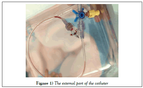
After completion of the operation, she was transferred to the postresuscitation department for close observation. Two hours later, when continuous arterial pressure monitoring was no longer required, the nurse received the instruction to remove the arterial line. While trying to remove it, the patient was scared and retracted her hand sharply. The nurse felt the section of the catheter and removed the dissociated external part (Figure 1). She immediately used gauze to compress the wound and called the doctor. It was found that the removal arterial catheter was shortened at its origin with a length of less than 1 cm still attached to its hub (Figure 1).
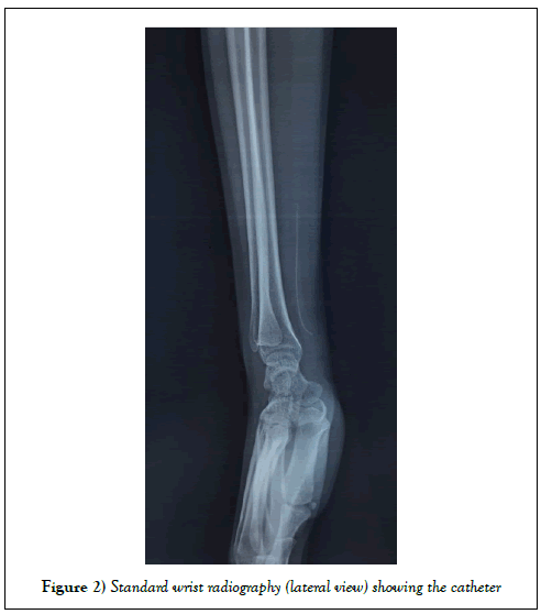
The resuscitator in the intensive care unit confirmed the presence of the intra-arterial catheter at the puncture site by clinical wrist examination, demonstrating a longitudinal seam below the skin. A standard x-ray of the wrist (lateral view) confirmed the presence of the fragment thanks to its radio-opaque character (Figure 2). The right radial pulse was present and symmetric. There was no sign of ischemia or other complication.
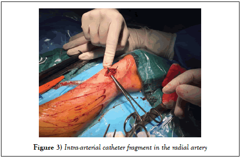
The patient and his family were informed of the incident and the retained fractured fragment in the body. She was reviewed by the vascular surgeon on duty that night and was advised to undergo immediate surgical exploration under local anaesthesia, to which she consented. It was found intraoperatively that the retained catheter fragment was still attached at its point of puncture in the radial artery (Figure 3).
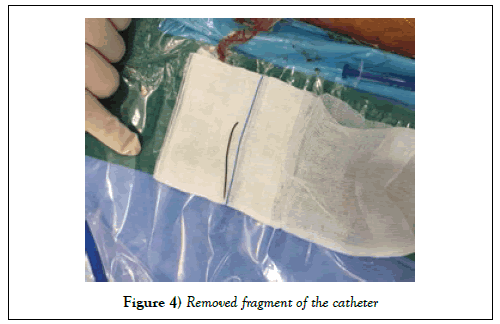
Its length having initially allowed its fixation to the vascular wall, then allowed us to simply withdraw the catheter by its external part (Figure 4). This was followed by a 2 minutes mechanical haemostasis and the wound was closed in layers. She was followed-up 4 weeks later, which showed that the wound had healed well. There were no residual sequelae.
Discussion
Arterial catheterisation is a common procedure in the care and management of critically ill patients and most commonly placed in the radial and femoral arteries [3]. The complications of arterial cannulation have been comprehensively reviewed [4,5]. The cut of arterial catheter has previously been reported, but rarely. Similar cases were reported by Shah et al. [6] in 1996, Mayne and Kharwar [7] in 1997, Ho et al. [8] in 2003, Ferguson et al. [9] in 2005, Chiao-Fen et al. [10] in 2010 and Suresh Babu Kale et al. [11] in 2017.
The management of this complication begins with the localization of the intra-corporal catheter fragment. Shah et al. [6], Ho et al. [8] and Suresh Babu Kale et al. [11] undertook surgical exploration without imaging proof. Mayne and Kharwar [7] and Ferguson et al. [9] used standard x-rays of the wrist to show the presence of a broken catheter in the radial artery. Chiao- Fen et al. [10] used an emergency computed tomography to reveal a 4.5 cm radio-opaque fractured fragment of the catheter anchored in the volar aspect of the distal radius. Ball et al. [12] suggested that real-time ultrasound imaging might be helpful by allowing rapid localization of the intraluminal arterial foreign body before surgical removal. Moody et al. [13] used ultrasound to guide localization and removal of a retained arterial cannula fragment. Application of ultrasound seems advantageous because of its rapidity, its cost and it does not expose the patient to radiation. But several factors led us to choose the standard x-ray of the wrist rather than ultrasound: first, the difficulty of getting an ultrasound machine in the middle of the night in our hospital and secondly the chance of using a radio-opaque catheter even though it exists catheters that are not in the same department. Its location was confirmed in the radiography with a radio-opaque linear material in front of the external anterior face of the inferior extremity of the radius. This observation combined with the clinical examination was sufficient to confirm the presence of the catheter and allowed for extremely rapid management. Chiao-Fen et al. [10] suggested that the three-dimensional CT was a useful tool to locate more precisely the retained arterial cannula fragment. Takada et al. [14] recommended the use of the three-dimensional CT to recognize an intravascular foreign body. In our situation, this examination was unnecessary and on the contrary we would have lost time, without even considering the long administrative process and the higher cost. We suggest that in the absence of an ultrasound machine at hand and if the material is radiopaque, standard radiography is the fastest and most useful way for emergency management.
The loss of a fragment of the catheter inside the artery could have had very serious consequences. To begin with distal thrombosis and embolization resulting in digital ischemia and subsequent infection might occur if surgical exploration is not performed in time as described by Bentz et al. [15]. Egwu et al. [16] reported the case of a fragment of an intra-arterial catheter that was retained in the patient’s radial artery for 4 weeks without being discovered and led to a pain in the wrist. These complications make us investigate how such accidents happen to prevent them in the future.
We can review the literature to find how these accidents happen. In Shah’s et al. [6] case, the arterial catheter was damaged by the surgical needle while sewing the catheter to the skin. In Mayne and Kharwar’s [7] case the cause was unknown but the breakage was noticed in the intensive care unit. In Ho’s et al. [8] case the cause was also unknown but the arterial line was noted to be shortened after they removed it. In Chiao-Fen et al. [10] case, the arterial catheter is suspected to have been severed accidentally by the scissors while trying to cut the tape to separate the intravenous catheter and the arterial catheter. Fergusson et al. [9] reported a similar complication of transection of an arterial cannula that resulted from cutting the securing stitch with a stitch cutter. We suggest that the use of scissors or other sharp objects for the removal of any catheter should always be extremely meticulous or even avoided. Furthermore, it is suggested that each catheter should be taped separately to the skin to avoid any difficulty when removing it. In our case, the rapid movement of the patient’s hand as the nurse removed the tape covering the entry point of the catheter caused its breakage. While the inner part remained in the radial artery, the outer part was detached from the hand while remaining glued to the plaster. The arterial entry point of the catheter could have increased, leading to significant haemorrhage. The weakness of the equipment causing its dislocation was its strength and avoided a more serious complication. This resulted in a slight bleeding quickly taken care of by the medical team. We suggest always taking your time with patients, especially in intensive care when removing sensitive material. Working with a previously reassured and enlightened patient would surely have prevented this complication. It is also extremely important to always check the integrity of the equipment once removed. It probably could have avoided the case described by the Egwu et al. [16].
Conclusion
It is a rare complication but can be very serious with risk of temporary or permanent occlusion of the radial artery, pseudoaneurysm, sepsis and paralysis of the median nerve. Simple and reproducible catheterization and removal techniques must be instituted to avoid its occurrence as much as possible, especially since it is almost always iatrogenic.
Conflict of Interest
The author(s) declare(s) that there is no conflict of interest regarding the publication of this article.
Patient Consent
The patient gave her full permission for the publication, reproduction, broadcast and other use of photographs and textual material (case histories) in all editions of the Journal of Vascular Surgery Publications and in any other publication (including books, journals, CD-ROMs, online and internet), as well as in any advertising or promotional material for such product or publications.
REFERENCES
- Tegtmeyer K, Brady G, Lai S, et al. Placement of an arterial line. N Engl J Med. 2006;354:e13.
- Scheer B, Perel A, Pfeiffer UJ. Clinical review: Complications and risk factors of peripheral arterial catheters used for haemodynamic monitoring in anaesthesia and intensive care medicine. Crit Care 2002;6:199−204.
- Wu CY, Fu JY, Feng PH, et al. Catheter fracture of intravenous ports and its management. World J Surg. 2011;35:2403–410.
- Cousins TR, O’Donnell JM. Arterial cannulation: A critical review. AANA J. 2004;72:267−271.
- Durie M, Beckmann U, Gillies DM. Incidents relating to arterial cannulation as identified in 7,525 reports submitted to the Australian Incident Monitoring Study (AIMS-ICU). Anaesth Intensive Care. 2002;30:60−65.
- Shah US, Downing R, Davis I. An iatrogenic arterial foreign body. Br J Anaesth. 1996;77:430−431.
- Mayne D, Kharwar F. Another complication of radial artery cannulation. Anaesthesia 1997;52:89−90.
- Ho KS, Chia KH, Teh LY. An unusual complication of radial artery cannulation and its management: A case report. J Oral Maxillofac Surg. 2003;61:955−957.
- Ferguson E, Baumgartner P, Hamilton M. Accidental transection of radial (correction of radical) artery cannula. Anaesth Intensive Care. 2005;33:142−143.
- Luo CF, Mao C, Su B, et al. An iatrogenic complication of radial artery cannulation. Acta Anaesthesiol Taiwan. 2010;48(3):145−147.
- Kale SB, Ramaligam S. Spontaneous arterial catheter fracture and embolisation: Unpredicted complication. Indian J Anaesth. 2017;61(6):505–507.
- Ball DR, Stott SA, Rouse R. Locating an arterial foreign body by ultrasonography. Br J Anaesth. 1996;77:811.
- Moody C, Bhimarasetty C, Deshmukh S. Ultrasound guided location and removal of retained arterial cannula fragment. Anaesthesia. 2009;64:338−339.
- Takada M, Kashiwagi R, Sakane M, et al. 3D-CT diagnosis for ingested foreign bodies. Am J Emerg Med. 2000;18:192−193.
- Bentz ML, Jones NF. Foreign body arterial embolus to the hand. J Hand Surg. 1992;17:484−486.
- Egwu VN, Sadr B. Radial arterial foreign body as a cause of wrist pain. Anesth Analg. 1987;66:1058−1059.


