Incidence of wormian bones in the dried skull of Nigerian males
Received: 14-Feb-2018 Accepted Date: Feb 27, 2018; Published: 07-Mar-2018, DOI: 10.37532/1308-4038.18.11.32
Citation: Uchewa OO, Egwu OA, Egwu AJ, et al. Incidence of wormian bones in the dried skull of Nigerian males. Int J Anat Var. 2018;11(1):032-034.
This open-access article is distributed under the terms of the Creative Commons Attribution Non-Commercial License (CC BY-NC) (http://creativecommons.org/licenses/by-nc/4.0/), which permits reuse, distribution and reproduction of the article, provided that the original work is properly cited and the reuse is restricted to noncommercial purposes. For commercial reuse, contact reprints@pulsus.com
Summary
Sutural (Wormian) bones are small bones that appear between or close to the cranial sutures. Various authors expressed their opinions about formation of these bones but most of them believe they are formed due to additional ossification centers in or close to the cranial sutures. They are considered to be a supplementary point of ossification without any vital role in supporting the cranium. In this present study we examined 22 skulls prepared from dissected male cadavers over a period of 5 years at the Department of Anatomy, Faculty of Basic Medical Sciences, Federal University Ndufu-Alike, Ikwo (FUNAI). There were 45.46% incidences of wormian bones as discovered in this study while 54.55% of the skulls had no sutural (wormian) bones present in them. 36.36% eight skulls out of the 22 skulls have wormian bones at the lambdoid suture. The incidence of epipteric bones at pterion was 9.09% as observed. We did not record any case of wormian bone in the coronal sutures, none in the squamosal sutures and also none in the sagital sutures of the skulls. There have been very few studies reporting the occurrence of sutural bones in the coronal, sagittal, and squamosal suture. Sutural (wormian) bones are seen in the study accounting for about 45.46% of the dissected cadavers.
Keywords
Skull; Wormian bones; Dissection; Coronal; Lambda; Incidence
Introduction
The skull is the bone that envelopes the brain. It is composed of 28 separate bones and most of them are paired. These bones provide attachment for the numerous muscles of facial expression. Sometimes, there usually appear small irregular ossicular bones located at the cranial sutures in the skull.
According to Standring et al., [1] sutural (Wormian) bones develop as a result of the appearance of an additional ossification center which occurs in or close to the sutures. In 1643, sutural bones were named Wormian bones after a Danish anatomist and a medical doctor at the University of Copenhagen, Olaus Wormius who made the first detailed description of the presence of the bone in the skull [2,3]. Some other authors reported that these bones may be as a result of hydrocephalus [4-6]. It is believed that what affects the prevalence and mechanisms of wormian bones formation in the skull are still unknown [2]. The wormian (sutural) bones wherever present may be misleading in the results of X-ray diagnosis and most often mistaken for fractures [2]. According to Burgener and Kormano 1997 [7] and Shantharam et al., [8] they reported that Wormian bones are also found in healthy individuals, however a higher incidence of multiple wormian bones have been recorded in a variety of congenital disorders like osteogenesis imperfecta, cretinism (hypothyroidism), cleidocranial dysostosis, progeria, hypophosphatasia, rickets etc. The morphological knowledge of wormian bones is very vital in the diagnosis of these disorders [9,10].
Based on the report of Gumusburun et al. [11], the incidence of sutural bones are well suited for comparative studies as an anthropological marker or an indicator of population distance. The knowledge of the presence of these small bones is very important to human anatomists, physical anthropologists, forensic medical imaging personnel [12,13]. These bones are studied and reported as ethnic variables in the population because their presence is very important. They have been reports of very high frequencies of wormian bones in some population and their absence in others which are geographically close or are subject to the same environmental stresses [5,14] suggesting a genetic mechanism of formation. Khan et al. [15], reported incidence is variable, ranging from around 10% (Caucasian), 40% (Indians), to 80% (Chinese). Gailland (2008) [16] also reported that Wormian bones are more prevalent in the male folks than the females. These bones are most common in the lambdoid suture followed by the epipteric centre (pterion ossicles) close to the site of antero-lateral fontanelle [15,17]. These bones can also be seen in other sutures such as the coronal, sagital and squamosal sutures but their presence are very rare. The present study is aimed at reporting the incidence of Wormian bones in the skull of 22 cadavers dissected in anatomy department, Faculty of Basic Medical Sciences Federal University Ndufu-Alike, Ikwo (FUNAI).
Materials and Methods
The study was carried out on the dried adult human skulls of 22 cadavers dissected in the department of human anatomy, Faculty of Basic Medical Sciences Federal University Ndufu-Alike, Ikwo (FUNAI), Ebonyi State, over a period of five (5) years. All the specimens (skulls) used were the male bodies dissected and the bones prepared and kept in the museum of the above department of the same University. The skulls were examined; the incidence recorded and the photograph of the presence of sutural (Wormian) bones taken before returning them to the same museum of the department.
Results
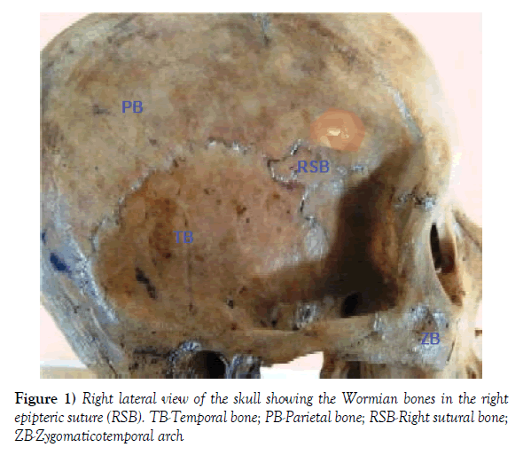
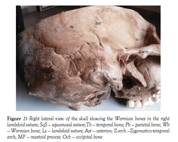
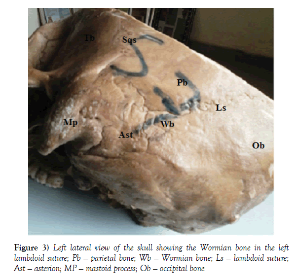
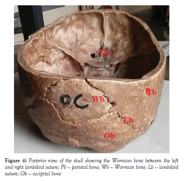
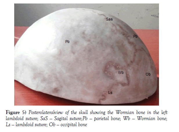
In this present study which we examined 22 human skulls dissected and prepared in the Department of Human Anatomy, Faculty of Basic Medical Sciences Federal University Ndufu-Alike, Ikwo (FUNAI), the Wormian bones were observed in 10 skulls of the examined skulls which represent 45.46%. The Wormian bones were not seen in the remaining 12 skull representing 54.55% of the population. We observed that 9.09% [2] skulls of the sutural bones are located in the epipteric suture, 36.36% [8] skull presented Lambdoid sutural bones. There were no Wormian (sutural) bones at the site of the coronal suture, bregma, and sagital suture in all the examined skulls. The result is presented in a summarized format in the table below (Figures 1-5).
Figure 1) Right lateral view of the skull showing the Wormian bones in the right epipteric suture (RSB). TB-Temporal bone; PB-Parietal bone; RSB-Right sutural bone; ZB-Zygomaticotemporal arch
Discussion
Sutural (Wormian) bones articulate with the surrounding bones by sutures and their dentations are more complex on the outside than on the internal surface of the skull. They are considered to be a supplementary points of ossification and do not have any vital role in the mechanism of supporting the cranium [10]. There can be another bone called the pre-interparietal bone or inca bone at the lambda [15]. These bones are of different shapes and sizes such as oval, oblong, round, quadrilateral, polygonal and triangular which can range from below 1 mm in diameter to as big as 5 x 9 cm [2].
There were 45.46% of Wormian bones as discovered in this study while 54.55% of the skulls had no sutural bones present in them. Saxena et al. [18] reported that 11.79% of Indian skulls and 5.06% Nigerians skulls had epipteric bone. It is still unclear why sutural bones are common in certain races.
In the present study, 36.36% [8] skulls out of the 22 skulls of cadavers dissected over a period of 5 years in FUNAI have Wormian bones at the lambdoid suture. This is in agreement with the study carried out by Bergman et al., [19] when they reported that 40% of skulls have sutural bones in the lambdoid suture. The above statements are based on the number of skulls observed in any study. The reason for the occurrence of the Wormian bones may be regulated by a genetic factor [20]. According to Nair et al., the gross incidence of epipteric or Wormian bones at pterion was 6% which is closely related to the 9.09% as observed in our present study. There can be another bone called the pre-interparietal bone or inca bone at the lambda [21]. Gaillard (2008) and Tripathi (2011) [16,22], reported that presence of wormian bones were associated with rickets, hypothyroidism, down syndrome, osteogenesis imperfecta, pycnodysostosis and cleidocranial dysplasia. Jeanty et al. [23] reported the presence of wormian bones in four foetuses, but none of them were associated with any anomalies. In the present study, out of 22 skulls observed, we found no case of wormian bone in the coronal sutures, none in the squamosal sutures, and also none in the sagital sutures of the skulls. This agrees with the study of Tewari et al. [24] when they studied 1500 skulls for the presence of sutural bones, but they failed to find a single case of Wormian bones in the coronal, squamosal, and sagital sutures.
Our findings disagrees with the study of Khan et al. [15] in which they reported that out of 25 skulls, 2 cases of sutural bones were in the coronal sutures, 2 in the squamosal sutures and 2 in the sagital sutures of the skulls and another study carried out by Berry and Berry 1967 [25,26] in their study on epigenetic variations in the human cranium, reported the presence of Wormian bones in the coronal and squamosal sutures. There have been very few studies reporting the occurrence of sutural bones in the coronal, sagittal and squamosal suture.
Conclusion
Wormian bones are actually more prevalent in the human skull than previously reported and surgeons or radiographers should be on the alert not to be confused during surgery or imaging of the skull.
REFERENCES
- Standring S. Skull and Mandible. In: Gray's Anatomy: Anatomical Basis of Clinical Practice Standring S(Ed). 2008.
- Reveron RR. Anatomical Classification of Sutural Bones. MOJ Anat &Physiol. 2017;3:101.
- Mohammed Ahad, Thenmozhi MS. Study on Asterion and Presence of Sutural Bones in South Indian Dry Skull. J Pharm Sci & Res. 2015;7:390-2.
- Hess L. Ossicula wormiana. Human Biol. 1946;18:61-80.
- Finkel DJ. Wormian bone formation in the skeletal population from Lachish. J Hum Evolut. 1976;5:291-5.
- Singh R. Incidence of sutural bones at asterion in adults Indian skulls. Int J Morphol. 2012;30:1182-6.
- Burgener FA, Kormano M. Bone and joint disorders, conventional radiologic differential diagnosis. New York: Thieme medical publishers. 1997;pp.130.
- Shantharam V, Manjunath KY. Occurrence of Sutural Bones in Adult Human Skulls. Nat J Basic Med Sci. 2017;7.
- Cremin B, Goodman H, Spranger J, et al. Wormian bones in osteogenesis imperfecta and other disorders. Skeletal Radiology. 1982;8:35-8.
- Murlimanju BV, Prabhu LV, Ashraf CM, et al. Morphological and topographical study of Wormian bones in cadaver dry skulls. J Morphol Sci. 2011;28:176-9.
- Gumusburun E, Sevim A, Katkici U, et al. A study of sutural bones in Anatolian-Ottoman skulls. Int J Anthropol. 1997;12:43-8.
- Hernandez FG, Echeverria GM. Frequency of skull bones with lambdoid in artificial deformation in Northern Chile. Int J Morphol. 2009;27:933-8.
- Hussain Saheb S, Haseena S, Prassana LC. Unusualwormian bones at Pterion-Three Case Reports. J Biomed Sci and Res. 2010;2:116-8.
- Satheesha Nayak B, Soumya KV. Unusual sutural bones at Pterion. Int J Anat var. 2008;1:19-20.
- Khan AA, Asari MA, Hassian A. Unusual presence of Wormian (sutural) bones in human skulls. Folia Morphol. 2011;70:4.
- Gaillard F. Case no. 5280. 2008.
- Raja SK, Siva NR, Sundara S. Incidence of sutural bones at pterion in south Indian dried skulls. Int J Anat Res. 2016;4:2099-101.
- Saxena SK, Jain SP, Chowdhary DS. A comparative study of pterion formation and its variations in the skulls of Nigerians and Indians. Anthropologischer Anzeiger. 1988;46:75-82.
- Bergman RA, Afifi AK, Miyauchi R. Skeletal systems: Cranium. In: Compendium of human anatomical variations. Urban and Schwarzenberg, Baltimore. 1988;pp. 197-205.
- El-Najjar M, Dawson GL. The effect of artificial cranial deformation on the incidence of Wormian bones in the lambdoidal suture. Am J Phys Anthropol. 1977;46:155-160.
- Berry AC. Factors affecting the incidence of non-metrical skeletal variants. J Anat. 1975;120:519-535.
- Tripathi A. Case no. 13352. 2011.
- Jeanty P, Silva SR, Turner C. Prenatal diagnosis of Wormian bones. J Ultrasound Med. 2000;19:863-9.
- Tewari PS, Malhotra VK, Agarwal SK, et al. Preinterparietal bone in man. Anat Anz. 1982;152:337-9.
- Berry CA, Berry RJ. Epigenetic variation in the human cranium. J Anat. 1967;101:361-79.
- Barberini F, Bruner E, Cartolari R, et al. An unusually-wide human bregmatic Wormian bone: anatomy, tomographic description and possible significance. Surgical and Radiologic Anatomy. 2008;30:683-7.


