Incomplete superficial palmar arch55
Mookambica RV1, Velayudham Nair1, Rema Nair1, Somayaji SN2, Narendra Pamidi2* and Venkata R Vollala2
1Sri Mookambica Institute of Medical Sciences, Kulasekharam, Kanyakumari, Tamilnadu, India
2Melaka Manipal Medical College (ICHS), Manipal, Karnataka, India
- *Corresponding Author:
- Narendra Pamidi
Senior Grade Lecturer, Department of Anatomy, Melaka Manipal Medical College (Manipal Campus), International Centre for Health Sciences Manipal, 576104, Karnataka, India
Tel: +91 (820) 2922642
E-mail: pommidi_narendra@yahoo.co.in
Date of Received: September 22nd, 2009
Date of Accepted: March 15th, 2010
Published Online: April 27th, 2010
© Int J Anat Var (IJAV). 2010; 3: 65–66.
[ft_below_content] =>Keywords
superficial palmar arch, ulnar artery, collateral circulation, hand surgery
Introduction
Superficial palmar arch (SPA) is an arterial arcade and a dominant vascular structure of the palm. It is localized just deep to palmar aponeurosis and is superficial to digital branches of the median nerve, long flexor tendons of the forearm and lumbricals of the palm [1]. The arch is formed by superficial terminal branch of ulnar artery and can be completed on lateral side by superficial palmar branch of radial artery or arteria princeps pollicis (APP) or arteria radialis indicis (ARI) or median artery which accompanies the median nerve [2]. From the convexity of SPA three common palmar digital arteries will arise and each one divides into two proper palmar digital arteries. These run along the contiguous sides of all four medial fingers (Except the radial side of the index and ulnar side of the little fingers) to supply them. The palmar digital artery for the medial side of the little finger leaves the arch under palmaris brevis. The radial side of the index finger is supplied by ARI and the thumb is supplied by APP both of these are branches of radial artery [3]. The anastomoses between radial and ulnar arteries through superficial and deep palmar arches in the palm play a significant role through collateral circulation in the diseases of the palm. Knowledge of the frequency of anatomical variations of the arterial pattern of the hand is crucial for safe and successful hand surgeries [4].
Case Report
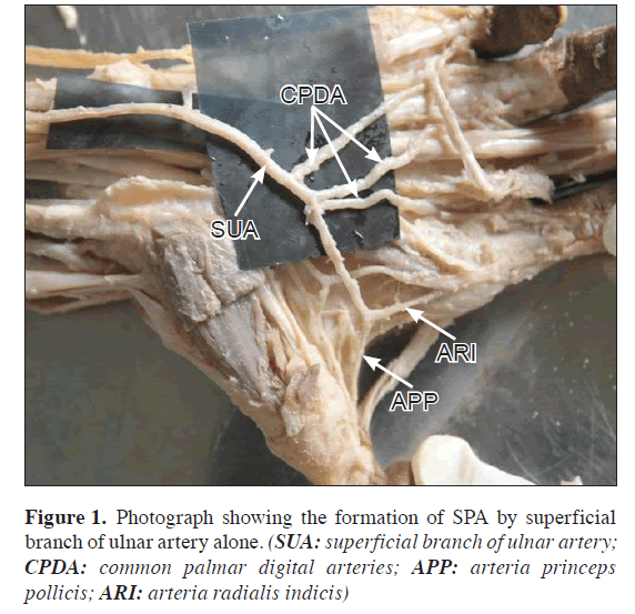
The present variation was observed during the routine dissection classes for the medical undergraduates in a arch55-year-old male cadaver. In the present case superficial palmar arch (SPA) was formed exclusively by superficial branch of the ulnar artery, without the contribution of any other vessel. Ulnar artery entered the palm by coursing in front of flexor retinaculum, just distal to the retinaculum it gave a deep branch and continued as SPA. But it was an incomplete arch, occupying almost normal position but it supplied palmar aspect of all the fingers. It gave a digital branch to the ulnar side of little finger, three common palmar digital branches to supply adjacent sides of medial four fingers, and then continued as the first common palmar digital artery to the interdigital cleft between index finger and the thumb, and this digital artery was dividing into APP and ARI (Figure 1). The superficial branch of radial artery was small and terminated by nourishing the thenar muscles. The only major communication between radial artery and deep branch of ulnar artery was completion of deep palmar arch which is the major route for collateral circulation.
Discussion
Gellman et al. classified the SPA into two categories as complete and incomplete. In complete arch there will be an anastomosis between vessels constituting it. There will be an absence of a communication or anastomosis between the vessels constituting it in incomplete arch. This classification is simple and understandable for many anatomists and researchers and is currently in use [5]. Ikeda et al. reported 3.6% incomplete forms in their observations [6], Coleman and Anson found 21.5% incomplete form out of 650 cases [7]. In a 500 hands study by Janevski et al., the complete arches were seen in 75% and incomplete arches in 25% of subjects [8]. SPA alone formed by ulnar artery (ulnar dominance) was reported by Coleman and Anson as 37% [7], Jelicic et al. as 10% [4] and by Ikeda et al. as 25.5% [6]. Tagil et al. noticed that the most consistent incomplete form was the ulnar artery alone forming SPA which was seen in 20% of subjects [9].
Erbil et al. described the SPA providing blood supply to the thumb and index fingers through the APP and ARI arteries in five cases in their study [10]. Gajisin and Zbrodowski did not refer to many branches from the SPA supplying the first web space out of 200 specimens study. They did not mention the nomenclature of APP and ARI to the arteries supplying thumb and index fingers, if they were not from the deep palmar arch [1]. There is a report of superficial palmar branch of the radial artery terminating in the thenar muscles without any contribution to the SPA [11]. Turk and Metcalf found that in addition to the common palmar digital arteries to the II., III. and IV. interdigital spaces, they found a branch from the SPA supplying the ulnar side of the thumb and the radial side of the index finger and they named it as the first common metacarpal artery [12]. The nomenclatures of the arteries originating from SPA supplying the thumb and index fingers have to be discussed because of their surgical importance.
In hand surgeries like vascular graft applications arterial repairs, free and/or pedicled flaps clinicians should be aware of these variations, because in most of the traumatic events and the surgical procedures of the hand the SPA plays an important role [9]. Techniques like Doppler ultrasound, modified Allen test, pulse oximetry and arterial angiography or a combination of the standard Allen test and ultrasonography can be used to identify the vascular pattern of the palm [13]. In the present case the only major communication between radial artery and deep branch of ulnar artery was completion of the deep palmar arch. In cases of ulnar skin flaps damage to ulnar artery may present a risk. Interference with an efficient blood supply may results in inefficient utility of the movements of fingers and the hand.
References
- Gajisin S, Zbrodowski A. Local vascular contribution of the superficial palmar arch. Acta. Anat (Basel). 1993; 147: 248–251.
- Datta AK. Essentials of human anatomy. Superior and inferior extremities. 2nd Ed., Calcutta, Current Books International. 2000; 99–100.
- Standring S, ed. Gray’s Anatomy: The anatomical basis of clinical practice. 39th Ed., Elsevier, Churchill Livingstone. 2005; 929–930.
- Jelicic N, Gajisin S, Zbrodowski A. Arcus palmaris superficialis. Acta Anat (Basel). 1988; 132: 187–190.
- Gellman H, Botte MJ, Shankwiler J, Gelberman RH. Arterial patterns of the deep and superficial palmar arches. Clin Orthop Relat Res. 2001; 383: 41–46.
- Ikeda A, Ugawa A, Kazihara Y, Hamada N. Arterial patterns in the hand based on a three-dimensional analysis of 220 cadaver hands. J Hand Surg Am. 1988; 13: 501–509.
- Coleman SS, Anson BJ. Arterial patterns in the hand based upon the study of 650 specimens. Surg Gynecol Obstet. 1961; 113: 409–424.
- Janevski BK. Angiography of the upper extremity. The Hague, Martinus Nijhoff. 1982; 73–122.
- Tagil SM, Cicekcibasi AE, Ogun TC, Buyukmumcu M, Salbacak A. Variations and clinical importance of the superficial palmar arch. S.D.U. Tip Fakultesi Dergisi. 2007; 14: 11–16.
- Erbil M, Aktekin M, Denk CC, Onderoglu S, Surucu HS. Arteries of the thumb originating from the superficial palmar arch: five cases. Surg Radiol Anat. 1999; 21: 217–220.
- Bataineh ZM, Moqattash ST. A complex variation in the superficial palmar arch. Folia Morphol (Warsz). 2006; 65: 406–409.
- Al-Turk M, Metcalf WK. A study of superficial palmar arteries using the Doppler Ultrasonic Flowmeter. J Anat. 1984; 138: 27–32.
- Pola P, Serricchio M, Flore R, Manasse E, Favuzzi A, Possati GF. Safe removal of the radial artery for myocardial revascularization: a Doppler study to prevent ischemic complications to the hand. J Thorac Cardiovasc Surg. 1996; 112: 737–744.
Mookambica RV1, Velayudham Nair1, Rema Nair1, Somayaji SN2, Narendra Pamidi2* and Venkata R Vollala2
1Sri Mookambica Institute of Medical Sciences, Kulasekharam, Kanyakumari, Tamilnadu, India
2Melaka Manipal Medical College (ICHS), Manipal, Karnataka, India
- *Corresponding Author:
- Narendra Pamidi
Senior Grade Lecturer, Department of Anatomy, Melaka Manipal Medical College (Manipal Campus), International Centre for Health Sciences Manipal, 576104, Karnataka, India
Tel: +91 (820) 2922642
E-mail: pommidi_narendra@yahoo.co.in
Date of Received: September 22nd, 2009
Date of Accepted: March 15th, 2010
Published Online: April 27th, 2010
© Int J Anat Var (IJAV). 2010; 3: 65–66.
Abstract
the anastomoses between the superficial branch of the ulnar artery and superficial palmar branch of the radial artery. Generally arteria radialis indicis and arteria princeps pollicis are the branches of deep palmar arch. The authors report a variation of superficial palmar arch alone formed by superficial branch of ulnar artery and giving origin to arteria radialis indicis and arteria princeps pollicis. In hand surgeries like vascular graft applications arterial repairs, free and/or pedicled flaps, clinicians should be aware of these variations.
-Keywords
superficial palmar arch, ulnar artery, collateral circulation, hand surgery
Introduction
Superficial palmar arch (SPA) is an arterial arcade and a dominant vascular structure of the palm. It is localized just deep to palmar aponeurosis and is superficial to digital branches of the median nerve, long flexor tendons of the forearm and lumbricals of the palm [1]. The arch is formed by superficial terminal branch of ulnar artery and can be completed on lateral side by superficial palmar branch of radial artery or arteria princeps pollicis (APP) or arteria radialis indicis (ARI) or median artery which accompanies the median nerve [2]. From the convexity of SPA three common palmar digital arteries will arise and each one divides into two proper palmar digital arteries. These run along the contiguous sides of all four medial fingers (Except the radial side of the index and ulnar side of the little fingers) to supply them. The palmar digital artery for the medial side of the little finger leaves the arch under palmaris brevis. The radial side of the index finger is supplied by ARI and the thumb is supplied by APP both of these are branches of radial artery [3]. The anastomoses between radial and ulnar arteries through superficial and deep palmar arches in the palm play a significant role through collateral circulation in the diseases of the palm. Knowledge of the frequency of anatomical variations of the arterial pattern of the hand is crucial for safe and successful hand surgeries [4].
Case Report
The present variation was observed during the routine dissection classes for the medical undergraduates in a arch55-year-old male cadaver. In the present case superficial palmar arch (SPA) was formed exclusively by superficial branch of the ulnar artery, without the contribution of any other vessel. Ulnar artery entered the palm by coursing in front of flexor retinaculum, just distal to the retinaculum it gave a deep branch and continued as SPA. But it was an incomplete arch, occupying almost normal position but it supplied palmar aspect of all the fingers. It gave a digital branch to the ulnar side of little finger, three common palmar digital branches to supply adjacent sides of medial four fingers, and then continued as the first common palmar digital artery to the interdigital cleft between index finger and the thumb, and this digital artery was dividing into APP and ARI (Figure 1). The superficial branch of radial artery was small and terminated by nourishing the thenar muscles. The only major communication between radial artery and deep branch of ulnar artery was completion of deep palmar arch which is the major route for collateral circulation.
Discussion
Gellman et al. classified the SPA into two categories as complete and incomplete. In complete arch there will be an anastomosis between vessels constituting it. There will be an absence of a communication or anastomosis between the vessels constituting it in incomplete arch. This classification is simple and understandable for many anatomists and researchers and is currently in use [5]. Ikeda et al. reported 3.6% incomplete forms in their observations [6], Coleman and Anson found 21.5% incomplete form out of 650 cases [7]. In a 500 hands study by Janevski et al., the complete arches were seen in 75% and incomplete arches in 25% of subjects [8]. SPA alone formed by ulnar artery (ulnar dominance) was reported by Coleman and Anson as 37% [7], Jelicic et al. as 10% [4] and by Ikeda et al. as 25.5% [6]. Tagil et al. noticed that the most consistent incomplete form was the ulnar artery alone forming SPA which was seen in 20% of subjects [9].
Erbil et al. described the SPA providing blood supply to the thumb and index fingers through the APP and ARI arteries in five cases in their study [10]. Gajisin and Zbrodowski did not refer to many branches from the SPA supplying the first web space out of 200 specimens study. They did not mention the nomenclature of APP and ARI to the arteries supplying thumb and index fingers, if they were not from the deep palmar arch [1]. There is a report of superficial palmar branch of the radial artery terminating in the thenar muscles without any contribution to the SPA [11]. Turk and Metcalf found that in addition to the common palmar digital arteries to the II., III. and IV. interdigital spaces, they found a branch from the SPA supplying the ulnar side of the thumb and the radial side of the index finger and they named it as the first common metacarpal artery [12]. The nomenclatures of the arteries originating from SPA supplying the thumb and index fingers have to be discussed because of their surgical importance.
In hand surgeries like vascular graft applications arterial repairs, free and/or pedicled flaps clinicians should be aware of these variations, because in most of the traumatic events and the surgical procedures of the hand the SPA plays an important role [9]. Techniques like Doppler ultrasound, modified Allen test, pulse oximetry and arterial angiography or a combination of the standard Allen test and ultrasonography can be used to identify the vascular pattern of the palm [13]. In the present case the only major communication between radial artery and deep branch of ulnar artery was completion of the deep palmar arch. In cases of ulnar skin flaps damage to ulnar artery may present a risk. Interference with an efficient blood supply may results in inefficient utility of the movements of fingers and the hand.
References
- Gajisin S, Zbrodowski A. Local vascular contribution of the superficial palmar arch. Acta. Anat (Basel). 1993; 147: 248–251.
- Datta AK. Essentials of human anatomy. Superior and inferior extremities. 2nd Ed., Calcutta, Current Books International. 2000; 99–100.
- Standring S, ed. Gray’s Anatomy: The anatomical basis of clinical practice. 39th Ed., Elsevier, Churchill Livingstone. 2005; 929–930.
- Jelicic N, Gajisin S, Zbrodowski A. Arcus palmaris superficialis. Acta Anat (Basel). 1988; 132: 187–190.
- Gellman H, Botte MJ, Shankwiler J, Gelberman RH. Arterial patterns of the deep and superficial palmar arches. Clin Orthop Relat Res. 2001; 383: 41–46.
- Ikeda A, Ugawa A, Kazihara Y, Hamada N. Arterial patterns in the hand based on a three-dimensional analysis of 220 cadaver hands. J Hand Surg Am. 1988; 13: 501–509.
- Coleman SS, Anson BJ. Arterial patterns in the hand based upon the study of 650 specimens. Surg Gynecol Obstet. 1961; 113: 409–424.
- Janevski BK. Angiography of the upper extremity. The Hague, Martinus Nijhoff. 1982; 73–122.
- Tagil SM, Cicekcibasi AE, Ogun TC, Buyukmumcu M, Salbacak A. Variations and clinical importance of the superficial palmar arch. S.D.U. Tip Fakultesi Dergisi. 2007; 14: 11–16.
- Erbil M, Aktekin M, Denk CC, Onderoglu S, Surucu HS. Arteries of the thumb originating from the superficial palmar arch: five cases. Surg Radiol Anat. 1999; 21: 217–220.
- Bataineh ZM, Moqattash ST. A complex variation in the superficial palmar arch. Folia Morphol (Warsz). 2006; 65: 406–409.
- Al-Turk M, Metcalf WK. A study of superficial palmar arteries using the Doppler Ultrasonic Flowmeter. J Anat. 1984; 138: 27–32.
- Pola P, Serricchio M, Flore R, Manasse E, Favuzzi A, Possati GF. Safe removal of the radial artery for myocardial revascularization: a Doppler study to prevent ischemic complications to the hand. J Thorac Cardiovasc Surg. 1996; 112: 737–744.


