Indolent CD8 positive lymphoid proliferation of the ear: A case report
2 Department of Cellular Pathology, Southmead Hospital, Bristol, England
3 Bristol Haematology and Oncology Centre, Bristol, England, Email: Matthew.beasley@uhbristol.nhs.uk
Received: 24-Jul-2018 Accepted Date: Aug 14, 2018; Published: 24-Aug-2018
Citation: Ormerod E, Murigu T, Pawade J, et al. Indolent CD8 positive lymphoid proliferation of the ear: A case report. J Dermatopathol. 2018;1(1):10-11.
This open-access article is distributed under the terms of the Creative Commons Attribution Non-Commercial License (CC BY-NC) (http://creativecommons.org/licenses/by-nc/4.0/), which permits reuse, distribution and reproduction of the article, provided that the original work is properly cited and the reuse is restricted to noncommercial purposes. For commercial reuse, contact reprints@pulsus.com
Abstract
Objective: The aim of this case is to highlight indolent CD8-positive lymphoproliferative disorder as a distinct type of cutaneous T-cell lymphoma, which has a benign course despite its histological appearance.
Methods: We report the case of a man with a 30-year history of intermittent ear swellings and discuss this in the context of previous similar cases of this condition.
Results: Skin biopsy from our patient showed a dense, diffuse proliferation of monomorphous medium-sized T cells throughout the dermis and subcutis, with no epidermotropism. Immunohistochemistry was positive for CD2, CD3, CD5 and CD8 and focally positive for CD7. He was treated with low dose radiotherapy and has been in remission since.
Conclusion: Indolent CD8-positive lymphoproliferative disorder of the ear is a rare, distinct type of cutaneous T-cell lymphomas. Despite its worrisome histological appearance it has an benign clinical course. It is therefore important to recognise this as a distinct entity from other more aggressive CD8-positive lymphomas, for which the management is very different
Keywords
CD8-positive; Lymphoproliferative; Cutaneous T-cell lymphoma
Introduction
Indolent CD8-positive lymph proliferative disorder as a distinct type of cutaneous T-cell lymphoma, which has a benign course despite its histological appearance. We report the case of a man with a 30-year history of intermittent ear swellings and discuss this in the context of previous similar cases of this condition. Skin biopsy from our patient showed a dense, diffuse proliferation of monomorphous medium-sized T cells throughout the dermis and sub cutis, with no epidermotropism. Immunohistochemistry was positive for CD2, CD3, CD5 and CD8 and focally positive for CD7. He was treated with low dose radiotherapy and has been in remission since. Indolent CD8-positive lymph proliferative disorder of the ear is a rare, distinct type of cutaneous T-cell lymphomas. Despite its worrisome histological appearance it has an benign clinical course. It is therefore important to recognize this as a distinct entity from other more aggressive CD8-positive lymphomas, for which the management is very different
Case Report
A 75-year-old man presented to dermatology with a 30-year-long history of intermittent bumps and swellings of the ears. These were present most of the time but sometimes resolved spontaneously over a period of weeks, then recurred either at the same location or at other points on the ear. They were occasionally tender. He was otherwise systemically well and there was no history of weight loss.
He had a background of prostate cancer (Gleason 3+4, stage T3b N0 M0) which was treated with radiotherapy and hormonal therapy, a superficial spreading melanoma (Breslow 1.24 mm, stage IB), pancreatic insufficiency, a non-ST-elevation myocardial infarction, and a tissue aortic valve replacement for severe aortic stenosis. His medications included leuprorelin, dexamethasone, aspirin, bisoprolol, atorvastatin and lansoprazole.
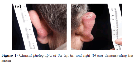
Examination revealed mildly discolored, swollen, tender nodules on the right upper and left mid pinna (Figure 1). There were no other suspicious lesions, no lymphadenopathy and no hepatosplenomegaly.
Results
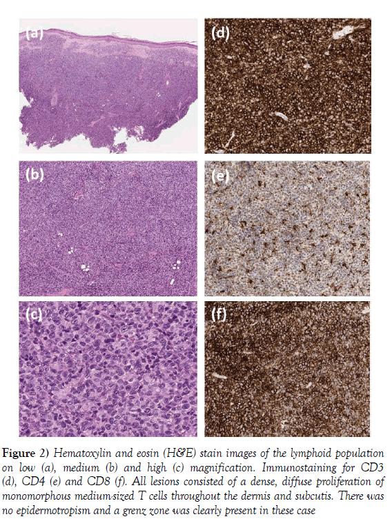
A biopsy was performed which showed a diffuse dermal lymphoid infiltration. This comprised large cells with complex folded nuclei but showing no blast excess. They were positive on immunohistochemistry for CD2, CD3, CD5 and CD8. CD7 was focally positive. CD4, PD1, TIA-1, Granzyme B, CD30 and CD56 were negative in the malignant lymphoid population (Figure 2). Ki 67 immunostaining was within the normal range for the majority of neoplastic cells, but had a slightly higher mitotic activity of up to 30% in the superficial aspect of the biopsy. The patient was treated with very low dose, localised radiotherapy (4 Gy in 2 fractions) to the right ear, and has since remained in remission for 18 months.
Figure 2) Hematoxylin and eosin (H&E) stain images of the lymphoid population on low (a), medium (b) and high (c) magnification. Immunostaining for CD3 (d), CD4 (e) and CD8 (f). All lesions consisted of a dense, diffuse proliferation of monomorphous medium-sized T cells throughout the dermis and subcutis. There was no epidermotropism and a grenz zone was clearly present in these case
Discussion
In 2007 Petrella [1] described a case series of 4 patients, each of whom presented with a slow-growing nodule of the ear, and had histology showing a clonally restricted, well-differentiated, nonepidermotropic, CD8-dominant lymphocytic infiltrate. They noted that these cases did not correspond to a recognized cutaneous T-cell lymphoma, and proposed a CD8 variant of primary cutaneous pleomorphic T-cell lymphoma.
Since then a further 25 similar cases have been described [2-5]. All lesions have involved the face with the most common site being the ear, and all have shown a relatively indolent clinical course. Typical histopathological features include the presence of a clear Grenz zone without epidermotropism, a diffuse dermal CD8-positive, granzyme B-negative lymphocytic infiltrate, a low proliferative index, and T-cell clonality [6-10].
These cases do not correspond clearly to any recognized category of cutaneous T-cell lymphoma (CTCL) described in the World Health Organization (WHO), or the European Organization for Research and Treatment of Cancer (EORTC) 2005 classification. Indeed they suggest an additional category of cutaneous indolent CD8-positive T-cell lymphoma, and differentiation between this and other more aggressive CD8-positive lymphomas is essential in avoiding excessive treatment and complications of therapy, for a condition that has a relatively benign clinical course.
Conclusion
Our case shows similar clinical and histological features to previous ones described, and fits with the notion that indolent CD8-positive lymphoproliferative disorder of the ear should be managed as a distinct clinical entity to other CD8-positive cutaneous T-cell lymphomas.
Conflict of Interest
None.
REFERENCES
- Petrella T, Maubec E, Cornillet-Lefebvre P, et al. Indolent CD8-positive lymphoid proliferation of the ear: a distinct primary cutaneous T-cell lymphoma? Am J Surg Pathol 2007;31:1887-92.
- Li JY, Guitart J, Pulitzer MP, et al. Multicenter case series of indolent small/medium-sized CD8+ lymphoid proliferations with predilection for the ear and face. Am J Dermatopathol 2014;36:402-8.
- Hagen JW, Magro CM. Indolent CD8+ lymphoid proliferation of the face with eyelid involvement. Am J Dermatopathol 2014;36:137-41.
- Greenblatt D, Ally M, Child F, et al. Indolent CD8(+) lymphoid proliferation of acral sites: a clinicopathologic study of six patients with some atypical features. J Cutan Pathol 2013;40:248-58.
- Swick BL, Baum CL, Venkat AP, Liu V. Indolent CD8+ lymphoid proliferation of the ear: report of two cases and review of the literature. J Cutan Pathol 2011;38:209-15.
- Beltraminelli H, Müllegger R, Cerroni L. Indolent CD8+ lymphoid proliferation of the ear: a phenotypic variant of the small-medium pleomorphic cutaneous T-cell lymphoma? J Cutan Pathol 2010;37:81-4.
- Girisha BS, Srinivas T, Noronha TM, et al. A case of indolent CD8-positive lymphoid proliferation of the ear. Indian J Dermatol 2018;63:342-5.
- Valois A, Bastien C, Granel-Broca F, et al. Indolent lymphoma of the ar. Ann Dermatol Venereol 2012;139:818-23.
- Petrella T. Indolent CD8+ lymphoid proliferation of the ear. Ann Dermatol Venereol 2012;139:789-90.
- Zeng W, Nava VE, Cohen P, Ozdemirli M. Indolent CD8-positive T-cell lymphoid proliferation of the ear: a report of two cases. J Cutan Pathol. 2012;39:696-700.


