Intraparenchymatous inferior parathyroid glands
Mario Pedro San Mauro*, Facundo Edgardo Patronelli, Daniel Alfredo Covello and Aldo José Cordero
Cátedra B de Anatomía, Facultad de Ciencias Médicas, Universidad Nacional de La Plata, Buenos Aires, Argentina
- *Corresponding Author:
- Mario Pedro San Mauro, MD, PhD
Associate Professor of Anatomy, Universidad Nacional de La Plata, Facultad de Ciencias Médicas, Cátedra B de Anatomía, Av 60 y 120 (1900) La Plata, Buenos Aires, Argentina
Tel: +54 (221) 4276726
E-mail: mariosanmauro@yahoo.com.ar
Date of Received: June 22nd, 2009
Date of Accepted: October 12th, 2009
Published Online: October 22nd, 2009
© IJAV. 2009; 2: 134–135.
[ft_below_content] =>Keywords
anatomy of parathyroids, intraparenchymatose parathyroids, anatomy of the neck
Introduction
Parathyroid glands are located in the neck, behind the thyroid gland. There are different opinions regarding their exact location. Superior parathyroid glands arise from the fourth pharyngeal arch, and inferior parathyroid glands from the third pharyngeal arch. This determines the anatomic variants of the inferior parathyroid glands due to the track they follow to their permanent location [1].
Frequently, inferior parathyroid glands move only towards the inferior border of thyroid gland, but they can descend with the thymus up to the thorax or not descend at all, remaining near the carotid junction [2].
Inferior thyroidal artery and its anastomoses between the superior thyroidal artery supply the parathyroid glands. Lymphatic vessels are associated with those of the thyroid gland and thymus. Innervation is sympathetic, from the middle or superior cervical ganglion.
Inferior parathyroid glands (IPG) are generally located behind the thyroid gland inferior lobe and there are a number of different descriptions for them. Granberg´s studies performed on more than 500 patients showed that in 60% of the cases parathyroid glands are close to the thyroid gland’s inferior end: behind it, in front of it, or next to it; in 38% of the cases they are within or next to the thyrothymic ligament; in 2 % of the cases they are within the mediastinum; and they are never intraparenchymatous [3].
Wang published cases with similar features and percentages to Granberg´s, and he did not mention intraparenchymatous parathyroid glands either [4].
Case Report
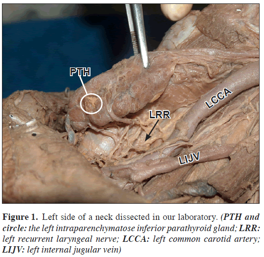
In 15 dissections performed on bodies fixed in 40% buffered formaldehyde, one intraparenchymatous inferior parathyroid gland was found in the mass of the thyroid gland’s left inferior lobe.
Initially, the IPG was searched on a surface of 1.5–2 cm around the junction between the inferior thyroidal artery and the recurrent laryngeal nerve. Not being able to recognize the IPG in this area, it was proceeded to visualize and dissect the branches of the inferior thyroidal artery as described by Gough [5].
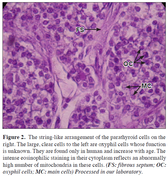
When dissecting one of them, whose track became intrathyroidal, a tissue different in colour and consistence from the thyroid gland was found 3 mm deep the gland. A sample was taken and histological confirmation was obtained that it certainly was the parathyroid gland (Figures 1,2).
Figure 2: The string-like arrangement of the parathyroid cells on the right. The large, clear cells to the left are oxyphil cells whose function is unknown. They are found only in human and increase with age. The intense eosinophilic staining in their cytoplasm reflects an abnormally high number of mitochondria in these cells. (FS: fibrous septum; OC: oxyphil cells; MC: main cells) Processed in our laboratory.
Discussion
Low concluded that in 13.8% of 866 cases, ectopic parathyroid glands were found, 6.7% of them were intrathyroidal [6]. Gough found 8 intraparenchymatous inferior parathyroid glands and 2 intraparenchymatous superior parathyroid glands in a total of 50 cases [5]. Even though samples of both studies are not comparable with each other, the number of intraparenchymatous inferior parathyroid glands is not insignificant. This does not coincide with what is described in classic anatomy books, where only superior intrathyroidal parathyroid glands are mentioned [6].
In a vast majority of thyroid gland surgery it is important to recognise and preserve at least one parathyroid gland. According to Santos Gomes et al. 80% of the patients have 4 glands, 14.3% have supernumerary parathyroid glands and 5.7% have only 3 [7].
Given the variability in their location and the fact that they are not always 4, it is of major importance to be aware that IPG can be intrathyroidal, similar to superior parathyroid glands.
Acknowledgement
To Miss Maria Clara del Río for translating the manuscript into English.
References
- Orts Llorca Francisco. Anatomía Humana. Tercera edición. Tomo III. Barcelona, Editorial Científico Médica. 1967; 454–461.
- Langman J. Embriología Médica. 7th Ed. Mexico, Panamericana. 1996; 299–301.
- Granberg PO, Cedermark B, Farnebo LO, Hamberger B, Werner S. Parathyroid Tumors. Curr Probl Cancer. 1985; 9: 1-52.
- Wang CA. Surgical management of primary hyperparathyroidism. Curr Probl Surg. 1985; 22: 1–50.
- Gough I. Preoperative parathyroid surgery: the importance of ectopic location and multigland disease. ANZ J Surg. 2006; 76: 1048–1050.
- Low A, Katz AD. Parathyroidectomy via bilateral cervical exploration: A retrospective review of 866 cases. Head Neck. 1998; 20: 583–587.
- Gomes EM, Nunes RC, Lacativa PG, Almeida MH, Franco FM, Leal CT, Patricio Filho PJ, Farias ML, Goncalves MD. Ectopic and extranumerary parathyroid glands location in patients with hyperparathyroidism secondary to end stage renal disease. Acta Cir Bras. 2007; 22: 105–109.
Mario Pedro San Mauro*, Facundo Edgardo Patronelli, Daniel Alfredo Covello and Aldo José Cordero
Cátedra B de Anatomía, Facultad de Ciencias Médicas, Universidad Nacional de La Plata, Buenos Aires, Argentina
- *Corresponding Author:
- Mario Pedro San Mauro, MD, PhD
Associate Professor of Anatomy, Universidad Nacional de La Plata, Facultad de Ciencias Médicas, Cátedra B de Anatomía, Av 60 y 120 (1900) La Plata, Buenos Aires, Argentina
Tel: +54 (221) 4276726
E-mail: mariosanmauro@yahoo.com.ar
Date of Received: June 22nd, 2009
Date of Accepted: October 12th, 2009
Published Online: October 22nd, 2009
© IJAV. 2009; 2: 134–135.
Abstract
Parathyroid glands are located in the neck, behind the thyroid gland. Inferior glands are described behind the thyroid´s inferior lobe but there are a number of different descriptions of them. We publish an atypical location as intraparenchymatous gland.
-Keywords
anatomy of parathyroids, intraparenchymatose parathyroids, anatomy of the neck
Introduction
Parathyroid glands are located in the neck, behind the thyroid gland. There are different opinions regarding their exact location. Superior parathyroid glands arise from the fourth pharyngeal arch, and inferior parathyroid glands from the third pharyngeal arch. This determines the anatomic variants of the inferior parathyroid glands due to the track they follow to their permanent location [1].
Frequently, inferior parathyroid glands move only towards the inferior border of thyroid gland, but they can descend with the thymus up to the thorax or not descend at all, remaining near the carotid junction [2].
Inferior thyroidal artery and its anastomoses between the superior thyroidal artery supply the parathyroid glands. Lymphatic vessels are associated with those of the thyroid gland and thymus. Innervation is sympathetic, from the middle or superior cervical ganglion.
Inferior parathyroid glands (IPG) are generally located behind the thyroid gland inferior lobe and there are a number of different descriptions for them. Granberg´s studies performed on more than 500 patients showed that in 60% of the cases parathyroid glands are close to the thyroid gland’s inferior end: behind it, in front of it, or next to it; in 38% of the cases they are within or next to the thyrothymic ligament; in 2 % of the cases they are within the mediastinum; and they are never intraparenchymatous [3].
Wang published cases with similar features and percentages to Granberg´s, and he did not mention intraparenchymatous parathyroid glands either [4].
Case Report
In 15 dissections performed on bodies fixed in 40% buffered formaldehyde, one intraparenchymatous inferior parathyroid gland was found in the mass of the thyroid gland’s left inferior lobe.
Initially, the IPG was searched on a surface of 1.5–2 cm around the junction between the inferior thyroidal artery and the recurrent laryngeal nerve. Not being able to recognize the IPG in this area, it was proceeded to visualize and dissect the branches of the inferior thyroidal artery as described by Gough [5].
When dissecting one of them, whose track became intrathyroidal, a tissue different in colour and consistence from the thyroid gland was found 3 mm deep the gland. A sample was taken and histological confirmation was obtained that it certainly was the parathyroid gland (Figures 1,2).
Figure 2: The string-like arrangement of the parathyroid cells on the right. The large, clear cells to the left are oxyphil cells whose function is unknown. They are found only in human and increase with age. The intense eosinophilic staining in their cytoplasm reflects an abnormally high number of mitochondria in these cells. (FS: fibrous septum; OC: oxyphil cells; MC: main cells) Processed in our laboratory.
Discussion
Low concluded that in 13.8% of 866 cases, ectopic parathyroid glands were found, 6.7% of them were intrathyroidal [6]. Gough found 8 intraparenchymatous inferior parathyroid glands and 2 intraparenchymatous superior parathyroid glands in a total of 50 cases [5]. Even though samples of both studies are not comparable with each other, the number of intraparenchymatous inferior parathyroid glands is not insignificant. This does not coincide with what is described in classic anatomy books, where only superior intrathyroidal parathyroid glands are mentioned [6].
In a vast majority of thyroid gland surgery it is important to recognise and preserve at least one parathyroid gland. According to Santos Gomes et al. 80% of the patients have 4 glands, 14.3% have supernumerary parathyroid glands and 5.7% have only 3 [7].
Given the variability in their location and the fact that they are not always 4, it is of major importance to be aware that IPG can be intrathyroidal, similar to superior parathyroid glands.
Acknowledgement
To Miss Maria Clara del Río for translating the manuscript into English.
References
- Orts Llorca Francisco. Anatomía Humana. Tercera edición. Tomo III. Barcelona, Editorial Científico Médica. 1967; 454–461.
- Langman J. Embriología Médica. 7th Ed. Mexico, Panamericana. 1996; 299–301.
- Granberg PO, Cedermark B, Farnebo LO, Hamberger B, Werner S. Parathyroid Tumors. Curr Probl Cancer. 1985; 9: 1-52.
- Wang CA. Surgical management of primary hyperparathyroidism. Curr Probl Surg. 1985; 22: 1–50.
- Gough I. Preoperative parathyroid surgery: the importance of ectopic location and multigland disease. ANZ J Surg. 2006; 76: 1048–1050.
- Low A, Katz AD. Parathyroidectomy via bilateral cervical exploration: A retrospective review of 866 cases. Head Neck. 1998; 20: 583–587.
- Gomes EM, Nunes RC, Lacativa PG, Almeida MH, Franco FM, Leal CT, Patricio Filho PJ, Farias ML, Goncalves MD. Ectopic and extranumerary parathyroid glands location in patients with hyperparathyroidism secondary to end stage renal disease. Acta Cir Bras. 2007; 22: 105–109.


