Langer’s axillary arch (axillopectoral muscle): a variation of latissimus dorsi muscleos ilium. The axillary arch is a variant muscular slip of this muscle
Sinan Bakirci, Ilker Mustafa Kafa*, Murat Uysal and Erdogan Sendemir
Department of Anatomy, Faculty of Medicine, Uludag University, Bursa, Turkey
- *Corresponding Author:
- Ilker Mustafa Kafa, MD
Department of Anatomy, Uludag University, Faculty of Medicine, Gorukle, 16059, Bursa, Turkey
Tel: +90 (224) 295 23 16
E-mail: imkafa@uludag.edu.tr
Date of Received: November 20th, 2009
Date of Accepted: June 21st, 2010
Published Online: June 30th, 2010
© IJAV. 2010; 3: 91–92.
[ft_below_content] =>Keywords
axillary arch, Langer’s muscle, latissimus dorsi muscle, axillopectoral muscle, axillary fossa
Introduction
Best-known variant structure of the axillary components of men is a muscular or fibro-muscular slip extending from the latissimus dorsi muscle to the tendons, muscles or fasciae of the superior part of the humerus. This variation had first described by Ramsay in 1795 and confirmed by Langer in 1864 and named as axillary arch of Langer. Although different frequencies have been reported from cadaver investigations and clinical studies, among different populations this variation occurs unilaterally up to 7%; its bilateral incidence is unknown. Among the variety of terminology that describes this variant structure as “Achselbogen”, “axillopectoral muscle”, “axillary arch”, “Langer’s axillary arch” or “muscular axillary arch” [1–3], we prefer the term “axillary arch” (“arcus axillaris” in Latin) in this report.
The latissimus dorsi muscle originates from sacrum, iliac crest and along the five lumbar and lower six thoracic vertebrae. It is a flat triangular muscle which covers lumbar regions and is gradually contracted into a narrow fasciculus before its insertion. The quadrilateral tendon of latissimus dorsi muscle, which is about 7 cm long, lies in front of the tendon of the teres major muscle and is inserted into the intertubercular groove of the humerus. Variations of latissimus dorsi can be seen at its origins where the number of dorsal vertebrae to which it attached, vary from four to seven or eight, as well as the varying number of costal attachments and muscle fibers that may or may not be reaching to the crest of the muscleos ilium. The axillary arch is a variant muscular slip of this muscle and is about 7 to 10 cm in length, splits from the upper edge of the latissimus dorsi and crosses the axilla in front of the axillary vessels and nerves [4]. This variant muscle slip joins inferiorly to the tendon of the pectoralis major, the coracobrachialis or the fascia over the biceps brachii. Latissimus dorsi muscle is supplied by the sixth, seventh and eighth cervical spinal nerves through the thoracodorsal (long subscapular) nerve. Axillary arch can receive nerve fibers from the lateral pectoral nerve, medial pectoral nerve, intercostobrachial nerve or thoracodorsal nerve [1]. Its embryonic origin is not clear and some anatomists consider muscular arches of the axilla as rudimentary phylogenetic remnants of the panniculus carnosus [3].
Anatomic variations of axilla are important for physicians and surgeons who perform axillary examination or surgery. Axillary arch, which crosses the axillary artery and vein just above the region, might usually be selected for the application of a ligature, and may mislead the surgeons during the operation [4].
Case Report
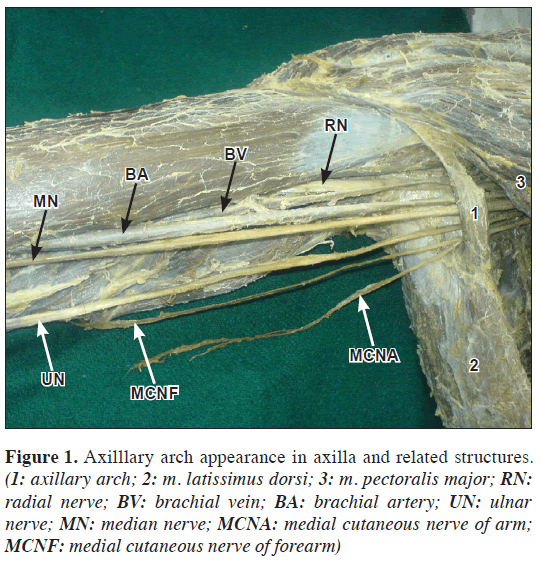
During routine dissections at the Uludag University, Faculty of Medicine, Department of Anatomy, we encountered a muscular variation opening to the right axillary fossa of 40-45 years old, black, male cadaver (Figure 1). An unusual muscular slip originating from the upper border of the latissimus dorsi muscle inserted into the deep surface of the tendon of pectoralis major muscle, also forming a bridge that constituted an axillary arch.
Figure 1: Axilllary arch appearance in axilla and related structures. (1: axillary arch; 2: m. latissimus dorsi; 3: m. pectoralis major; RN: radial nerve; BV: brachial vein; BA: brachial artery; UN: ulnar nerve; MN: median nerve; MCNA: medial cutaneous nerve of arm; MCNF: medial cutaneous nerve of forearm)
Its dimensions were 5.6 cm in length and 1.3 cm in width. The axillary artery, veins and nerves of the brachial plexus were lying under this muscular arch and the muscle slip was supplied by the thoracodorsal nerve. It was located in the mid-axillar region and crossed the axillary artery at its end. There were no additional variations of muscles or vessels at the same region. Left axillary fossa of the cadaver showed no structural variation.
Discussion
Ramsay initially mentioned muscular variations located in the axillary fossa in 1795 [2]. Carl Langer described the fibrous thickening of the medial edge of the axillary fascia between the borders of the pectoralis major and the latissimus dorsi muscle as “Achselbogen” [3]. Consequently, Testut called it as “axillary arch of Langer” [1]. Sochetella used the term “axillopectoral muscle” in 1977 [5], followed by Sisley in 1987 [6], and Turgut et al. in 2005 [7].
Prevalence of this variation appears to be higher in dissected cadavers than surgical examinations. In Japanese, the prevalence of axillary arch is found to be 9.1% [8], and 5.3% [9] in two different studies on 176 and 94 body halves respectively. Prevalence of this variation in Turkish population reported as 1.9% in 26 cadavers [7], where in Bulgarian population reported as 3.6% in 56 cadavers [2] and in Spanish population it is reported as 3% from a study of 50 cadavers [10].
Arch-shaped variations in the axils could be considered in two groups, muscular form (type I) and tendinous form (type II), accompanying different subtypes based on their nerve supplies and site of their attachment points [9]. However, clinical classification of the axillary arches could be defined as superficial and deep arch groups. Superficial group arches cross in front of the vessels and nerves, and the veins could be affected primarily within this variation which may play a role in intermittent obstruction of the axillary vein [1]. Deep group arches occur deeply on the posterior or lateral walls of the axilla. These arches usually cross only parts of the neurovascular bundle and axillary or radial nerves could possibly be affected. During mammography interpretations it should be kept in mind that the radiographic signs of a muscular slip related to latissimus dorsi muscle could be seen in this area. CT or MRI is furthermore helpful for definitive evaluation of differentiation.
This variation is important for axillary surgery, and the surgeon must recall its possible presence and must be cautious during dissection. If a muscular slip related to the latissimus dorsi muscle is present during routine axillary lymphadenectomy, it is possible that some lymph nodes could be localized posteriorly and laterally to the arch. It may also bear various surgical and medical problems such as axillary vein entrapment syndrome, development of lymph edema of the upper limb following breast surgery, upper limb neurovascular symptoms or it is likely presenting as an axillary mass, which confuses with axillary lymph nodes [11].
References
- Jelev L, Georgiev GP, Surchev L. Axillary arch in human: common morphology and variety. Definition of “clinical” axillary arch and its classification. Ann Anat. 2007. 189: 473–481.
- Georgiev GP, Jelev L, Surchev L. Axillary arch in Bulgarian population: clinical significance of the arches. Clin Anat. 2007; 20: 286–291.
- Besana-Ciani I, Greenall MJ. Langer’s axillary arch: anatomy, embryological features and surgical implications. Surgeon. 2005; 3: 325–327.
- Williams PL, Warwick R, Dyson M, Bannister LH. Gray’s Anatomy. 37th Ed., Churchill Livingstone, Edinburgh, London, Melbourne, New York. 1989; 610.
- Sachatello CR. The axillopectoral muscle (Langer’s axillary arch): a cause of axillary vein obstruction. Surgery. 1977; 81: 610–612.
- Sisley JF. The axillopectoral muscle. Surg Gynecol Obstet. 1987; 165: 73.
- Turgut HB, Peker T, Gulekon N, Anil A, Karakose M. Axillopectoral muscle (Langer’s muscle). Clin Anat. 2005; 183: 220–223.
- Kasai T, Chiba S. [True nature of the muscular arch of the axilla and its nerve supply.] Kaibogaku Zasshi. 1977; 52: 309–336. (Japanese)
- Takafuji T, Igarashi J, Kanbayashi T, Yokoyama T, Moriya A, Azuma S, Sato Y. [The muscular arch of the axilla and its nerve supply in Japanese adults.] Kaibogaku Zasshi. 1991; 66: 511–523. (Japanese)
- Miguel M, Llusa M, Ortiz JC, Porta N, Lorente M, Gotzens V. The axillopectoral muscle (of Langer): report of three cases. Surg Radiol Anat. 2001; 23: 341–343.
- Daniels IR, della Rovere GQ. The axillary arch of Langer--the most common muscular variation in the axilla. Breast Cancer Res Treat. 2000; 59: 77–80.
Sinan Bakirci, Ilker Mustafa Kafa*, Murat Uysal and Erdogan Sendemir
Department of Anatomy, Faculty of Medicine, Uludag University, Bursa, Turkey
- *Corresponding Author:
- Ilker Mustafa Kafa, MD
Department of Anatomy, Uludag University, Faculty of Medicine, Gorukle, 16059, Bursa, Turkey
Tel: +90 (224) 295 23 16
E-mail: imkafa@uludag.edu.tr
Date of Received: November 20th, 2009
Date of Accepted: June 21st, 2010
Published Online: June 30th, 2010
© IJAV. 2010; 3: 91–92.
Abstract
Langer’s axillary arch (axillopectoral muscle) is a variant muscular structure of the axilla which was described under various names as Langer’s muscle, axillary arch or muscular axillary arch by different authors. During routine dissections, we found a muscular slip on the right axillary fossa that originated from latissimus dorsi muscle and attached to the deep surface of the tendon of pectoralis major muscle, and described it as Langer’s axillary arch. Arterial, venous and nervous structures passed under this muscular slip which constitutes an arch in the axillary fossa. Although axillary arch is not very rare, it is generally neglected and not explored or described well. It has immense clinical and morphologic importance for surgical operations performed on axillary region; thus, surgeons should well be aware of its possible existence.
-Keywords
axillary arch, Langer’s muscle, latissimus dorsi muscle, axillopectoral muscle, axillary fossa
Introduction
Best-known variant structure of the axillary components of men is a muscular or fibro-muscular slip extending from the latissimus dorsi muscle to the tendons, muscles or fasciae of the superior part of the humerus. This variation had first described by Ramsay in 1795 and confirmed by Langer in 1864 and named as axillary arch of Langer. Although different frequencies have been reported from cadaver investigations and clinical studies, among different populations this variation occurs unilaterally up to 7%; its bilateral incidence is unknown. Among the variety of terminology that describes this variant structure as “Achselbogen”, “axillopectoral muscle”, “axillary arch”, “Langer’s axillary arch” or “muscular axillary arch” [1–3], we prefer the term “axillary arch” (“arcus axillaris” in Latin) in this report.
The latissimus dorsi muscle originates from sacrum, iliac crest and along the five lumbar and lower six thoracic vertebrae. It is a flat triangular muscle which covers lumbar regions and is gradually contracted into a narrow fasciculus before its insertion. The quadrilateral tendon of latissimus dorsi muscle, which is about 7 cm long, lies in front of the tendon of the teres major muscle and is inserted into the intertubercular groove of the humerus. Variations of latissimus dorsi can be seen at its origins where the number of dorsal vertebrae to which it attached, vary from four to seven or eight, as well as the varying number of costal attachments and muscle fibers that may or may not be reaching to the crest of the muscleos ilium. The axillary arch is a variant muscular slip of this muscle and is about 7 to 10 cm in length, splits from the upper edge of the latissimus dorsi and crosses the axilla in front of the axillary vessels and nerves [4]. This variant muscle slip joins inferiorly to the tendon of the pectoralis major, the coracobrachialis or the fascia over the biceps brachii. Latissimus dorsi muscle is supplied by the sixth, seventh and eighth cervical spinal nerves through the thoracodorsal (long subscapular) nerve. Axillary arch can receive nerve fibers from the lateral pectoral nerve, medial pectoral nerve, intercostobrachial nerve or thoracodorsal nerve [1]. Its embryonic origin is not clear and some anatomists consider muscular arches of the axilla as rudimentary phylogenetic remnants of the panniculus carnosus [3].
Anatomic variations of axilla are important for physicians and surgeons who perform axillary examination or surgery. Axillary arch, which crosses the axillary artery and vein just above the region, might usually be selected for the application of a ligature, and may mislead the surgeons during the operation [4].
Case Report
During routine dissections at the Uludag University, Faculty of Medicine, Department of Anatomy, we encountered a muscular variation opening to the right axillary fossa of 40-45 years old, black, male cadaver (Figure 1). An unusual muscular slip originating from the upper border of the latissimus dorsi muscle inserted into the deep surface of the tendon of pectoralis major muscle, also forming a bridge that constituted an axillary arch.
Figure 1: Axilllary arch appearance in axilla and related structures. (1: axillary arch; 2: m. latissimus dorsi; 3: m. pectoralis major; RN: radial nerve; BV: brachial vein; BA: brachial artery; UN: ulnar nerve; MN: median nerve; MCNA: medial cutaneous nerve of arm; MCNF: medial cutaneous nerve of forearm)
Its dimensions were 5.6 cm in length and 1.3 cm in width. The axillary artery, veins and nerves of the brachial plexus were lying under this muscular arch and the muscle slip was supplied by the thoracodorsal nerve. It was located in the mid-axillar region and crossed the axillary artery at its end. There were no additional variations of muscles or vessels at the same region. Left axillary fossa of the cadaver showed no structural variation.
Discussion
Ramsay initially mentioned muscular variations located in the axillary fossa in 1795 [2]. Carl Langer described the fibrous thickening of the medial edge of the axillary fascia between the borders of the pectoralis major and the latissimus dorsi muscle as “Achselbogen” [3]. Consequently, Testut called it as “axillary arch of Langer” [1]. Sochetella used the term “axillopectoral muscle” in 1977 [5], followed by Sisley in 1987 [6], and Turgut et al. in 2005 [7].
Prevalence of this variation appears to be higher in dissected cadavers than surgical examinations. In Japanese, the prevalence of axillary arch is found to be 9.1% [8], and 5.3% [9] in two different studies on 176 and 94 body halves respectively. Prevalence of this variation in Turkish population reported as 1.9% in 26 cadavers [7], where in Bulgarian population reported as 3.6% in 56 cadavers [2] and in Spanish population it is reported as 3% from a study of 50 cadavers [10].
Arch-shaped variations in the axils could be considered in two groups, muscular form (type I) and tendinous form (type II), accompanying different subtypes based on their nerve supplies and site of their attachment points [9]. However, clinical classification of the axillary arches could be defined as superficial and deep arch groups. Superficial group arches cross in front of the vessels and nerves, and the veins could be affected primarily within this variation which may play a role in intermittent obstruction of the axillary vein [1]. Deep group arches occur deeply on the posterior or lateral walls of the axilla. These arches usually cross only parts of the neurovascular bundle and axillary or radial nerves could possibly be affected. During mammography interpretations it should be kept in mind that the radiographic signs of a muscular slip related to latissimus dorsi muscle could be seen in this area. CT or MRI is furthermore helpful for definitive evaluation of differentiation.
This variation is important for axillary surgery, and the surgeon must recall its possible presence and must be cautious during dissection. If a muscular slip related to the latissimus dorsi muscle is present during routine axillary lymphadenectomy, it is possible that some lymph nodes could be localized posteriorly and laterally to the arch. It may also bear various surgical and medical problems such as axillary vein entrapment syndrome, development of lymph edema of the upper limb following breast surgery, upper limb neurovascular symptoms or it is likely presenting as an axillary mass, which confuses with axillary lymph nodes [11].
References
- Jelev L, Georgiev GP, Surchev L. Axillary arch in human: common morphology and variety. Definition of “clinical” axillary arch and its classification. Ann Anat. 2007. 189: 473–481.
- Georgiev GP, Jelev L, Surchev L. Axillary arch in Bulgarian population: clinical significance of the arches. Clin Anat. 2007; 20: 286–291.
- Besana-Ciani I, Greenall MJ. Langer’s axillary arch: anatomy, embryological features and surgical implications. Surgeon. 2005; 3: 325–327.
- Williams PL, Warwick R, Dyson M, Bannister LH. Gray’s Anatomy. 37th Ed., Churchill Livingstone, Edinburgh, London, Melbourne, New York. 1989; 610.
- Sachatello CR. The axillopectoral muscle (Langer’s axillary arch): a cause of axillary vein obstruction. Surgery. 1977; 81: 610–612.
- Sisley JF. The axillopectoral muscle. Surg Gynecol Obstet. 1987; 165: 73.
- Turgut HB, Peker T, Gulekon N, Anil A, Karakose M. Axillopectoral muscle (Langer’s muscle). Clin Anat. 2005; 183: 220–223.
- Kasai T, Chiba S. [True nature of the muscular arch of the axilla and its nerve supply.] Kaibogaku Zasshi. 1977; 52: 309–336. (Japanese)
- Takafuji T, Igarashi J, Kanbayashi T, Yokoyama T, Moriya A, Azuma S, Sato Y. [The muscular arch of the axilla and its nerve supply in Japanese adults.] Kaibogaku Zasshi. 1991; 66: 511–523. (Japanese)
- Miguel M, Llusa M, Ortiz JC, Porta N, Lorente M, Gotzens V. The axillopectoral muscle (of Langer): report of three cases. Surg Radiol Anat. 2001; 23: 341–343.
- Daniels IR, della Rovere GQ. The axillary arch of Langer--the most common muscular variation in the axilla. Breast Cancer Res Treat. 2000; 59: 77–80.


