Left dominant arterial morphology with hypoplastic right vertebral artery, P1- and A1-segments of cerebral arteries, and unusual right accessory posterior cerebral artery
Received: 04-Jun-2023, Manuscript No. ijav-23-6520; Editor assigned: 05-Jun-2023, Pre QC No. ijav-23-6520 (PQ); Accepted Date: Jun 23, 2023; Reviewed: 19-Jun-2023 QC No. ijav-23-6520; Revised: 23-Jun-2023, Manuscript No. ijav-23-6520 (R); Published: 30-Jun-2023, DOI: 10.37532/1308-4038.16(6).270
Citation: Tessema CB. Left dominant arterial morphology with hypoplastic right vertebral artery, P1- and A1-segments of cerebral arteries, and unusual right accessory posterior cerebral artery. Int J Anat Var. 2023;16(6):315-316.
This open-access article is distributed under the terms of the Creative Commons Attribution Non-Commercial License (CC BY-NC) (http://creativecommons.org/licenses/by-nc/4.0/), which permits reuse, distribution and reproduction of the article, provided that the original work is properly cited and the reuse is restricted to noncommercial purposes. For commercial reuse, contact reprints@pulsus.com
Abstract
A hypoplastic right vertebral artery (VA), a complete circle of Willis with hypoplastic right P1- and A1-segments, and left dominant arterial configuration were incidentally detected in a 75-year-old male donor. The P1- segment of the left posterior cerebral artery (PCA) gave branch to an accessory right posterior cerebral artery (APCA). The hypoplastic right A1-segment arose from the right middle cerebral artery (MCA). The right posterior communicating artery (PCoA) from the smaller right internal carotid artery (ICA) continued as the right P2-segment. The larger left ICA gave branch to the dominant left anterior cerebral artery (ACA) from which the anterior communicating artery (ACoA) and both the left and right A2 –segments arose. Due to the well stablished close association of vascular dominance and variations with cerebrovascular diseases, the knowledge of the variable vascular architecture at the base of the brain, would be imperative in the various diagnostic and therapeutic procedures.
Keywords
Circle of willis; Cerebral artery; Hypoplastic segment; Accessory posterior cerebral artery; Vertebral artery
INTRODUCTION
Intracranial vascular variations are frequent findings in the general population [1]. These variations involve both the internal carotid (anterior) and vertebro-basilar (posterior) brain circulations interconnected by the circle of Willis and their immediate branches. Since such variations are risk factors in the development of aneurysm and ischemia that can result in transient ischemic attack or stroke, they are of immense clinical significance [1]. It was also known that these clinical conditions are closely associated with right or left dominance of these arteries. As a previous study noted, adult extracranial and intracranial vessels are primarily left dominant and there is a left side predilection for cerebrovascular pathology [2]. The commonly encountered variations both in the anterior and posterior circulations were morphologically categorized into four groups that include variations in vessel origin, number, morphology and pathway [3].
In the posterior circulation, it is well recognized that hypoplastic vertebral artery is a common vascular variant and asymmetry of the vertebral arteries with left dominance and hypoplastic right vertebral artery is the most frequent finding [3], [4]. Several clinical studies have also shown that vertebral artery dominance is a risk factor for posterior circulation ischemic stroke [4] but has no significant relationship to hemispheric dominance or handedness [5]. Depending on the comparative diameter of the P1-segment of the PCA and the PCoA, the terminal configuration of the branches of the posterior circulation at the circle of Willis was classified as adult type (most frequent type with larger P1-segment than PCoA), transitional type (where P1-segment and PCoA are of equal size) and fetal type (with P1-segment smaller than the PCoA and PCA arises from the ICA) [6]. The fetal type, which is believed to be prone to ischemic stroke that involves ICA, is more frequently associated with hypoplasia of the VA on the same side, while absence or hypoplasia of the PCoA is more common in subjects with normal (non-hypoplastic) VA and are less frequent in those with VA hypoplasia [6]. Furthermore, it was also noted that the fetal- type is closely associated with ipsilateral A1 hypoplasia in the anterior circulation [6] [7].
Very few previous reports noted the existence of APCA, describing it as a hyperplastic anterior choroidal artery supplying the territories of the posterior cerebral artery [8].
The well-known anatomical variations that involve the anterior circulation include the unilateral or bilateral absence of the internal carotid arteries (ICA’s) [9] and variations of their branches in the form of absence, duplication, and hypoplasia of ACoA as well as the hypoplasia, fenestration, duplication and increased length of ACA [10]. Regarding A1 asymmetry, Wu T-C et al 2020 [11] clearly stated that the ICA diameter of the dominant A1 is significantly greater than the contralateral side. Therefore, the knowledge of such variant arteries supplying the brain is very crucial in the diagnosis and treatment of acute stroke [12]).
METHOD
After opening of the skull and removal of the brain in a 75-year-old male donor, the arteries on the basal aspect of the brain including the cranial part of VA, basilar artery, the two internal carotid arteries, circle of Willis and the branches thereof, were carefully dissected to look for their interconnections and distribution patterns. All the normal and variant arteries were exposed during the dissection, painted red and photographs were taken for illustrations
CASE REPORT
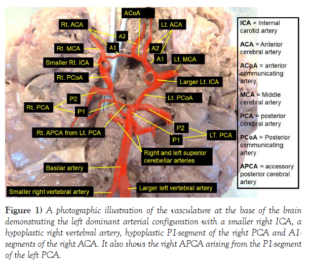
According to observation in this donor, there was an obvious asymmetry of most of the vasculature at the base of the brain with left dominance and a complete formation of the circle of Willis but with hypoplastic right A1- and P1-segments, hypoplasia of the right VA and an APCA. In general, the following variant arteries illustrated by the figure below were observed. 1. A smaller/hypoplastic right VA joined a larger left VA just caudal to the pontomedullary junction to form the basilar artery.
2. The basilar artery, formed by the major contribution of the larger left VA, terminally divided into a fetal-type right PCA with a hypoplastic P1- segment and an adult-type left PCA. The P1-segment of the left PCA gave branch to an APCA that crossed to the right ventral to the cerebral peduncles to enter the right occipital lobe.
3. The larger left ICA gave branch to the left PCoA, left MCA and a comparatively larger left ACA. The A1-segment of the left ACA formed the major stem that divided into the left A2-segment and the ACoA. The ACoA was then joined by the hypoplastic A1-segment of the right ACA and continued through the interhemispheric fissure as the right A2-segment.
4. The generally smaller right ICA gave branch to the right larger and dominant PCA (the only dominant artery of the right side), which was joined by the hypoplastic P1-segment of the right PCA and turned posterolaterally to the right and became the P2-segment that entered the right occipital lobe. The right MCA arose from the right ICA and gave branch to the hypoplastic A1-segment of the right ACA (Figure 1).
Figure 1) A photographic illustration of the vasculature at the base of the brain demonstrating the left dominant arterial configuration with a smaller right ICA, a hypoplastic right vertebral artery, hypoplastic P1-segment of the right PCA and A1- segments of the right ACA. It also shows the right APCA arising from the P1-segment of the left PCA.
DISCUSSION
It is a well-known fact that variation of the anterior and posterior brain circulations including the circle of Willis is a risk factor in the development of aneurysm, transient ischemic attack or stroke [1]. A previous study showed that both neonatal and adult strokes are more common in the left than the right cerebral hemisphere. With regard to arterial caliber and cortical directed blood flow, the adult type was found to be left dominant; supporting the left side predilection of cerebrovascular pathology described earlier [2]. The observation in the present case report, where almost all the arteries on the left side are larger in size (except the right PCoA) and their branches covered the territories of the left PCA, MCA, ACA, ACoA, A2-segment of the right ACA and contributed to the supply of the right PCA territory by its APCA branch, theoretically confirms the result of this previous study. According to Hakim A et al 2018[3] some of the most commonly encountered variation in any of these arteries are persistent primitive fetal arteries, hypoplasia and aplasia (agenesis) of arterial segment, which were also observed in this case report as hypoplastic P1- and A1-segments of the right PCA and ACA respectively. It is also well documented that hypoplastic VA is a common vascular variant and asymmetry of the VA’s with left dominance and hypoplastic right VA is the most usual finding (50%), while right dominance with hypoplastic left VA and symmetric codominance of both arteries constitute about 25% each [3], [4]. Several clinical studies have shown that VA dominance is a risk factor for posterior circulation ischemic stroke [4] but has no significant relationship to hemispheric dominance [5]. Even though this current case report is based on the observation in a single donor, the presence of hypoplastic right vertebral artery attests with the finding of the two authors above. As relates to the earlier classification of the contribution of the vertebro-basilar circulation to the arterial supply of the brain and its configuration into adult type, transitional type and fetal type [6, 7], the current case report does not exactly fit into the these three classification, rather it is a combination of adult type (left) with a large P1-segment of the left PCA than the left PCoA and fetal type (right) with the presence of hypoplastic VA, P1-segment, and A1-segment with Additional presence of APCA. As noted in previous studies and reports, APCA is considered to be a hyperplastic anterior choroidal artery, supplying all of the territories of the posterior cerebral artery and a recent paper by Rusu MC et al 2023 [8] elaborated that APCA and hyperplastic anterior choroidal artery describe the same morphology. However, the right APCA observed in the current case report differs from this earlier description in that it originated from the left PCA and crossed to the right ventral to the cerebral peduncles to accompany the right P2-segment formed by the right PCoA with no relation to the anterior choroidal artery. Despite the exhaustive search of available literature on the different variations of the brain circulation, there are no adequate resources that describe the existence of accessory right posterior cerebral artery (APCA) originating from the left PCA. Therefore, to the best of my knowledge, this is the first of its kind, which has not been described before.
The well-known anatomical variations that involve ICA at the base of the brain include their unilateral or bilateral absence [9] or absence, duplication, and hypoplasia of the ACoA as well as the hypoplasia, fenestrations, duplications and increased length of the ACA in the anterior portion of the circle of Willis [10]. Regarding A1-asymmetry, the ICA diameter of the dominant A1 is significantly greater than the contralateral side [11]. The finding in this current case report is more or less consistent with the descriptions above but the hypoplastic A1-segment arising from the right MCA and a larger and dominant right PCoA. According to the observation in this current donor the significant contribution of the right ICA to the anterior cerebral circulation is only via the right MCA and the P2-segment of the right PCA, while the rest is a territory of the left ICA. The vertebrobasilar circulation also appears to be diverted to the left through the large PCA. Therefore, as stated by earlier authors [12], the lack of knowledge of the presence of such variants, may play a detrimental role in the diagnosis and management of acute stroke.
CONCLUSION
The knowledge of such normal variations in vascular architecture at the base of the brain with right or left dominance and the implicated close associations with cerebrovascular diseases, would be valuable in the various clinical and radiological diagnostic assessment of patients with such diseases and in decision making to take appropriate therapeutic measures.
ACKNOWLEDGEMENT
As usual, I am thankful to the donor and his families for their invaluable donation and consent for education, research and publication. I would also like to express my gratitude to the department of biomedical sciences for the encouragement and uninterrupted support. Similarly, I am also grateful to Denelle Kees and John Opland for their immense assistance during the dissection of this cadaver in the gross anatomy lab.
REFERENCES
- Kovac JD, Stankovic A, Stankovic D, Kovac B, Saranovic D, et al. Intracranial arterial variations: A comprehensive evaluation using CT angiography. Med Sci Monit. 2014; 20:420-427.
- Van Vuuren AJ, Saling MM, Ameen O, Naidoo N, Solms M, et al. Hand preference is selectively related to common and internal carotid arterial asymmetry. Laterality. 2017; 22(4):377-398.
- Hakim A, Gralla J, Rozeik C, Morddasini P, Leidolt L, et al. Anomalies and normal variants of the cerebral arterial supply: A comprehensive pictorial review with a proposed workflow for classification and significance. J. Neuroimaging. 2018; 28: 14-35.
- Sun Y, Shi Y-M, Xu P. The clinical research progress of vertebral artery dominance and posterior circulation ischemic stroke. Cardiovasc Dis. 2022; 51:553-556.
- Vural A, Cicek ED. Is asymmetry between vertebral arteries related to cerebral dominance? Turk J Med Sci. 2019; 49: 1721-1726.
- Gaigalaite V, Dementaviciene J, Vilimas A, Kalibatiene D. Association between the posterior part of the circle of Willis and vertebral artery hypoplasia. PLoS ONE. 2019; 14(9): e0213-226.
- Mujagic S, Kozic D, Huseinagic H, Smajlovic D. Symmetry, asymmetry and hypoplasia of intracranial internal carotid artery on magnetic resonance angiography. Acta Med Acad. 2016; 45:1- 9.
- Rusu MC, Vrapclu AD, Lazar M. A rare variant of accessory cerebral artery. Surg Radiol Anat. 2023; 45(5):523-526.
- Mani K. Absent internal carotid artery in the circle of Willis. IOSR-JDMS. 2015; 14(11): 38-40.
- Dumitrescu Am, Cobzaru RG, Ripa C, TanaseMD, Sorodoc V, et al. Anatomical variation of the anterior part of the circle of Willis- an Autopsic study. J Univers Surg. 2021; 9(4): 21.
- Wu T-C, Chen T-Y, Ko C-C, Chen J-H, Lin C-P, et al. Correlation of internal carotid artery diameter and coronary flow with asymmetry of the circle of Willis. BMC Neurology. 2020; 20:251.
- Barbato F, Allocca R, Bosso G, Numis FG. Anatomy of cerebral arteries with clinical aspects in patients with ischemic stroke. Anatomia. 2022; 1:152-169.
Indexed at, Google Scholar, Crossref
Indexed at, Google Scholar, Crossref
Indexed at, Google Scholar, Crossref
Indexed at, Google Scholar, Crossref
Indexed at, Google Scholar, Crossref
Indexed at, Google Scholar, Crossref
Indexed at, Google Scholar, Crossref
Indexed at, Google Scholar, Crossref
Indexed at, Google Scholar, Crossref
Indexed at, Google Scholar, Crossref


