Left side Variant additional renal artery
Zelalem Animaw*, Biniam Ewnete
Department of Biomedical Sciences, College of Health Sciences, Debre Tabor University, Ethiopia.
- *Corresponding Author:
- Zelalem Animaw
Department of Biomedical Sciences College of Health Sciences
Debre Tabor University, Ethiopia
Tel: +251-913-122352
E-mail: zelalem.a01@gmail.com
Published Online : 01 February 2017
Citation: Animaw Z, Ewnete B. Left side variant additional renal artery. Int J Anat Var. 2017;10(1):006-7.
© This open-access article is distributed under the terms of the Creative Commons Attribution Non-Commercial License (CC BY-NC) (http:// creativecommons.org/licenses/by-nc/4.0/), which permits reuse, distribution and reproduction of the article, provided that the original work is properly cited and the reuse is restricted to noncommercial purposes. For commercial reuse, contact reprints@pulsus.com
[ft_below_content] =>Keywords
Renal artery, Renal vasculature, Embryological process
Renal arteries are paired end arteries branched from abdominal aorta supplying the kidneys. They consume around 20% of the total cardiac output. Due to embryological and racial reasons there are a lot of structural variations of renal arteries among individuals where variant additional renal arteries being the commonest [1,2].
Case Report
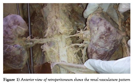
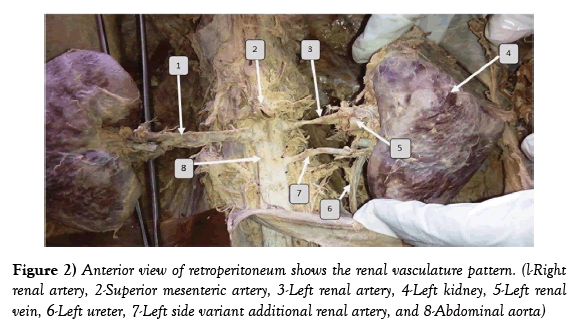
A case of unilateral variant additional renal artery was observed during a routine cadaveric dissection session of retroperitoneal region for second year medical students. In this male cadaver a variant additional renal artery was found on the left side of the body measuring 4.2 cm. It rises from abdominal aorta 1.5 cm below the left renal artery. It entered in to the kidney parenchyma just inferior to hailum of the left kidney; anterior to the upper part of left ureter (Figure 1). No branching of this variant artery is visualized.
Discussion
A case of variant additional renal artery can be explained through its embryological process. Between the 6th and 9th week of embryonic period, the kidneys ascend from the pelvis to the retro-peritoneum. During their path, they obtain blood supply from temporary arteries which arise from the developing abdominal aorta; later these arteries will be degenerated and replaced by new arteries above till the final paired definitive renal arteries. When these transient arteries failed to degenerate and remain persistent due to incomplete apoptosis, they will appear as variant additional renal arteries [1,2].
These variant renal arteries are the commonest anatomical variations occurring in renal vasculature which is presumed to be found about one third of the time; usually found unilaterally. In favor of current case, Studies showed that left is a common side for variant additional renal arteries to be found [2-4]. In spite of this, there are contradicting reports from Brazil and India revealed variant additional renal arteries were found on the right side in most of the cases [5-7].
Even though the common site of origin for such kinds of variant artery is abdominal aorta similar with current case, there are other rare extra aortic origins identified in different literatures, such as from common iliac, inferior mesenteric, main renal artery and celiac trunk [3,8,9]. As in this case Variant additional renal arteries are commonly found below the renal arteries explained by kidneys ascending path from the pelvic to retroperitoneal region during embryonic life where the most likely additional arteries fail to degenerate and persist will remain in the lower part of the renal artery [8,9].
Number of additional arteries may vary from person to person. But, up to 5 additional renal arteries might be found unlike our case which is limited only to 1 [10].
Conclusion
In conclusion, renal vasculature can be found varying from the commonly existing arrangement. Among these variant additional arteries are the commonest. These variant arteries can found in different number and pattern. Therefore due to the currently advanced renal vasculature surgery including renal transplantation, it is important to state the vascular distribution and variation of renal arteries among different population (Figure 2).
Acknowledgment
We would like to thank technical assistants of Debre Tabor University who participated in the dissection process. Our gratitude also goes to staffs of Debre Tabor University department of Biomedical Sciences.
References
- Natsis K, Paraskevas G, Panagouli E, et al. A morphometric study of multiple renal arteries in Greek population and a systematic review. Rom J Morphol Embryol. 2015;55(3 Suppl):1111-22.
- Saldarriaga B, Pérez A, Ballesteros L. A direct anatomical study of additional renal arteries in a Colombian mestizo population. Folia Morphol. 2008;67:129-34.
- Johnson PB, Cawich SO, Shah SD, et al. Accessory renal arteries in a Caribbean population: a computed tomography based study. Springer Plus. 2013;2:1.
- Sasikala P, Sulochana S, Rajan T, et al. Comparative Study of Anatomy of Renal Artery in Correlation with the Computed Tomography Angiogram. World J Med Sci. 2013;8:300-5.
- Budhiraja V, Rastogi R, Asthana A. Renal artery variations: embryological basis and surgical correlation. Rom J Morphol Embryol. 2010;51:533-6.
- Santos Soares TR, Soares Ferraz J, Buziquia Dartibale C, et al. Variations in human renal arteries. Acta Scientiarum: Biological Sciences. 2016;35.
- Nagato A, Rocha C, Bandeira A, et al. Morphometric and quantitative analysis of the afferent renal artery variation. J Morphol. 2013;30:82-5.
- He B, Hamdorf J. Clinical importance of anatomical variations of renal vasculature during laparoscopic donor nephrectomy. OA Anatomy. 2013;18:25-31.
- Ankolekar V, Sengupta R. Renal artery variations: a cadaveric study with clinical relevance. International J Curr Res Rev. 2013;5:154.
- Mishra A, Sharma P, Manik P, et al. Bilateral multiple renal arteries: A case report. 2014.
Zelalem Animaw*, Biniam Ewnete
Department of Biomedical Sciences, College of Health Sciences, Debre Tabor University, Ethiopia.
- *Corresponding Author:
- Zelalem Animaw
Department of Biomedical Sciences College of Health Sciences
Debre Tabor University, Ethiopia
Tel: +251-913-122352
E-mail: zelalem.a01@gmail.com
Published Online : 01 February 2017
Citation: Animaw Z, Ewnete B. Left side variant additional renal artery. Int J Anat Var. 2017;10(1):006-7.
© This open-access article is distributed under the terms of the Creative Commons Attribution Non-Commercial License (CC BY-NC) (http:// creativecommons.org/licenses/by-nc/4.0/), which permits reuse, distribution and reproduction of the article, provided that the original work is properly cited and the reuse is restricted to noncommercial purposes. For commercial reuse, contact reprints@pulsus.com
Abstract
During development of renal vasculature, there are different variations manifested in adult anatomy. Among these, variant additional renal arteries are frequently observed. This case study describes a unilateral variant additional renal artery in a male cadaver during a routine dissection session. Stating renal vasculature pattern is clinically important to anticipate proper management during renal vasculature procedure.
-Keywords
Renal artery, Renal vasculature, Embryological process
Renal arteries are paired end arteries branched from abdominal aorta supplying the kidneys. They consume around 20% of the total cardiac output. Due to embryological and racial reasons there are a lot of structural variations of renal arteries among individuals where variant additional renal arteries being the commonest [1,2].
Case Report
A case of unilateral variant additional renal artery was observed during a routine cadaveric dissection session of retroperitoneal region for second year medical students. In this male cadaver a variant additional renal artery was found on the left side of the body measuring 4.2 cm. It rises from abdominal aorta 1.5 cm below the left renal artery. It entered in to the kidney parenchyma just inferior to hailum of the left kidney; anterior to the upper part of left ureter (Figure 1). No branching of this variant artery is visualized.
Discussion
A case of variant additional renal artery can be explained through its embryological process. Between the 6th and 9th week of embryonic period, the kidneys ascend from the pelvis to the retro-peritoneum. During their path, they obtain blood supply from temporary arteries which arise from the developing abdominal aorta; later these arteries will be degenerated and replaced by new arteries above till the final paired definitive renal arteries. When these transient arteries failed to degenerate and remain persistent due to incomplete apoptosis, they will appear as variant additional renal arteries [1,2].
These variant renal arteries are the commonest anatomical variations occurring in renal vasculature which is presumed to be found about one third of the time; usually found unilaterally. In favor of current case, Studies showed that left is a common side for variant additional renal arteries to be found [2-4]. In spite of this, there are contradicting reports from Brazil and India revealed variant additional renal arteries were found on the right side in most of the cases [5-7].
Even though the common site of origin for such kinds of variant artery is abdominal aorta similar with current case, there are other rare extra aortic origins identified in different literatures, such as from common iliac, inferior mesenteric, main renal artery and celiac trunk [3,8,9]. As in this case Variant additional renal arteries are commonly found below the renal arteries explained by kidneys ascending path from the pelvic to retroperitoneal region during embryonic life where the most likely additional arteries fail to degenerate and persist will remain in the lower part of the renal artery [8,9].
Number of additional arteries may vary from person to person. But, up to 5 additional renal arteries might be found unlike our case which is limited only to 1 [10].
Conclusion
In conclusion, renal vasculature can be found varying from the commonly existing arrangement. Among these variant additional arteries are the commonest. These variant arteries can found in different number and pattern. Therefore due to the currently advanced renal vasculature surgery including renal transplantation, it is important to state the vascular distribution and variation of renal arteries among different population (Figure 2).
Acknowledgment
We would like to thank technical assistants of Debre Tabor University who participated in the dissection process. Our gratitude also goes to staffs of Debre Tabor University department of Biomedical Sciences.
References
- Natsis K, Paraskevas G, Panagouli E, et al. A morphometric study of multiple renal arteries in Greek population and a systematic review. Rom J Morphol Embryol. 2015;55(3 Suppl):1111-22.
- Saldarriaga B, Pérez A, Ballesteros L. A direct anatomical study of additional renal arteries in a Colombian mestizo population. Folia Morphol. 2008;67:129-34.
- Johnson PB, Cawich SO, Shah SD, et al. Accessory renal arteries in a Caribbean population: a computed tomography based study. Springer Plus. 2013;2:1.
- Sasikala P, Sulochana S, Rajan T, et al. Comparative Study of Anatomy of Renal Artery in Correlation with the Computed Tomography Angiogram. World J Med Sci. 2013;8:300-5.
- Budhiraja V, Rastogi R, Asthana A. Renal artery variations: embryological basis and surgical correlation. Rom J Morphol Embryol. 2010;51:533-6.
- Santos Soares TR, Soares Ferraz J, Buziquia Dartibale C, et al. Variations in human renal arteries. Acta Scientiarum: Biological Sciences. 2016;35.
- Nagato A, Rocha C, Bandeira A, et al. Morphometric and quantitative analysis of the afferent renal artery variation. J Morphol. 2013;30:82-5.
- He B, Hamdorf J. Clinical importance of anatomical variations of renal vasculature during laparoscopic donor nephrectomy. OA Anatomy. 2013;18:25-31.
- Ankolekar V, Sengupta R. Renal artery variations: a cadaveric study with clinical relevance. International J Curr Res Rev. 2013;5:154.
- Mishra A, Sharma P, Manik P, et al. Bilateral multiple renal arteries: A case report. 2014.


