Liver Variant: A Report on Diaphragmatic Slips and Hepatic Sulci
2 Faculty of Medical Sciences, University of the West Indies, Mona Campus, Jamaica
3 Manchester Royal Infirmary, Oxford Road, Manchester, M13 9WL, United Kingdom
4 Direttore Chirugia Generale e Oncologica, Ospedale Giglie Cefalu, Italia, Italy
Received: 16-Oct-2020 Accepted Date: Jan 20, 2021; Published: 27-Jan-2021, DOI: 10.37532/1308-4038.14(1).146-147
This open-access article is distributed under the terms of the Creative Commons Attribution Non-Commercial License (CC BY-NC) (http://creativecommons.org/licenses/by-nc/4.0/), which permits reuse, distribution and reproduction of the article, provided that the original work is properly cited and the reuse is restricted to noncommercial purposes. For commercial reuse, contact reprints@pulsus.com
Abstract
Background: Hepatic sulci are morphologic anomalies appearing as prominent depressions on the diaphragmatic surface of the liver. Although there are many theories to explain their origin in humans, it remains debated in medical literature. Case Report: We report on the case of a 60 year-old woman diagnosed with pancreatic head adenocarcinoma on CT scans. This patient had wellpronounced diaphragmatic slips associated with deep hepatic sulci. We had the opportunity to correlate the radiologic and operative findings in this patient. The radiologic findings were readily confirmed. Conclusion: The findings in this case report lend some support to the diaphragmatic-slip theory for hepatic surface groove formation. We acknowledge that thiscorrelation in a single patient is weak evidence in support of the diaphragmatic-slip theory. Larger clinical studies are required to confirm this relationship in living persons.
Keywords
Liver; Variant; Vein; Hepatic; Surgery
Introduction
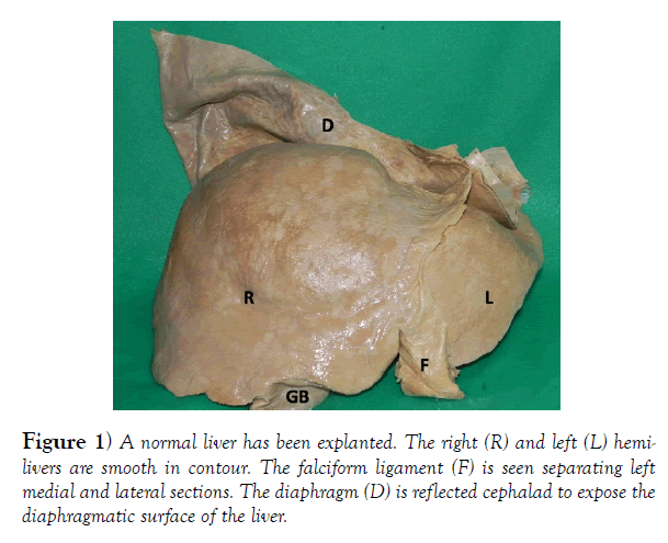
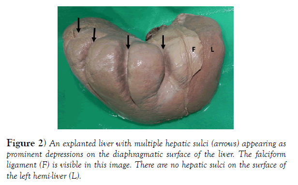
In classic descriptions of normal liver anatomy, the diaphragmatic surface of the liver is smooth and rounded (Figure 1). Hepatic sulci are morphologic anomalies [1], appearing as prominent depressions on the diaphragmatic surface of the liver (Figure 2). They range widely in their prevalence across the globe, from 5% to 40% [2,3].
There are many theories to explain their origin in humans, but this remains debated in medical literature. We report on a case we encountered where well-pronounced diaphragmatic slips were associated with deep surface grooves.
Report of a Case
A 60 year-old woman with no chronic medical diseases presented to the emergency room complaining of epigastria pain and vomiting. The abdomen was soft, but tender in the epigastrium with localized peritonitis. The serum amylase was markedly elevated at 2100 IU/L (normal 30-110 IU/L). She was diagnosed with acute pancreatitis. Her APACHE II score predicted a mild clinical course. In keeping with this, she settled promptly with medical management.
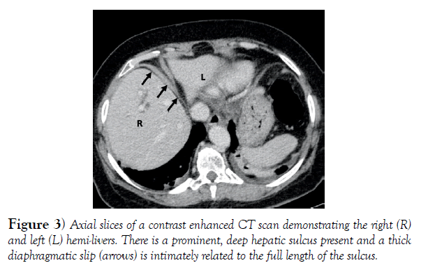
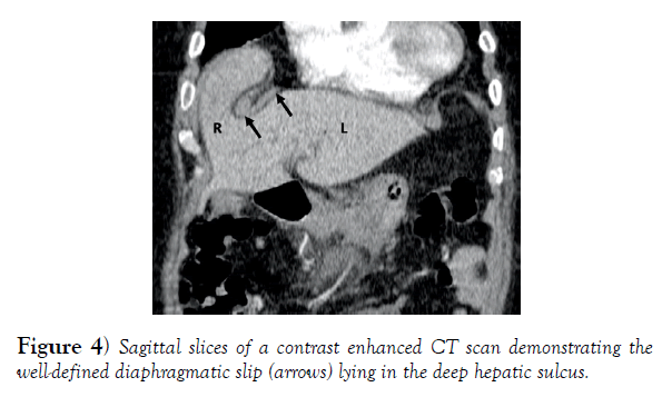
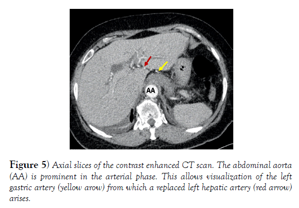
A contrast-enhanced CT scan was requested to exclude neoplastic disease. This revealed a pancreatic head mass with dilated biliary and pancreatic ductal systems creating a double-duct sign. In addition, there was a very deep hepatic surface groove at hepatic segments IV/VII/VIII. Lying in the hepatic surface groove was a thick, well-developed diaphragmatic slip (Figures 3 & 4). There was also anomalous arterial supply to the liver – a replaced left hepatic artery was arising from the left gastric artery (Figure 5).
This patient was prepared for anesthesia and taken for a Whipple’s pancreatico-duodenectomy. Her recovery was uncomplicated. Histopathology confirmed the presence of pancreatic adenocarcinoma.
Discussion
When present, hepatic surface grooves are interesting anomalies. They also have clinical significance, because they can mimic liver lacerations and liver metastases on cross sectional imaging [1,2].
Many theories have been touted to explain their presence. Some authorities suggest that these are only post-mortem changes that occur when the ribs continuously compress a single area on the diaphragmatic surface after death, leading to localized extravasation of blood in the related parenchyma to create a corresponding depression [4,5]. Supporters of this theory make the argument that this accounts for the apparent increase in prevalence on cadaveric studies [5,6]. However, the images presented in this case directly challenge the post-mortem compression theory, as they are seen in a living patient where the constant movement of the diaphragm during respiratory oscillations do not allow continuous contact pressure from the ribs.
Others have postulated that the grooves result from pressure on the hepatic surface from pulmonary emphysema [7]. However, as a part of oncologic staging in this patient with a pancreatic head carcinoma, a complete CT scan of the chest was also done and did not reveal any evidence of pulmonary emphysema – not lending support to the emphysema-theory.
Diaphragmatic slips have also been offered as a potential cause for the grooves [8]. Macchi et al [8] proposed the existence of “weak zones” on the liver surface that were amenable to compression with minimal external pressure.
Subsequently, many proposed that there were projections from the diaphragm that they termed diaphragmatic slips that exerted pressure on the “weak zones” leading to a corresponding depression [8-10]. In support of this, Newell et al [5] noted that diaphragmatic slips were oriented with a course roughly parallel to the falciform ligament corresponding with the orientation of the surface grooves.
However, we recently published a cadaveric study that evaluated specimens with hepatic surface grooves [1] and we noted that in all cases, the diaphragm appeared normal. There were no fibrotic bands, thickened diaphragmatic slips, areas of focal muscular hypertrophy, diaphragmatic scars or abnormal hepatic ligaments present in these specimens. Therefore, our conclusion was “these data did not support diaphragmatic slips as the theory to explain HSG formation.” [1]. This case, however, challenges our prior stance because there is a well-defined diaphragmatic slip present and it is clearly related to the very deep surface groove, in length and direction. Furthermore, there are no additional grooves or diaphragmatic slips.
We also had the opportunity to correlate the radiologic and operative findings in this patient. The radiologic findings were readily confirmed. It was interesting that this patient had full neuromuscular blockade to allow muscle paralysis for the surgeons to complete a Whipple’s pancreaticoduodenectomy. This should have prevented contraction of muscular diaphragmatic fibres intra-operatively, although the prominent diaphragmatic slips remained visible during our operation. There is a possibility that there was incomplete neuromuscular blockade intra-operatively, accounting for the persistence of the slips at surgery. Alternatively, the slips may have been fibrotic in nature, but this could not be histologically confirmed in our case.
We do acknowledge that this correlation in a single patient is weak (level IV) evidence in support of the diaphragmatic-slip theory. But, having observed thisrelationship in a living patient, we are forced to retract our prior statement. We must now at least re-think the diaphragmatic slip theory.
Conclusion
The findings in this case report lend some support to the diaphragmatic-slip theory for hepatic surface groove formation. However, larger clinical studies are required to confirm this relationship in living persons.
Acknowledgements
There are no additional acknowledgements.
Grant Support
No funding or research grants were made available to facilitate this research.
Presentation at meetings
This research has not been presented at any scientific meetings
Conflicts of Interest / Competing Interests
The authors have no competing interests to declare that may serve as conflicts of interest.
REFERENCES
- Gardner MT, Cawich SO, Shetty R, et al. Hepatic Surface Grooves in an Afro-Caribbean Population: A Cadaver Study. Italian J Anat Embryol. 2015;120:117-26.
- Othman FB, Latiff AA, Suhaimi FH, et al. Accessory Sulci of the Liver: An Anatomical Study with Clinical Implications. Saudi Med J. 2008;29:1247-9.
- Macchi V, Feltrin G, Parenti A, et al. Diaphragmatic sulci and portal fissures. J Anat. 2003;202:303-308.
- Auh YH, Rubenstein WA, Zirinsky K, et al. Accessory Fissures of the liver: CT and sonographic appearance. Am. J Roentgenol. 1984;3: 565- 72.
- Newell RLM, Morgan-Jones R. Grooves in the superior surface of the liver. Clin Anat. 1993;6:333-6.
- Muktyaz H, Nema U, Suniti MR, et al. Anatomical study of Accessory Sulci of Liver and its Clinical Significance in North Indian Population. Int J Med Health Sci. 2013;2:222-9.
- Schumaker U. Groovy livers. Clin Anat. 1997;10:144-5.
- Macchi V, Feltrin G, Parenti A, et al. Diaphragmatic sulci and portal fissures. J Anat. 2003;202:303-8.
- Joshi SD, Joshi SS, Athavale SA. Some interesting observations on the sur- face features of the liver and their clinical implications. Singapore Med J. 2009;50:715-9.
- Yang DM, Kim HS, Cho SW, et al. Various causes of hepatic capsular retraction: CT and MR findings: Pictorial review. Br J Radiol. 2002;75:994-1002.


