Massive gastric submucosal hematoma: An unusual complication after diagnostic gastroscopy
Received: 05-Jan-2018 Accepted Date: Jan 08, 2018; Published: 15-Jan-2018
This open-access article is distributed under the terms of the Creative Commons Attribution Non-Commercial License (CC BY-NC) (http://creativecommons.org/licenses/by-nc/4.0/), which permits reuse, distribution and reproduction of the article, provided that the original work is properly cited and the reuse is restricted to noncommercial purposes. For commercial reuse, contact reprints@pulsus.com
Abstract
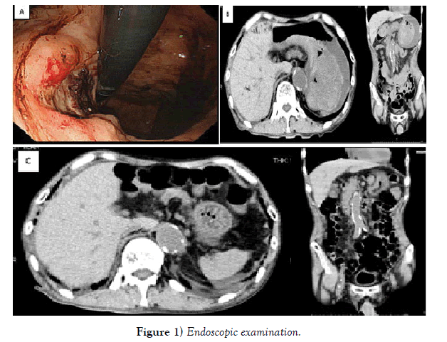
Laboratory studies were notable for a red-cell count of 1,580,000 per ml, a hemoglobin level of 4.8 g per deciliter, and hematocrit level of 15.1%. Emergency endoscopic examination revealed an active gastric ulcer (Figure 1, Panel A). Two days later, mild epigastric tenderness was present. Computed tomography of the abdomen revealed hepatic portal venous gas and massive gastric submucosal hematoma (Figure 1, Panel B); however, there was no evidence of perforation. Four days after conservative treatment, on follow-up computed tomography, neither gastric submucosal hematoma nor hepatic portal venous gas was observed (Figure 1, Panel C). Recovery was uneventful and the patient was well discharged on the 20 th hospital day.
Laboratory studies were notable for a red-cell count of 1,580,000 per ml, a hemoglobin level of 4.8 g per deciliter, and hematocrit level of 15.1%. Emergency endoscopic examination revealed an active gastric ulcer (Figure 1, Panel A). Two days later, mild epigastric tenderness was present. Computed tomography of the abdomen revealed hepatic portal venous gas and massive gastric submucosal hematoma (Figure 1, Panel B); however, there was no evidence of perforation. Four days after conservative treatment, on follow-up computed tomography, neither gastric submucosal hematoma nor hepatic portal venous gas was observed (Figure 1, Panel C). Recovery was uneventful and the patient was well discharged on the 20th hospital day.


