MDCT angiographic evaluation of multiple renal arteries – a case report
Chittapuram Srinivasan Ramesh Babu1* and Om Prakash Gupta2
1Department of Anatomy, Muzaffarnagar Medical College, Muzaffarnagar (UP), India
2Dr. O. P. Gupta Imaging Center, Bachcha Park, Meerut, India
- *Corresponding Author:
- C. S. Ramesh Babu
Associate Professor, Department of Anatomy, Muzaffarnagar Medical College, Muzaffarnagar (UP), India
Tel: +91 (121) 2768805
E-mail: csrameshb@gmail.com
Date of Received: December 21st, 2011
Date of Accepted: September 9th, 2012
Published Online: December 31st, 2012
© Int J Anat Var (IJAV). 2012; 5: 137–140.
[ft_below_content] =>Keywords
multiple renal arteries, precaval renal artery, MDCT angiography
Introduction
Variations of renal arteries occur frequently in general population with a reported prevalence of about 28–30% [1]. Multi-detector computed tomography (MDCT) angiography is a fast, reliable, non-invasive, cost effective, preferable imaging modality for comprehensive evaluation of renal vascular anatomy as it depicts exquisite anatomical details [2–4]. Two types of numerical variations of renal artery are encountered: multiple renal arteries and early prehilar branching into segmental arteries. Multiple renal arteries are of two types: hilar which enter hilum along with main renal artery; and polar, which enter the renal parenchyma directly from the capsule [5]. Variant origin of renal artery is also seen where the main renal artery or extra renal arteries arise from superior mesenteric, inferior mesenteric or common iliac arteries.
Case Report
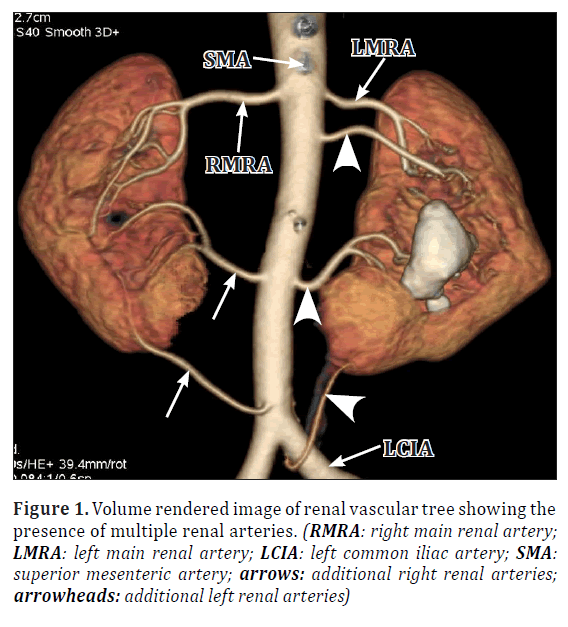
A 58-year-old male patient was referred for evaluation of a suspected mass in the right upper quadrant of the abdomen. MDCT triphasic angiography was performed using 128 slice Optima 600 scanner (GE). About 85 ml contrast (Omnipaque) was infused at the rate of 5 ml per second through controlled pressure injector. MDCT angiography revealed the presence of a malignant mass with lymph node involvement in the right lobe of liver that proved to be adenocarcinoma. A detailed study of the axial and volume rendered images revealed the presence of bilateral multiple renal arteries (Figure 1). The patient also had a replaced left hepatic artery originating from the left gastric artery.
Figure 1. Volume rendered image of renal vascular tree showing the presence of multiple renal arteries. (RMRA: right main renal artery; LMRA: left main renal artery; LCIA: left common iliac artery; SMA: superior mesenteric artery; arrows: additional right renal arteries; arrowheads: additional left renal arteries)
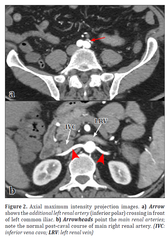
Both kidneys were malrotated with the hilum facing anteromedially, and a large calculus was present in the renal pelvis of the left kidney. Left kidney was supplied by four renal arteries. The main left renal artery arose from the lateral aspect of abdominal aorta just below the origin of superior mesenteric artery and showed prehilar branching into two segmental branches. Two hilar additional renal arteries, one arising below the origin of main renal artery and the second below the origin of inferior mesenteric artery were seen. The second additional renal artery showed prehilar branching into two segmental arteries. The lower pole of the left kidney received an inferior polar artery arising from posteromedial aspect of left common iliac artery and reached lower pole crossing anterior to left common iliac artery (Figures 1, 2).
Figure 2. Axial maximum intensity projection images. a) Arrow shows the additional left renal artery (inferior polar) crossing in front of left common iliac. b) Arrowheads point the main renal arteries; note the normal post-caval course of main right renal artery. (IVC: inferior vena cava; LRV: left renal vein)
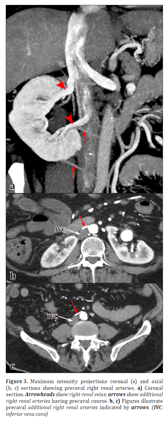
The right kidney received three arteries of aortic origin, one main renal artery and two hilar additional renal arteries. The main right renal artery originated just below the origin of superior mesenteric artery, and exhibited prehilar branching into three segmental arteries. The first hilar additional renal artery arose below the origin of inferior mesenteric artery and divided into two prehilar segmental branches. The second hilar additional renal artery arose from the anterior aspect of abdominal aorta just above its bifurcation (Figure 1). Both additional right renal arteries showed a precaval course passing anterior to inferior vena cava (Figure 3). Right kidney was drained by two renal veins (Figure 3).
Figure 3. Maximum intensity projections coronal (a) and axial (b, c) sections showing precaval right renal arteries. a) Coronal section. Arrowheads show right renal veins; arrows show additional right renal arteries having precaval course. b, c) Figures illustrate precaval additional right renal arteries indicated by arrows. (IVC: inferior vena cava)
Discussion
Study of renal vascular variations has gained importance with the increased incidence of minimally invasive surgical and radiological procedures. Successful conduction of both open and minimally invasive surgeries depends upon the familiarity of anatomical variants. Several reports indicate that multiple renal arteries are more common on the left side [6–7]. We are reporting here an incidental finding of bilateral multiple renal arteries, three on the right and four on the left, in a male patient.
Ozkan et al. studied 855 angiograms and reported an incidence of three right renal arteries in 1%, and four left renal arteries in 0.2% cases [5]. Pollak et al. reported presence of triple renal arteries in 4%, and quadruple renal arteries in 1% cases [6]. MDCT angiographic evaluation of a 36-year-old male patient revealed presence of 7 renal arteries, 4 on the right side and 3 on the left side. Out of 4 renal arteries on the right side, three were of aortic origin and one originated from right common iliac artery. On the left side, 2 originated from aorta and one arose from inferior mesenteric artery [8]. In an angiographic study of 215 adults, 2 additional renal arteries were observed in 5.6% and 3 additional arteries in 0.4% cases [9]. Evaluation of 102 live kidney donors indicated the presence of 3 and 4 renal arteries on right side in 1 case each and 5 renal arteries on the left side in 1 case [7]. Jetti et al. reported the origin of an additional renal artery supplying the lower pole of left kidney from left common iliac artery in a male cadaver similar to our case [10].
Petit reported the prevalence of precaval right renal artery to be 0.8% in a series of 380 cases [11]. Yeh et al. have reported the presence of 52 precaval right renal arteries in 48 cases, out of which 35% originated from anterior aspect of aorta [12]. In our case also one of the precaval right renal artery originated from the anterior aspect of the aorta just above its bifurcation. Such an anterior origin may be mistaken for a ventral branch of aorta and can also pose problems during endovascular embolization or stent placement procedures. Moreover, a right renal artery passing in front of inferior vena cava is also important for proper preoperative planning to avoid inadvertent damage to it.
Four precaval right renal arteries of aortic origin were also reported [13]. Three cases of precaval renal arteries, all additional vessels to the lower pole, were identified during laparoscopic and endourological procedures [14]. In a study of 50 cadavers, precaval right renal arteries were found in 6% cases out of which 3% were dominant and 3% were accessory arteries [15]. We have also observed additional right renal arteries having precaval course. Gupta et al. have also reported the presence of double precaval right renal arteries associated with four left renal arteries, all of aortic origin, in a male cadaver [16].
Knowledge of the variant renal vascular anatomy is most essential for radiologists performing various interventional procedures, surgeons performing renal transplantation, laparoscopic nephrectomy and other retroperitoneal surgeries and vascular surgeons. Accurate preoperative knowledge of renal vascular anatomy is required for proper selection of kidney and surgical planning. MDCT angiography is increasingly being employed as the preferable imaging tool for screening renal vascular anatomy, which is of paramount importance for adequate preoperative planning. This method is particularly useful for the detection of additional renal arteries, their origin and course, prehilar early branching as well as renal vein variations with an overall accuracy rate ranging from 89–100%.
References
- Satyapal KS, Haffejee AA, Singh B, Ramsaroop L, Robbs JV, Kalideen JM. Additional renal arteries: incidence and morphometry. Surg Radiol Anat. 2001; 23: 33–38.
- Hazirolan T, Oz M, Turkbey B, Karaosmanoglu AD, Oguz BS, Canyigit M. CT angiography of the renal arteries and veins: normal anatomy and variants. Diagn Interv Radiol. 2011; 17: 67–73.
- Urban BA, Ratner LE, Fishman EK. Three dimensional volume-rendered CT angiography of the renal arteries and veins: normal anatomy, variants and clinical applications. Radiographics. 2001; 21: 373–386.
- Kulkarni S, Emre S, Arvelakis A, Asch W, Bia M, Formica R, Israel G. Multidetector CT angiography in living donor renal transplantation: accuracy and discrepancies in right venous anatomy. Clin Transplant. 2011; 25: 77–82.
- Ozkan U, Oguzkurt L, Tercan F, Kizilkilic O, Koc Z, Koca N. Renal artery origins and variations: angiographic evaluation of 855 consecutive patients. Diagn Interv Radiol. 2006; 12: 183–186.
- Pollak R, Prusak BF, Mozes MF. Anatomic abnormalities of cadaver kidneys procured for purposes of transplantation. Am Surg. 1986; 52: 233–235.
- Patil UD, Ragavan A, Nadaraj, Murthy K, Shankar R, Bastani B, Ballal SH. Helical CT angiography in evaluation of live kidney donors. Nephrol Dial Transplant. 2001; 16: 1900–1904.
- Koplay M, Onbas O, Alper F, Gulcan E, Kantarci M. Multiple renal arteries: variations demonstrated by multidetector computed tomography angiography. Med Prin Pract. 2010; 19: 412–414.
- Papaloucas C, Fiska A, Pistevou-Gombaki K, Kouloulias VE, Brountzos EN, Argyriou P, Demetriou T. Angiographic evaluation of renal artery variation amongst Greeks. Aristotle Univ Med J. 2007; 34: 43–47.
- Jetti R, Jevoor P, Vollala VR, Potu BK, Ravishankar M, Virupaxi R. Multiple variations of the urogenital vascular system in a single cadaver: a case report. Cases J. 2008; 1: 344.
- Petit P, Chagnaud C, Champsaur P, Faure F. Precaval right renal artery: have you seen this? AJR Am J Roentgenol. 1997; 169: 317–318.
- Yeh BM, Coakley FV, Meng MV, Breiman RS, Stoller ML. Precaval right renal arteries: prevalence and morphologic associations at spiral CT. Radiology. 2004; 230: 429–433.
- Madhyastha S, Suresh R, Rao R. Multiple variations of renal vessels and ureter. Ind J Urol. 2001; 17: 164–165.
- Meng MV, Yeh BM, Breiman RS, Coakley FV, Schwartz BF, Stoller ML. Precaval right renal artery: description and embryologic origin. Urology. 2002; 60: 402–405.
- Gupta A, Gupta R, Singhal R. Precaval right renal artery: A cadaveric study. Incidence and clinical applications. Int J Biol Med Res. 2011; 2: 1195–1197.
- Gupta A, Kumar P, Soni G, Shukla L. Double precaval right renal artery associated with multiple left renal arteries: a rare case report. Int J Anat Var (IJAV). 2011; 4: 137–138.
Chittapuram Srinivasan Ramesh Babu1* and Om Prakash Gupta2
1Department of Anatomy, Muzaffarnagar Medical College, Muzaffarnagar (UP), India
2Dr. O. P. Gupta Imaging Center, Bachcha Park, Meerut, India
- *Corresponding Author:
- C. S. Ramesh Babu
Associate Professor, Department of Anatomy, Muzaffarnagar Medical College, Muzaffarnagar (UP), India
Tel: +91 (121) 2768805
E-mail: csrameshb@gmail.com
Date of Received: December 21st, 2011
Date of Accepted: September 9th, 2012
Published Online: December 31st, 2012
© Int J Anat Var (IJAV). 2012; 5: 137–140.
Abstract
The present article reports an incidental finding of multiple renal arteries, three on the right side and four on the left side in a male patient who was evaluated for a suspected mass in the right lobe of liver. On the right side we observed the presence of two precaval additional renal arteries of aortic origin and on the left side an additional renal artery entering the lower pole from left common iliac artery.
-Keywords
multiple renal arteries, precaval renal artery, MDCT angiography
Introduction
Variations of renal arteries occur frequently in general population with a reported prevalence of about 28–30% [1]. Multi-detector computed tomography (MDCT) angiography is a fast, reliable, non-invasive, cost effective, preferable imaging modality for comprehensive evaluation of renal vascular anatomy as it depicts exquisite anatomical details [2–4]. Two types of numerical variations of renal artery are encountered: multiple renal arteries and early prehilar branching into segmental arteries. Multiple renal arteries are of two types: hilar which enter hilum along with main renal artery; and polar, which enter the renal parenchyma directly from the capsule [5]. Variant origin of renal artery is also seen where the main renal artery or extra renal arteries arise from superior mesenteric, inferior mesenteric or common iliac arteries.
Case Report
A 58-year-old male patient was referred for evaluation of a suspected mass in the right upper quadrant of the abdomen. MDCT triphasic angiography was performed using 128 slice Optima 600 scanner (GE). About 85 ml contrast (Omnipaque) was infused at the rate of 5 ml per second through controlled pressure injector. MDCT angiography revealed the presence of a malignant mass with lymph node involvement in the right lobe of liver that proved to be adenocarcinoma. A detailed study of the axial and volume rendered images revealed the presence of bilateral multiple renal arteries (Figure 1). The patient also had a replaced left hepatic artery originating from the left gastric artery.
Figure 1. Volume rendered image of renal vascular tree showing the presence of multiple renal arteries. (RMRA: right main renal artery; LMRA: left main renal artery; LCIA: left common iliac artery; SMA: superior mesenteric artery; arrows: additional right renal arteries; arrowheads: additional left renal arteries)
Both kidneys were malrotated with the hilum facing anteromedially, and a large calculus was present in the renal pelvis of the left kidney. Left kidney was supplied by four renal arteries. The main left renal artery arose from the lateral aspect of abdominal aorta just below the origin of superior mesenteric artery and showed prehilar branching into two segmental branches. Two hilar additional renal arteries, one arising below the origin of main renal artery and the second below the origin of inferior mesenteric artery were seen. The second additional renal artery showed prehilar branching into two segmental arteries. The lower pole of the left kidney received an inferior polar artery arising from posteromedial aspect of left common iliac artery and reached lower pole crossing anterior to left common iliac artery (Figures 1, 2).
Figure 2. Axial maximum intensity projection images. a) Arrow shows the additional left renal artery (inferior polar) crossing in front of left common iliac. b) Arrowheads point the main renal arteries; note the normal post-caval course of main right renal artery. (IVC: inferior vena cava; LRV: left renal vein)
The right kidney received three arteries of aortic origin, one main renal artery and two hilar additional renal arteries. The main right renal artery originated just below the origin of superior mesenteric artery, and exhibited prehilar branching into three segmental arteries. The first hilar additional renal artery arose below the origin of inferior mesenteric artery and divided into two prehilar segmental branches. The second hilar additional renal artery arose from the anterior aspect of abdominal aorta just above its bifurcation (Figure 1). Both additional right renal arteries showed a precaval course passing anterior to inferior vena cava (Figure 3). Right kidney was drained by two renal veins (Figure 3).
Figure 3. Maximum intensity projections coronal (a) and axial (b, c) sections showing precaval right renal arteries. a) Coronal section. Arrowheads show right renal veins; arrows show additional right renal arteries having precaval course. b, c) Figures illustrate precaval additional right renal arteries indicated by arrows. (IVC: inferior vena cava)
Discussion
Study of renal vascular variations has gained importance with the increased incidence of minimally invasive surgical and radiological procedures. Successful conduction of both open and minimally invasive surgeries depends upon the familiarity of anatomical variants. Several reports indicate that multiple renal arteries are more common on the left side [6–7]. We are reporting here an incidental finding of bilateral multiple renal arteries, three on the right and four on the left, in a male patient.
Ozkan et al. studied 855 angiograms and reported an incidence of three right renal arteries in 1%, and four left renal arteries in 0.2% cases [5]. Pollak et al. reported presence of triple renal arteries in 4%, and quadruple renal arteries in 1% cases [6]. MDCT angiographic evaluation of a 36-year-old male patient revealed presence of 7 renal arteries, 4 on the right side and 3 on the left side. Out of 4 renal arteries on the right side, three were of aortic origin and one originated from right common iliac artery. On the left side, 2 originated from aorta and one arose from inferior mesenteric artery [8]. In an angiographic study of 215 adults, 2 additional renal arteries were observed in 5.6% and 3 additional arteries in 0.4% cases [9]. Evaluation of 102 live kidney donors indicated the presence of 3 and 4 renal arteries on right side in 1 case each and 5 renal arteries on the left side in 1 case [7]. Jetti et al. reported the origin of an additional renal artery supplying the lower pole of left kidney from left common iliac artery in a male cadaver similar to our case [10].
Petit reported the prevalence of precaval right renal artery to be 0.8% in a series of 380 cases [11]. Yeh et al. have reported the presence of 52 precaval right renal arteries in 48 cases, out of which 35% originated from anterior aspect of aorta [12]. In our case also one of the precaval right renal artery originated from the anterior aspect of the aorta just above its bifurcation. Such an anterior origin may be mistaken for a ventral branch of aorta and can also pose problems during endovascular embolization or stent placement procedures. Moreover, a right renal artery passing in front of inferior vena cava is also important for proper preoperative planning to avoid inadvertent damage to it.
Four precaval right renal arteries of aortic origin were also reported [13]. Three cases of precaval renal arteries, all additional vessels to the lower pole, were identified during laparoscopic and endourological procedures [14]. In a study of 50 cadavers, precaval right renal arteries were found in 6% cases out of which 3% were dominant and 3% were accessory arteries [15]. We have also observed additional right renal arteries having precaval course. Gupta et al. have also reported the presence of double precaval right renal arteries associated with four left renal arteries, all of aortic origin, in a male cadaver [16].
Knowledge of the variant renal vascular anatomy is most essential for radiologists performing various interventional procedures, surgeons performing renal transplantation, laparoscopic nephrectomy and other retroperitoneal surgeries and vascular surgeons. Accurate preoperative knowledge of renal vascular anatomy is required for proper selection of kidney and surgical planning. MDCT angiography is increasingly being employed as the preferable imaging tool for screening renal vascular anatomy, which is of paramount importance for adequate preoperative planning. This method is particularly useful for the detection of additional renal arteries, their origin and course, prehilar early branching as well as renal vein variations with an overall accuracy rate ranging from 89–100%.
References
- Satyapal KS, Haffejee AA, Singh B, Ramsaroop L, Robbs JV, Kalideen JM. Additional renal arteries: incidence and morphometry. Surg Radiol Anat. 2001; 23: 33–38.
- Hazirolan T, Oz M, Turkbey B, Karaosmanoglu AD, Oguz BS, Canyigit M. CT angiography of the renal arteries and veins: normal anatomy and variants. Diagn Interv Radiol. 2011; 17: 67–73.
- Urban BA, Ratner LE, Fishman EK. Three dimensional volume-rendered CT angiography of the renal arteries and veins: normal anatomy, variants and clinical applications. Radiographics. 2001; 21: 373–386.
- Kulkarni S, Emre S, Arvelakis A, Asch W, Bia M, Formica R, Israel G. Multidetector CT angiography in living donor renal transplantation: accuracy and discrepancies in right venous anatomy. Clin Transplant. 2011; 25: 77–82.
- Ozkan U, Oguzkurt L, Tercan F, Kizilkilic O, Koc Z, Koca N. Renal artery origins and variations: angiographic evaluation of 855 consecutive patients. Diagn Interv Radiol. 2006; 12: 183–186.
- Pollak R, Prusak BF, Mozes MF. Anatomic abnormalities of cadaver kidneys procured for purposes of transplantation. Am Surg. 1986; 52: 233–235.
- Patil UD, Ragavan A, Nadaraj, Murthy K, Shankar R, Bastani B, Ballal SH. Helical CT angiography in evaluation of live kidney donors. Nephrol Dial Transplant. 2001; 16: 1900–1904.
- Koplay M, Onbas O, Alper F, Gulcan E, Kantarci M. Multiple renal arteries: variations demonstrated by multidetector computed tomography angiography. Med Prin Pract. 2010; 19: 412–414.
- Papaloucas C, Fiska A, Pistevou-Gombaki K, Kouloulias VE, Brountzos EN, Argyriou P, Demetriou T. Angiographic evaluation of renal artery variation amongst Greeks. Aristotle Univ Med J. 2007; 34: 43–47.
- Jetti R, Jevoor P, Vollala VR, Potu BK, Ravishankar M, Virupaxi R. Multiple variations of the urogenital vascular system in a single cadaver: a case report. Cases J. 2008; 1: 344.
- Petit P, Chagnaud C, Champsaur P, Faure F. Precaval right renal artery: have you seen this? AJR Am J Roentgenol. 1997; 169: 317–318.
- Yeh BM, Coakley FV, Meng MV, Breiman RS, Stoller ML. Precaval right renal arteries: prevalence and morphologic associations at spiral CT. Radiology. 2004; 230: 429–433.
- Madhyastha S, Suresh R, Rao R. Multiple variations of renal vessels and ureter. Ind J Urol. 2001; 17: 164–165.
- Meng MV, Yeh BM, Breiman RS, Coakley FV, Schwartz BF, Stoller ML. Precaval right renal artery: description and embryologic origin. Urology. 2002; 60: 402–405.
- Gupta A, Gupta R, Singhal R. Precaval right renal artery: A cadaveric study. Incidence and clinical applications. Int J Biol Med Res. 2011; 2: 1195–1197.
- Gupta A, Kumar P, Soni G, Shukla L. Double precaval right renal artery associated with multiple left renal arteries: a rare case report. Int J Anat Var (IJAV). 2011; 4: 137–138.


