Morphological and histopathological analysis of cutaneous horn
Received: 31-Oct-2018 Accepted Date: Nov 22, 2018; Published: 30-Nov-2018
Citation: Deguchi M. Morphological and histopathological analysis of cutaneous horn. J Histol Histopathol Res 2018;2(2): 39-40.
This open-access article is distributed under the terms of the Creative Commons Attribution Non-Commercial License (CC BY-NC) (http://creativecommons.org/licenses/by-nc/4.0/), which permits reuse, distribution and reproduction of the article, provided that the original work is properly cited and the reuse is restricted to noncommercial purposes. For commercial reuse, contact reprints@pulsus.com
Abstract
Background: Cutaneous horn is a macroscopical designation to describe so many benign and malignant epidermal tumors which share unique morphological characteristics in common; hyperkeratotic colonial excrescence in which the height of the tumor must exceed at least one-half of its greatest diameter. There are some noticeable reports showing that cutaneous horn lesions with a wide tumor base or a low height-to-base ratio have a tendency to be malignant or premalignant in nature.
Objectives: To examine the possibility of discrimination between benign cutaneous horn cases and malignant or premalignant ones by using their height-to-base ratio, such cases were compared from the standpoints of the morphological data as well as their ages.
Methods: Surgically excised 14 specimens out of 13 patients of cutaneous horn were entered, and were divided into two groups; benign tumor group and the other one consisting of malignant or premalignant lesions. The two groups were compared from the standpoints of their height-to-base ratio and the ages.
Results: (p<0.05) The average age of malignant or premalignant tumor group was higher than that of benign tumor group. There was no significant difference of the greatest diameters between the benign group and malignant one. No significant difference was found between the height-to-base ratio of benign and malignant groups.
Conclusion: The discrepancy of the data between the literatures and ours may be due the difference of the numbers of patients. However, in the light of some cases of malignancy showing high height-to-base ratio, definite pathological diagnosis should be made to avoid misdiagnosis.
Keywords
Benign skin tumor; Malignant skin tumor; Actinic keratosis; Bowen disease; Squamous cell carcinoma
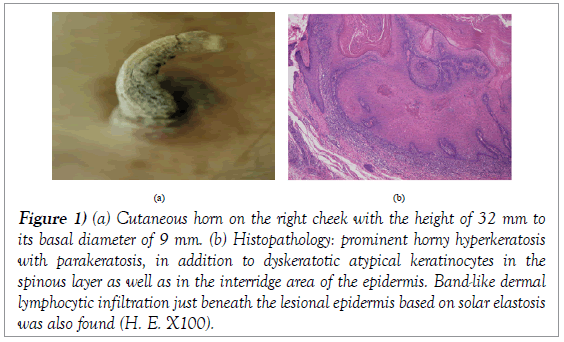
Cutaneous horn is a macroscopical designation to describe so many benign and malignant epidermal tumors which share unique morphological characteristics in common; hyperkeratotic colonial excrescence in which the height of the tumor must exceed at least one-half of its greatest diameter [1]. The term itself does not imply either malignancy or benign nature by definition. This entity includes verruca vulgaris, seborrheic keratosis, inverted follicular keratosis, trichilemmoma, keratoacanthoma, actinic keratosis, Bowen disease as well as squamaous cell carcinoma [1-5]. There are some noticeable reports showing that cutaneous horn lesions with a wide tumor base or a low height-to-base ratio have a tendency to be malignant or premalignant in nature [5,6]. On the other hand, recently a case of squamaous cell carcinoma manifested as a cutaneous horn with the larger height than the diameter of its base was reported [7]. In addition, more recently, we experienced a case of 99-year-old female presenting a cutaneous horn on the right cheek with the height of 32 mm to its basal diameter of 9 mm (Figure 1a). The histopathological findings of surgical specimen (Figure 1b) revealed prominent horny hyperkeratosis associated with parakeratosis, and dyskeratotic atypical keratinocytes were found in the spinous layer as well as in the interridge area of the epidermis. Additional findings include band-like dermal lymphocytic infiltration just beneath the lesional epidermis based on solar elastosis. These were histologically pathognomonic characteristics of actinic keratosis. Such controversial evidences on the morphology of epidermal tumors prompt us to examine the possibility of discrimination between benign cutaneous horn cases and malignant or premalignant ones by using their height-to-base ratio.
Figure 1: (a) Cutaneous horn on the right cheek with the height of 32 mm to its basal diameter of 9 mm. (b) Histopathology: prominent horny hyperkeratosis with parakeratosis, in addition to dyskeratotic atypical keratinocytes in the spinous layer as well as in the interridge area of the epidermis. Band-like dermal lymphocytic infiltration just beneath the lesional epidermis based on solar elastosis was also found (H. E. X100).
Methods
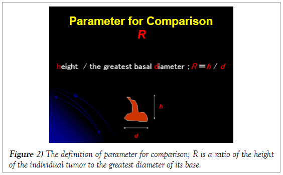
Surgically excised 14 specimens of cutaneous horn out of 13 patients for two years in our institute were entered into this examination (Table 1). To summarize, the patients ranged in age from 51 years to 90, with the average of 71.4 years. All the lesions arosed out of sun-exposed zones. The height of the tumors varied from 5 mm to 25 mm. These samples were divided into two groups according to the histopathological diagnosis; benign tumor group (benign group) (4 cases of verruca vulgaris, 1 case of seborrheic keratosis) and the other one consisting of malignant or premalignant lesions (malignant group) (7 cases of actinic keratosis, 1 case of Bowen disease, 1 case of squamous cell carcinoma in situ). The two groups were compared from the standpoints of their age, the greatest diameter of the base of the tumors, and their height-to-base ratio (R; ratio=h; height/d; the greatest diameter of the base) (Figure 2) by using Mann-Whitney U test (Dr. SPSS II for Windows, SPSS Co., Ltd Japan).
| Case # | Age | Gender | Lesional site | d | R | Pathological diagnosis |
|---|---|---|---|---|---|---|
| #1. | 90 | Male | Frontal scalp | 5 mm | 0.6 | Actinic keratosis |
| #2. | 63 | Male | Cheek | 6 mm | 0.5 | Seborrheic keratosis |
| #3. | 80 | Male | Clavicle | 5 mm | 1 | Bowen disease |
| #4. | 84 | Male | Cheek | 3 mm | 2.67 | Actinic keratosis |
| #5. | 63 | Female | Occipital | 4 mm | 1.75 | Verruca vulgaris |
| #6. | 73 | Female | Forearm | 3 mm | 1.66 | Verruca vulgaris |
| #7. | 70 | Male | Periocular | 5 mm | 2 | Actinic keratosis |
| #8. | 82 | Female | Cheek | 4 mm | 6.25 | Actinic keratosis |
| #9. | 51 | Male | Cheek | 5 mm | 1 | Verruca vulgaris |
| #10. | 53 | Male | Eyebrow | 3 mm | 2.33 | Squamous cell carcinoma in situ |
| #11. | 55 | Female | Inferior palpebra | 3 mm | 1.66 | Verruca vulgaris |
| #12. | 63 | Male | Cheek | 4 mm | 1.25 | Actinic keratosis |
| #13. | 86 | Female | Cheek | 4 mm | 1.5 | Actinic keratosis |
| #14 | 86 | Female | Eyebrow | 8 mm | 1.5 | Actinic keratosis |
Case #13 and #14 were from the same patient.
Table 1: The profiles of the patients.
Figure 2: The definition of parameter for comparison; R is a ratio of the height of the individual tumor to the greatest diameter of its base.
Results
In comparison between malignant group and benign group:
#1. Patients of malignant group were older than those in benign group (p<0.05).
#2. There was no significant difference between the greatest diameters of the base of tumors in the two groups (p<0.05).
#3. No significant difference was found between R of malignant group and that of benign group (p<0.05).
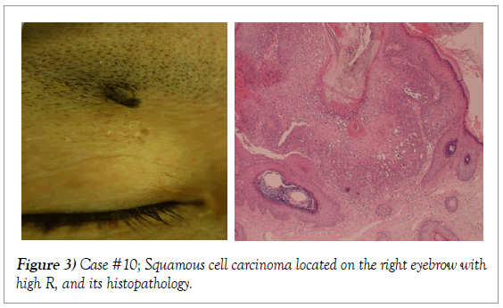
Among the cases of the present study, it is notable that case #10 presented higher R value, 2.33, despite its malignant nature (Figure 3).
Discussion
So many kinds of epidermal tumors can present cutaneous horn anywhere on the sun-exposed area, showing the peculiar appearance at the gross level. Therefore, it often needs to be discrimination between benign and malignant in origin. Among such trials, it is noteworthy that some authors proposed the hypothesis about the correlation between cutaneous horn lesions with a wide tumor base or a low height-to-base ratio and the tendency to be malignant or premalignant in nature [5,6]. On the other hand, however, controversy still remains [7]. Our present study showed no significant difference of the greatest basal diameters between benign group and malignant group. Another negative study includes the no significant difference of R between the two groups. The discrepancy of the data between those of Yu et al [5] and Pyne et al. [6] versus ours may be due to the large scales of entry patients in the formers in contrast to the small one of ours.
However, it always needs to be noted the exceptional cases out of such rules, and cutaneous horns at the gross level should prompt consideration of biopsy for the definite diagnosis. Our case #10 also seems to support such speculation.
REFERENCES
- Bart RS, Andrade R, Kopf AW. Cutaneous horns. A clinical and histopathologic study. Acta Derm Venereol. 1968;48:507-15.
- Mehrengan AH. Cutaneous horn: a clinicopathologic study. Dermatol Digest.1965;4:45-4.
- Hubler WR. Horrendous cutaneous horns. Cutis 1978;22:592-3.
- Vano-Galvan S, Sanchez-Olaso A. Images in clinical medicine. Squamous cell carcinoma manifesting as a cutaneous horn. N Engl J Med 2008;359:e10.
- Yu RC, Pryce DW, Macfarlane AW, et al. A histopathological study of 643 cutaneous horns. Br J Dermatol 1991;124: 449-52.
- Pyne J, Sapkota D, Wong JC. Cutaneous horns: clues to invasive squamaous cell carcinoma being present in the horn base. Dermatol Pract Concept 2013;3:3-7.
- Kimura R, Sugita K, Goto H, et al. Squamous cell carcinoma manifested as a cutaneous horn: A key to early detection. Yonago Acta Med2018;61:140-1


