Morphological Variations in Gall Bladders of Cadavers
Received: 28-Feb-2021 Accepted Date: May 17, 2021; Published: 24-May-2021, DOI: 10.37532/1308-4038.14(5).120-121
Citation: Parmar P, Rathee M, Rathee SK, et al. Morphological variations in gall bladders of cadavers. Int J Anat Var. 2021;14(5):89-88.
This open-access article is distributed under the terms of the Creative Commons Attribution Non-Commercial License (CC BY-NC) (http://creativecommons.org/licenses/by-nc/4.0/), which permits reuse, distribution and reproduction of the article, provided that the original work is properly cited and the reuse is restricted to noncommercial purposes. For commercial reuse, contact reprints@pulsus.com
Abstract
Gall bladder is a pear-shaped pouch located in right hypochondrium, in a shallow fossa on the quadrate lobe of the liver. There are very common variations associated with gall bladder and it is surgeon’s duty to be familiar with them so as to avoid surgical errors.
Aim: To describe external morphology, incidence of different shapes of gall bladder and to study variations in gross appearance of gall bladder
Material and method: A total number of 50 cadavers were dissected, analyzed using SPSS and tested at 5% level of significance.
Results: Out of 50 specimens studied, 92% gall bladders were found to be pear shape, 4% cylindrical, 2% flask shape and 2% irregular.
Conclusion: The knowledge of different shapes, variations and anomalies of gall bladder could be useful for radiologists and surgeons to prevent intraoperative hazards.
Keywords
Gall Bladder; Flask shape; Cylindrical
Introduction
The study and research in the field of human anatomy has a fundamental relationship and a lot of contribution in the practice of medicine and in particular to surgery. It is believed that human beings are similar in their general anatomical construction but when one investigates any particular region with more details, there found the variations in many aspects. The gall bladder is a pear-shaped pouch measuring 7×5×1 centimeter. It is situated in the right hypochondrium, in a shallow fossa on the quadrate lobe of liver. The fundus touches the anterior abdominal wall and continues with the body and the neck that narrows into cystic duct [1].
According to some eminent workers, a common pattern of variations may be found associated with gall bladder. These variations are due to arrest or deviation in normal embryological development. This knowledge of different variations and anomalies is necessary for surgeons during laparoscopic surgeries to prevent iatrogenic injuries those can leads to increase morbidity and mortality.
The present study describes variations seen in external morphology of gall bladder along with incidence and differences in dimensions and shapes.
Aim and Objectives
1.To study the incidence of different shapes of gall bladder.
2.To describe variations and anomalies associated with external morphology of gall bladder.
3.To analyze dimensions (length and breadth) of gall bladder.
Method and Materials
The present study was carried out in the Department of Anatomy in collaboration with the department of Forensic Medicine and Surgery department, PGIMS, Rohtak, Haryana, India. A total number of 50 adult human cadavers of both gender and age ranging from 18 years to 60 years were taken during medico-legal autopsies done in the department of Forensic Medicine after taking informed consent. The ethical clearance was taken from Biomedical Research Ethics Committee, Pt. B. D. Sharma Post Graduate Institute of Medical Sciences, UHS, Rohtak.
Specimens were collected as block dissection of the liver along with its associated structures and were fixed in 10% formalin for 24 hours. Gall bladder was dissected carefully and its shape, size and any associated variations were noted. Data was analyzed using SPSS 20.0 version and the result was tested at 5% level of significance. The present study was a part of larger study where extrahepatic biliary apparatus was taken into account.
Results
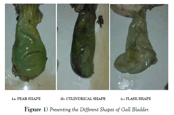
In the present study, out of total 50 cases studied, pear shaped gall bladder (Figure 1) was observed in 92% of the samples whereas cylindrical shaped (Figure 1), flask shaped (Figure 1) and irregular shaped gall bladders constituted 4%, 2% and 2% of the samples respectively. Most commonly observed shape was pear shape (as shown in Table 1).
Shapes |
No. of specimens | % (percentage) |
|---|---|---|
| Pear | 46 | 92% |
| Cylindrical | 2 | 4% |
| Flask | 1 | 2% |
| Irregular | 1 | 2% |
Table 1. Frequency of different shapes of gall bladder.
Mean length of the gall bladder was 6.47 ± 1.59 cm whereas mean breadth was 3.19 ± 0.66 cm. The length was observed to be ranging between 4 and 9cm whereas breadth of gall bladder was varying between 2 and 5.5 cm. A significant positive correlation was noted between length and breadth of gall bladder with Pearson’s correlation coefficient as 0.520 and p value as <0.01 (as shown in Table 2).
| Dimensions | Mean ± SD | Range |
|---|---|---|
| Length(cm) | 6.47 ± 1.59 | 4-9 cm |
| Breadth(cm) | 3.19 ± 0.66 | 2-5.5 cm |
| (Pearson’s correlation coefficient) r =0.520p value <0.01 | ||
Table 2. Dimensions of the gall bladder.
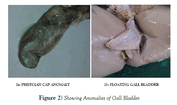
Out of total 50 cases, 3 Phrygian cap (Figure 2) and 3 Floating (Figure 2) gall bladder was observed with incidence of 6% each (Tables 3 & 4).
| Anomalies | No. of specimens | % (percentage) | |
|---|---|---|---|
| Gall bladder | Phrygian cap | 3 | 6% |
| Floating gall bladder | 3 | 6% | |
Table 3. Anomalies associated with the gall bladder.
| Authors | Length of gall bladder | Breadth of gall bladder | Shapes of gall bladder |
|---|---|---|---|
| Gregor et al6 | 7.5-10 cm | - | - |
| Turner& Fulcher7 | 10cm | 3-5cm | Elliptical (% not mentioned) |
| Moore & Dalley5 | 7-10cm | - | Pear shaped (% not mentioned) |
| Chiari & Shah2 | 7-10cm | 2-5cm | Pear shaped (% not mentioned) |
| Vakili & Pomfret8 | 7-10cm | 4cm | Piriform (% not mentioned) |
| Standring9 | 7-10cm | - | Flask shaped (% not mentioned) |
| Rajguru et al3 | 5-12cm | 2.5-5cm | Pear (85%), flask (5%), cylindrical (3.33%), hourglass & retort or irregular (1.67% each) |
| Anjankar et al10 | 7-10cm | 2-5cm | Pear (commonest) (82.22%) |
| Present study | 4-9cm | 2-5.5cm | Pear (92%), flask (2%), cylindrical (4%), irregular (2%) (Fig. 1a,b,c) |
Table 4. Comparison of dimensions and shapes of gall bladder as reported by various authors.
Discussion
The gall bladder varies greatly in size and shape and it may be impossible sometimes to distinguish between various parts described. The smallest and largest gall bladder observed in the present study was of 4 cm and 9 cm in length respectively. The length of gall bladder in the present study was slightly less than what had been reported by others. The breadth of the gall bladders in the present study ranged between 2-5.5 cm and it was similar to that reported by Chiari & Shah [2] and Rajguru et al. [3]. Gore et al. [4] stated that the size might increase after vagotomy, in diabetes because of autoimmune neuropathy, in pregnancy, in patients with sickle cell disease, after cystic duct obstruction and in extreme obese people whereas micro gallbladder was usually seen in association with cystic fibrosis. In the present study this point could not be discussed because this type of history was not taken during sample collection.
The pear shape of the gall bladder was found in most of the specimens (92%) in the present study and cylindrical, irregular and flask shaped gall bladders were also observed in 4%, 2% and another 2% of the cases respectively. These observations were in agreement with the findings of Rajguru et al [3]. But, no hourglass or retort shape of gall bladder was observed in the present study as reported by Rajguru et al. Moore & Dalley [5] and Chiari & Shah [2] also reported pear shaped gall bladder in most of the cases but percentage frequency of this type of gall bladder was not mentioned [6-10].
In the present study folded fundus (Phrygian cap) was found in three (6%) specimens. Similar findings were reported by Lichtenstein & Nicosia [11], Gore et al. [4] and Rajguru et al. [3] whereas Deutsch et al. reported this anomaly only in 0.33% cases [12].
Several authors such as Mayo and Kendrick [13], Goiney et al [14], Barnes et al [15], Bose & Sastry [16] in their studies had reported ectopic location of gall bladder, double and triple gall bladder and agenesis of gall bladder but no such findings were observed in the present study.
A wandering or floating gall bladder is suspended from a long mesentery and hanging freely from the liver bed. It is susceptible to torsion and consequent gangrene and may herniate through the foramen of Winslow into the lesser sac.
Floating gall bladder has been cited in literature in the form of numerous case reports. Kabarounders et al [17] reported a case of floating gall bladder with hypoplasia of right lobe of liver and Maeda [18] also reported a case of floating gall bladder associated with left hepatic lobe hypoplasia. In the present study, floating gall bladder was observed in 3 cases (6%).
Conclusion
The present study describes variations seen in external morphology of gall bladder along with incidence and differences in dimensions and shapes. The knowledge of different shapes, variations and anomalies of gall bladder could be useful for radiologists and surgeons to avoid surgical errors.
REFERENCES
- Ibingra CBR. Gross Anatomical variations and congenital anomalies of Surgical importance in hepatobiliary surgery in Uganda. ECAJS. 2006;12:93-8.
- Chiari RS, Shah SA. Sabiston Textbook of Surgery, in Biliary system. St. Louis: WB Saunders. 2008:1474-514.
- Rajguru J, Khare S, Jain S, et al. Variations in the external morphology of Gall Bladder. J Anat Soc Ind. 2012;61:9-12.
- Gore RM, Fulcher AS, Taylor AJ, et al. Textbook of gastrointestinal radiology, in anomalies and anatomic variants of the gallbladder and biliary tract. 2nded. Philadelphia: WB Saunders Co; 2000:1305-20.
- Moore KL, Dalley AF. Clinically oriented anatomy in abdomen. 5thed. Philadelphia: Lippincott Williams & Wilkins; 2006:302.
- Gregor MC, Decker GAG, Du Plessis DJ. Lee MC Gregor’s Synopsis of Surgical Anatomy in the liver & the biliary system. Bombay: KM Verghese& company. 1986:78-103.
- Turner MA, Fulcher AS, Gore RM, et al. Textbook of gastrointestinal radiology in gall bladder and biliary tract: normal anatomy and examination techniques. 2nd ed. Philadelphia: WB Saunders. 2000;2:1250-76.
- Vakili K, Pomfret EA. Biliary Anatomy and embryology. Surg Clin N Am. 2008;88:1159-74.
- Standring S. Gray’s anatomy: The anatomical basis of clinical practice, in gall bladder and biliary tree. 40th ed. Philadelphia: Elsevier Churchill Livingstone; 2008: 69;1177-81.
- Anjankar VP, Panshewdikar PN, Joshi DS, et al. A cadaveric study involving variations in external morphology of gall bladder. Int J Med Res Health Sci. 2013;2:239-42.
- Lichtenstein M, Nicosia AJ. The clinical significance of accessory hepato biliary ducts. Ann Surg. 1955;141:120-4.
- Deutsch AA, Englestein D, Cohen M, et al. Septum of the gall bladder, clinical implications and treatment. Postgrad Med J. 1986;62:453-6.
- Mayo CW, Kendrick DB. Anomalies of the gall bladder: report of a case of left sided floating gall bladder. Arch Surg. 1950;60:668-73.
- Goiney RC, Schoenecer SA, Cyr DA, et al. Sonography of gallbladder duplication and differential considerations. Am J Roentgenol. 1985;145:241-3.
- Barnes S, Nagar H, Levine C, et al. Triple gall bladder: preoperative sonographic diagnosis. J Ultrasound Med. 2004;23:1399-402.
- Bose SM, Sastry RA. Agenesis of gall bladder with choledocho-colonic fistula. Am J Gastroenterol. 1983;78:34-5
- Kabarounders A, Papaziogas B, Atmatzidis K, et al. Hypoplasia of the right hepatic lobe combined with floating gall bladder. Acta Chir Belg. 2003;103:425-7.
- Maeda N, Horie Y, Shiofa G, et al. Hypoplasia of the left lobe associated with floating gall bladder. 1998;45:100-3.


