Morphological variations of Cadaveric Livers in Dakshina Kannada population
Received: 01-Sep-2022, Manuscript No. PULIJAV-20-9358; Editor assigned: 05-Sep-2022, Pre QC No. ijav-20-9358 (PQ); Accepted Date: Sep 23, 2022; Reviewed: 19-Sep-2022 QC No. ijav-20-9358; Revised: 23-Sep-2022, Manuscript No. ijav-20-9358 (R); Published: 30-Sep-2022, DOI: 10.37532/1308-4038.15(9).219
Citation: Nallathamby R, Morphological variations of cadaveric livers in Dakshina Kannada population. Int J Anat Var 2022;15(9):223-227.
This open-access article is distributed under the terms of the Creative Commons Attribution Non-Commercial License (CC BY-NC) (http://creativecommons.org/licenses/by-nc/4.0/), which permits reuse, distribution and reproduction of the article, provided that the original work is properly cited and the reuse is restricted to noncommercial purposes. For commercial reuse, contact reprints@pulsus.com
Abstract
Liver is the largest gland in the body mainly situated in the right upper quadrant of the abdomen. Abnormalities of liver are rarely reported. Even though they are present in society, most of them are asymptomatic and remain undetected. The morphological abnormalities of liver can cause diagnostic confusion for physicians, surgeons, radiologist and anatomists.
Keywords
Morphological; liver; lobes; gall bladder
INTRODUCTION
A sound knowledge of the normal and variant liver anatomy is a prerequisite for a favorable surgical outcome. Knowledge of commonly occurring variations assumes more significance in the era of diagnostic imaging and minimally-invasive surgical approaches. Accessory lobe may be confused with tumor. Accessory fissure may mimic internal trauma at the time of the autopsy. In any operative procedure involving the liver, a surgeon’s knowledge of hepatic anatomy is vital in determining the outcome. Hence to alert clinicians, surgeons and radiologists and to add information to the data base of anatomists, this study is undertaken [1].
Objective
Although segmental anatomy of liver has been extensively researched, very few studies have dealt with the surface variations of liver. Aim of present study is to find out the morphological variation of liver lobes occurring in Dakshina Kannada population [2].
CASE STUDY
The materials used for present study comprises 60 adult livers with age ranging from 40 to 75 years which were dissected during routine dissection classes for medical undergraduate students over a period of 12 years in Department of Anatomy, Yenepoya Medical College, Mangalore. The Formalin fixed livers were carefully studied for the presence morphological variations such as presence of accessory lobes, accessory fissures atrophy of lobes, accessory fissures, elongated lobe, presence of Hartmann’s pouch, absence of cystic notch , accessory processes etc. Livers with features of cirrhosis or any damage or diseases were excluded. This was an observational study with no usage of any experimental instruments. Appropriate measurements were taken by calipers and measuring tape. The specimens were photographed and the findings were appropriately documented [3].
DISCUSSION
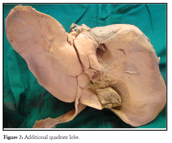
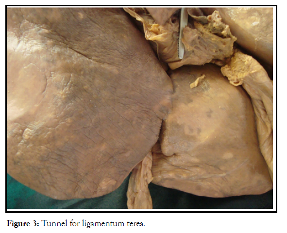
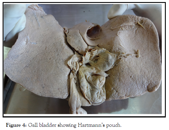
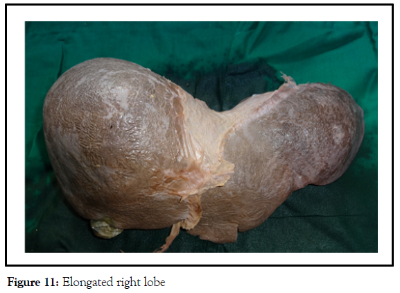
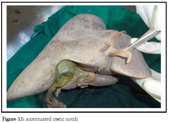
In our study out of 60 livers studied, we found morphological variations in 35 livers ie; 58.3%. We found Accessory liver lobes in 20 cases .i.e. 33.3%. Atrophy of left lobe of liver in 2 cases i.e. 3.3%, Accessory fissures (ranging from 1-5) in 23 cases i.e. 38.3%. Elongated right lobe in 2 cases i.e.3.3 %, interconnected left lobe and Quadrate lobe with absence of fissure for ligamentum teres in one case i.e. 1.6 % .and 1 case (1.6%) showed fissure for ligamentum teres not extending into the inferior border. In 13 cases ligamentum teres is found to run in a tunnel. (21.6%). 2 cases (3.3%) showed up with additional quadrate lobe and 7 cases (11.6%) showed accessory process from quadrate lobe. One liver (1.6%) showed additional caudate lobe and one liver (1.6%) had an unusually prominent caudate process [4]. 2 cases (3.3%) showed an exaggerated cystic notch whereas 2 livers (3.3%) had no cystic notches in their inferior borders with Gallbladder not extending into the inferior border. One case also showed the presence of Hartmann’s pouch [5].
According to hepatic anomalies can be divided into two categories, i.e. anomalies due to defective development and anomalies due to excessive development of the liver.
The liver tissue in the communicating with the main mass of liver is termed as accessory lobe while the liver tissue lying in the vicinity of the liver termed as ectopic liver [6]. Accessory lobe of the liver is very rare variation which may remain silent in many subjects. Accessory lobes, if present, are usually seen on the inferior surface of the liver, but also reported to be seen on the gall bladder surface and hepatogastric ligament [7-10]. In our study we found accessory lobes in33.3% of livers (Figure 1).
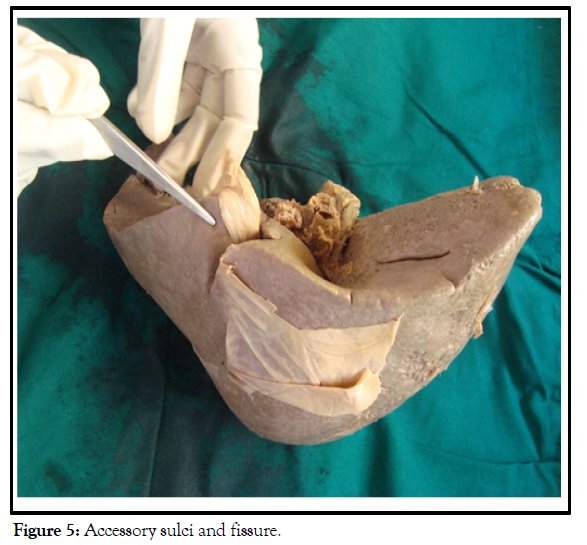
This study showed presence of accessory fissures in 23 cases i.e.; 38.3% of livers. Which inferior surface showed their presence in 7 cases, superior surface4 cases, caudate lobe 4 cases, anterior surface and posterior surface showed fissures in 1 case each (Figure 3).
Multiple accessory fissures may mimic pathologic liver nodules on CT and may be associated with diaphragmatic scalloping or eventration on the chest film (Figure 4).
When only parts of these fissures are seen sonographically, they may be mistaken for echogenic liver lesions [11]. Usually the diaphragm which is related to the superior surface may exert costal pressure to give rise to diaphragmatic fissures (Figure 5).
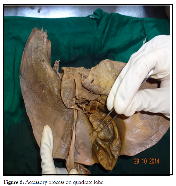
Any collection of fluid in these fissures may be mistaken for a liver cyst, intrahepatic hematoma or liver abscess [12]. Implantation of peritonealdisseminated tumor cells into these spaces may mimic intrahepatic focal lesions (Figure 6).
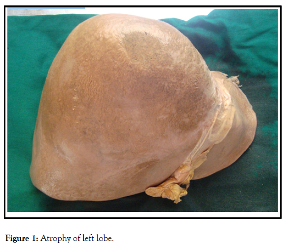
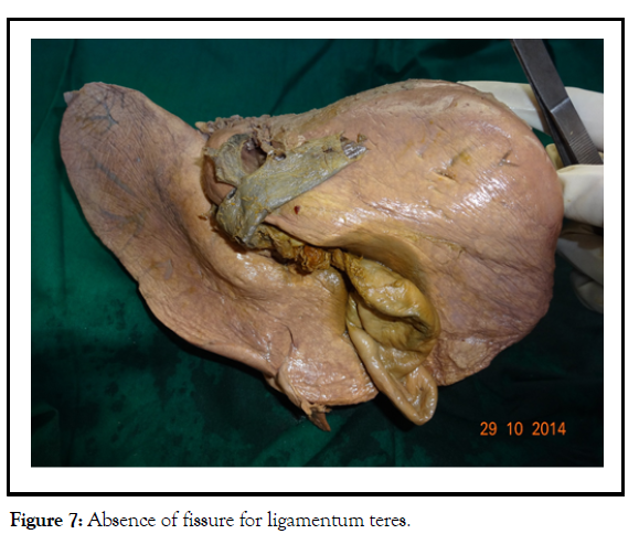
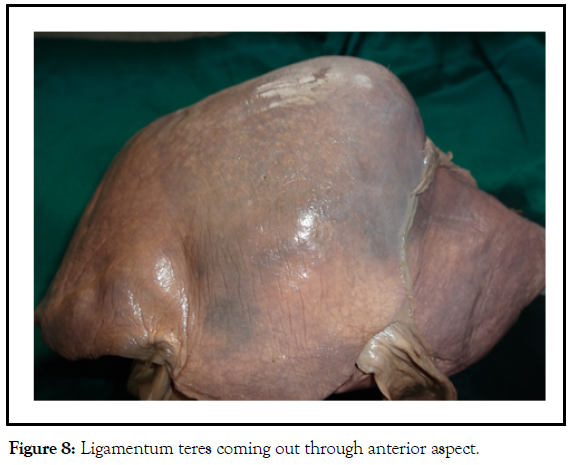
5 in her study revealed the presence of accessory lobes (6%) and accessory fissures (24%). found accessory liver lobes in 6 cadavers 14.6%, atrophy of left lobe in 2 cadavers 4.8%, accessory fissures in 5 cases 12.1%.6 In this study we got 3.3% of cases with atrophy of left lobe (Figures 7 and Figures 8).
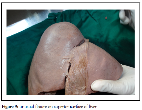
Lobar atrophy of the liver due to causes other than liver tumor or liver cirrhosis is a relatively rare pathological condition, and there are only a few reports in the literature.6. Hepatic lobar atrophy usually occurs in the setting of combined biliary and portal vein obstruction [13]. A significant correlation exists between hepatic lobar atrophy and ipsilateral portal vein obstruction (Figure 9).
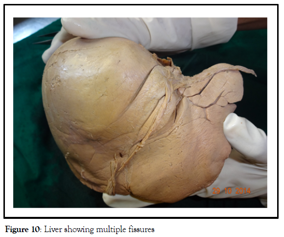
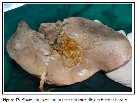
1 liver showed complete absence of fissure for ligamentum teres with hepatic tissue bridging where the ligamentum teres come out through the anterior surface and in 1 liver the fissure was not extending up to the inferior border (Figure 10). In 13 cases. (21.6%) ligamentum teres is found to run in a tunnel (Figure 11). Reports a case of liver with the presence of complete tunnel instead of fissure for ligamentum teres ( Figure 12) [14].
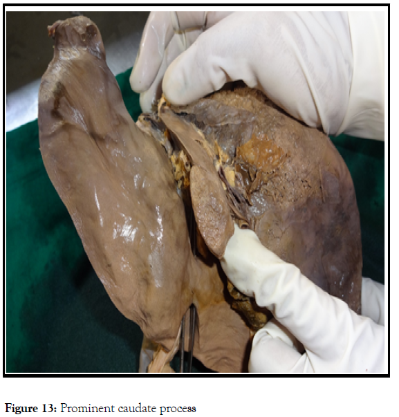
During development, the liver is separated into 2 lobes by the falciform and round ligaments by the second month of gestation, failure of which may cause the fusion of lobes to variable extent resulting in formation of a tunnel for ligamentum teres. When the patient lie in supine position, the fissure for ligamentum teres contains some air in case of pneumoperitoneum. This air is visible in radiographs as a verti¬cally directed area of hyperlucency which may be masked by the presence of hepatic tissue in case of a tunnel (12, 13). One liver (1.6%) showed additional caudate lobe and one liver (1.6%) had an unusually prominent caudate process (Figure13).
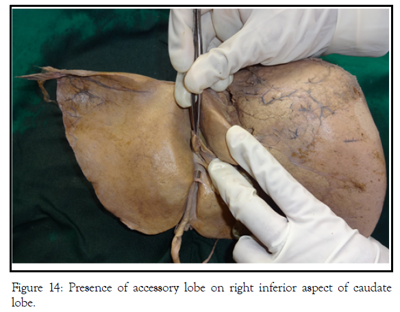
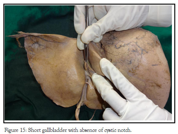
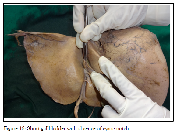
During the formation of caudate lobe, a small portion of caudate lobe may have become separated from it and included in mesentery of ductus venosus to form the accessory lobe (15). 2 cases (3.3%) showed up with additional quadrate lobe and 7 cases (11.6%) showed accessory process from quadrate lobe. Studied 90 livers where quadrate lobe was absent in 2 cases. (10) 2 cases (3.3%) showed an exaggerated cystic notch (Figure 14) whereas 2 livers (3.3%) had no cystic notches in their inferior borders with Gallbladder not extending into the inferior border (figure 15). One case also showed the presence of Hartmann’s pouch (figure 16).
The gall bladder is situated in the fossa for gall bladder on the inferior surface of Liver. Its fundus produces a cystic notch on the inferior border of the liver and projects beyond the inferior border and may touch the anterior abdominal wall near the tip of right ninth costal cartilage causing infective pathologies to spread easily into parietal peritoneum [15]. Short gall bladders hiding in their fossa, may lead to confusions in imaging and also in laparoscopic approaches. The observations are tabulated in table 1.
CONCLUSION
In this study we have described morphological variations of the liver lobes .Atrophy, presence of accessory fissure or lobe, can cause diagnostic confusion for surgeons during surgery and for physicians, radiologist and anatomist. Presence of variations in the liver may cause complications during transplantation surgeries and may present as incidental findings at autopsy creating confusions. This variations may complicate a liver transplantation surgery. The findings of study may be helpful to radiologists and surgeons respectively, to avoid possible errors in interpretations and subsequent misdiagnosis, and for planning appropriate surgical approaches.
REFERENCES
- Keith LM, Arthur FD, Anne MR. Clinically Oriented Anatomy. 6th ed. Lippincott Williams and Wilkins. 2010; 268-76.
- Champetier J, Yver R, Letoublon C. A general review of anomalies of hepatic morphology and their clinical implications. Anatomia Clinica. 1985; 7(4):285-299.
- Collan Y, Hakkiluoto A, Hästbacka J. Ectopic liver. In Annales Chirurgiae et Gynaecologiae. Clinica. 1978; 67(1): 27-29.
- Auh YH, Rubenstein WA, Zirinsky K. Accessory fissures of the liver: CT and sonographic appearance. AJR Am J Roentgenol. 1984; 143(3):565-72.
- Shailaja S, Lakshmi K, Jayanthi V. A Study of variant external features on cadaveric Livers. Anatomica Karnataka.2011; 5(3):12-16.
- Cullen TS. Accessory lobes of the liver: An accessory hepatic lobe springing from the surface of the gall bladder. Arch Surg.1925; 11(5): 718-64.
- Rendina E, Venuta F, Pescarmona E. Intrathoracic lobe of the liver: Case report and review of literature. Eur J Cardio thoracic Surg.1989; 3(1):75-78.
- Hansbrough ET, Lipin RJ. Intrathoracic accessory lobe of the liver. Annals of Surgery. 1957; 145(4):564.
- Auh YH, Lim JH, Kim KW. Loculated fluid collections in hepatic fissures and recesses: CT appearance and potential pitfalls. Radio graphics. 1994; 14(3):529-40.
- Joshi SD, Joshi SS, Athavale SA. Some interesting observations on the surface features of the liver and their clinical implications. Singapore Med J. 2009; 50(7):715.
- Ebby S, Ambike MV. Anatomical variation of ligamentum teres of liver—a case report. Webmed Cent Anat. 2012; 3(5).
- Sato S, Watanabe M, Nagasawa S. Laparoscopic observations of congenital anomalies of the liver. Gastrointest Endosc. 1998; 47(2):136-140.
- Cho KC, Baker SR. Air in the fissure for ligamentum teres: new sign of intraperitoneal air on plain radiographs. Radiology. 1991; 178(2):489-92.
- Nayak BS. A study on the anomalies of Liver in the South Indian cadavers. Int J Morphol. 2013; 31(2):658-661.
- Dodds WJ, Erickson SJ, Taylor AJ. Caudate lobe of the liver: Anatomy, embryology, and pathology. Am J Roentgenol. 1990; 154(1):87-93.
Indexed at, Google Scholar, Crossref
Indexed at, Google Scholar, Crossref
Indexed at, Google Scholar, Crossref
Indexed at, Google Scholar, Crossref
Indexed at, Google Scholar, Crossref
Indexed at, Google Scholar, Crossref
Indexed at, Google Scholar, Crossref
Indexed at, Google Scholar, Crossref
Indexed at, Google Scholar, Crossref
Indexed at, Google Scholar, Crossref
Indexed at, Google Scholar, Crossref
Indexed at, Google Scholar, Crossref
Indexed at, Google Scholar, Crossref


