Morphometric Study Of Kidney Donors By Intra Arterial Digital Subtraction Angiography (IADSA)
Received: 03-Oct-2022, Manuscript No. ijav-22-5423; Editor assigned: 05-Oct-2022, Pre QC No. ijav-22-5423 (PQ); Accepted Date: Oct 24, 2022; Reviewed: 19-Oct-2022 QC No. ijav-22-5423; Revised: 24-Oct-2022, Manuscript No. ijav-22-5423 (R); Published: 31-Oct-2022, DOI: 10.37532/1308-4038.15(10).220
Citation: Shaikh ST. Morphometric Study of Kidney Donors by Intra Arterial Digital Subtraction Angiography (IADSA). Int J Anat Var. 2022;15(10):223-224.
This open-access article is distributed under the terms of the Creative Commons Attribution Non-Commercial License (CC BY-NC) (http://creativecommons.org/licenses/by-nc/4.0/), which permits reuse, distribution and reproduction of the article, provided that the original work is properly cited and the reuse is restricted to noncommercial purposes. For commercial reuse, contact reprints@pulsus.com
Abstract
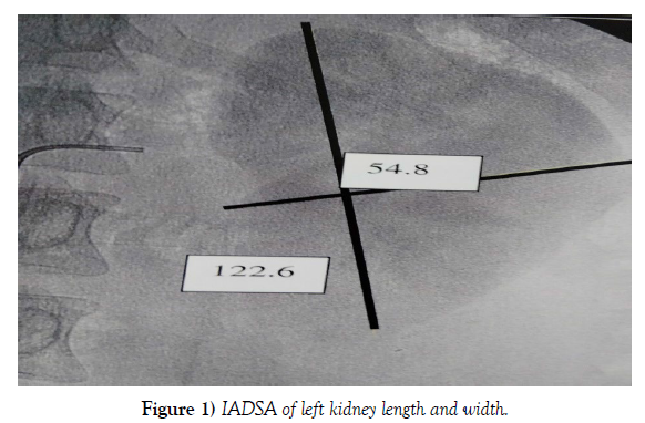
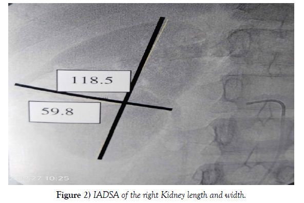
A morphometric study was done on 100 human kidneys by using IADSA in potential renal donors. Any donor with a pathology was excluded and their age ranged from 15 to 80 years. The result of renal measures showed the following averages: right kidney length 118.5mm (+/- 12mm), left kidney length 122.6mm (+/- 14mm). Thickness of right kidney was 59.8mm (+/- 4mm) and left kidney was 54.8mm (+/- 3mm). Left kidney presented usually greater length and the right kidney presented with greater width.
Keywords
Kidney length; Kidney thickness; IADSA; Donors
INTRODUCTION
To recognize anatomical variations it is necessary to establish the normal pattern of the human body. Both the body as a whole, and its internal organs and parts, show certain flexibility of size, form, structure and position. Such fluctuation within a commonly experienced range is considered as normal variations; nevertheless any departure beyond these limits is classified as anomalies or malformations [1]. Variations in the renal morphometry are no exception to this universal phenomenon.
Variations in the renal morphometry including their length and thickness are very common. It is not only ludicrous but also dangerous to suggest that both kidneys have the same dimensions.
Most of the anomalies are the persistent structures those do not disappear during the embryological process or occur due to a delay at the end of the development. As the kidneys ascend from the pelvis during the embryological development they receive their blood supply from the vascular structures close to them. In the ninth week of the intrauterine life the kidneys come in to contact with the suprarenal glands and the ascent stops. The kidneys receive their most cranial branches from the aorta. These are the permanent renal arteries. The permanent mesonephric arteries other than renal arteries are the middle supra renal, gonadal, and inferior phrenic arteries. This continuously changing blood supply of the kidneys as they ascend explains the high incidence of the variations in the blood supply to the kidneys [2].
Aim: To study the morphometric pattern of human kidney in potential Indian renal donors.
MATERIALS AND METHODS
Renal morphometry was studied in 50 subjects. The arteriograms were obtained from the department of Vascular and Interventional radiology of Seth G.S. Medical College & K.E.M. Hospital, Lower Parel, and Mumbai. The consent of the ethics committee was taken prior to the data collection and analysis. The total number of subjects studied was 50 out of which 24 were males and 26 were females. The total number of kidneys studied was 100. The subjects’ age ranged from 15 to 80 years (mean 49.5). Intra-arterial digital subtraction angiography was performed via per-cutaneous puncture through transfemoral approach.
OBSERVATION
Intra-arterial digital subtraction angiography was done in 50 subjects and the following result was obtained (Table 1-2) (Figure 1-2).
Right Kidney |
Left Kidney |
| 118.5mm | 122.6mm |
Table 1. Average length of the kidneys.
Right Kidney |
Left Kidney |
| 59.8mm | 54.8mm |
Table 2. Average thickness of the kidneys.
DISCUSSION
Variability is the law of life said Sir William Osler. Most variations are totally benign and some are errors of embryological development. The advent of more conservative methods in renal surgery has necessitated a more precise knowledge of renal dimensions. Many surgeons in the treatment of renal tuberculosis and calculus advocate partial nephrectomy. Familiarity with the normal renal anatomy and its vasculature including variants like presence of accessory arteries, early branching of artery, anomalous venous anatomy and ureteric abnormalities may influence the choice of the kidney for donor nephrectomy. This information is of paramount importance to the radiologists and surgeons for therapeutic interventions.
The differentiated observation of kidney sizes is of great significance clinically, as many diseases are associated with changes in kidney size [3]. The normal range is large [4], and what is “normal” depends on many factors. The influencing factors for size must be viewed individually to arrive at any relevant conclusions and information. In addition to kidney dimensions the renal vasculature can also be studied at a single go and hence potential renal donors may be included or excluded. The size of the kidney is important for transplantations. While the leading anatomy text describes the adult kidney as 12 cm long, 6 cm wide and 3 cm deep [2], further review of the literature shows that renal size varies with age, gender, body mass index, pregnancy and co-morbid conditions [5-6]. Renal size may be an indicator for the loss of kidney mass and therefore, kidney function [5-6]. It is valuable in monitoring unilateral kidney disease through comparison with the other, compensatorily increased side [7] and for the discrimination between upper and lower urinary tract infections8. Renal infections/inflammations, nephrologic disorders, diabetes mellitus and hypertension are the most important co-morbid conditions affecting renal size [8-9]. Since the renal size is affected by various factors, it is necessary to first establish the normal values. The information available in the West may not be extrapolated to our population since the renal size may differ between ethnic groups and according to body size [10- 12]. While population-based studies are needed to establish the normal values for Indian individuals, in our study we determined the renal size in a group of individuals with no known renal disease and compared our findings with the literature.
Renal arteriography may be performed either for diagnostic purposes or as a baseline before an interventional procedure. Detection of any dimensional changes is required, such as in cases of renal transplantation and Reno vascular hypertension.
CONCLUSION
In summary, normal values for kidney measurements are dependent on age, sex and body mass index. This has to be considered by the radiologists. Aberrations from these values can give valuable general clues and confirmations in the diagnosis of particular diseases. A slightly small right kidney may be considered as normal and a reference table has to be developed for routine evaluation. For the Indian population, normal kidney measurement values need to be developed by means of population-based studies.
ACKNOWLEDGEMENT
Author is thankful to Dean Dr. Varsha Phadke Madam for her support and encouragement. Author is also thankful to Dr. Arif A. Faruqui for his help. Author also acknowledges the immense help received from the scholars whose articles are cited and included in references of this manuscript. The author is also grateful to authors / editors / publishers of all those articles, journals and books from where the literature for this article has been reviewed and discussed.
REFERENCES
- Sanudo JR, Puerta VJR. Meaning and clinical interest of the anatomical variations in the 21st century. Eur J Anat. 2003; 7(1):1-3.
- Standring S. Grays Anatomy The anatomical basis of clinical practice. ELSEVIER. 2005:12-74.
- Emamian SA, Nielsen MB, Pedersen JF, Ytte L et al. Kidney dimensions at sonography: correlation with age, sex, and habitus in 665 adult volunteers. AJR. 1993; 160:83-86.
- Hodson CJ. Hypertension of Renal Origin. In Modern Trends in Diagnostic Radiology. New York 3 Series edition. Edited by McLaren JW, Hoeber PB. 1969; 124-134.
- Shcherbak Al. Angriographic criteria in the determination of indications for organ preserving surgery in renal artery occlusion. Klin Khir. 1989; 2:5.
- Guzman RP, Zierler RE, Isaacson J. Renal atrophy and arterial stenosis. A prospective study with duplex ultrasound. Hypertension. 1994; 23:346-347.
- Yamaguchi S, Fujii H, Kaneko S. Ultrasonographic study in patients with chronic renal failure. The Jpn J Neurosurg. 1990; 81:1175-77.
- Dinkel E, Ortit S, Dittrich M. Renal sonography in the differentiation of upper front lower urinary tract infection. Am J Roentgenol. 1986; 146:775-777.
- Yamada-H, Hishida-A, Kumagai-H. Effects of age, renal diseases and diabetes mellitus on the renal size reduction accompanied by the decrease of renal function. Nippon Jinzo Gakkai Shi. 1992; 34:1071-75.
- Emamian SA, Nielsen MB, Pedersen IF. Kidney dimensions at sonography. Correlation with age, sex and habitus in 665 adult’s volunteers. Am J Rocntgenol. 1993; 160:83-84.
- Odita JC, Ugbodaga Cl. Roentgenologic estimation of kidney size in adult Nigerians. Trop. Geogr Med. 1982; 34: 177-79.
- Wang F, Cheok SP, Kuan BB. Renal size in healthy Malaysian adults by ultrasonography. Med J Malaysia. 1989; 44:45-46.
Indexed at, Google Scholar, Crossref
Indexed at, Google Scholar, Crossref
Indexed at, Google Scholar, Crossref
Indexed at, Google Scholar, Crossref
Indexed at, Google Scholar, Crossref
Indexed at, Google Scholar, Crossref


