Multiple Arterial, Neural and Muscular Variations in the Root of the Neck, Axilla and Arm of Single Cadaver
Received: 26-Mar-2021 Accepted Date: Apr 10, 2021; Published: 17-May-2021, DOI: 10.37532/1308-4038.14(5).122-123
Citation: Tessema CB. Multiple Arterial, Neural and Muscular Variations in the Root of the Neck, Axilla and Arm of Single Cadaver. Int J Anat Var. 2021;14(5):86-88.
This open-access article is distributed under the terms of the Creative Commons Attribution Non-Commercial License (CC BY-NC) (http://creativecommons.org/licenses/by-nc/4.0/), which permits reuse, distribution and reproduction of the article, provided that the original work is properly cited and the reuse is restricted to noncommercial purposes. For commercial reuse, contact reprints@pulsus.com
Abstract
During the dissection of the root of the neck, axillae and upper limbs of a 77-year-old female cadaver multiple vascular, neural and muscular variations were detected. These variations included the internal thoracic artery in the left root of the neck, and the thoracoacromial artery, lateral thoracic artery and subscapular artery in the right axilla and the brachial artery in the right arm. Variations in the origin of the subscapular nerves in the right axilla, communicating branches between musculocutaneous and median nerves and a three headed biceps brachii muscle are the other variant findings in the right arm.
These findings demonstrate the concept that no two parts of the same individual are anatomically exactly alike. Therefore, serious attention should be paid to such major variations during dissection for a better anatomy teaching and during clinical procedures to avoid unintended damage and injury of variant structures for a positive clinical outcome.
Keywords
Arterial variation, Neural variation, Muscular variation, Common stem, Communicating branch
Introduction
The internal thoracic artery, which is the source of anterior intercostals, pericardiacophrenic, musculophrenic and superior epigastric arteries branches from the inferior aspect of the first part of the subclavian artery. Instead, this artery can also arise from the thyrocervical trunk, the third part of subclavian artery [1] or from the aortic arch [2]. In addition to its normal anatomical distribution to the thorax and anterior abdominal wall, this artery can be used clinically in cardiac surgery for coronary artery by-pass grafting (CABG) to treat coronary heart disease.
The thoracoacromial artery (trunk) is a branch from the second part of the axillary artery and provides pectoral, clavicular, deltoid and acromial branches in variable patterns [3]. This artery can be absence in which case all its branches arise directly from the second part of the axillary artery [4].
The lateral thoracic and subscapular arteries arise from the second and third parts of the axillary artery respectively. However, the lateral thoracic artery shows morphological variability in its origin, the commonest being from the thoracoacromial artery [5]. The subscapular artery also shows variability in its origin by arising independently from the second part of the axillary artery or via a common trunk with lateral thoracic artery [5].
Three subscapular nerves (upper, middle or thoracodorsal and lower) are traditionally known to originate from the posterior cord of the brachial plexus. However, there are reports that showed upper subscapular nerve, accessory subscapular and middle subscapular arising from the posterior cord of the brachial plexus with the lower subscapular nerves branching from the axillary nerve [6]. It is also known that all three subscapular nerves can arise from axillary nerve [7].
The brachial artery is a single artery which is the continuation of the axillary artery from the level of the inferior (lateral) border of teres major muscle to the cubital fossa and provides branches to the arm, shoulder and elbow joints. However, Superficial (accessory) brachial artery that arises from the second or third part of the axillary artery or from the brachial artery itself, either unilaterally or bilaterally was previously noted in various studies [8].
Median nerve that arises by medial and lateral root from the respective cords of the brachial plexus is a nerve to the forearm and hand and provides no branches in the arm except few articular twigs to the elbow joint. While musculocutaneous nerve that arises from the lateral cord of the brachialplexus is a nerve to the anterior compartment of the arm and to the skin of the radial side of the forearm. It is well stablished that these two nerves form variable number of communications between them along their course to the arm and forearm [9].
The biceps brachii is a two headed largest muscle of the anterior arm compartment; the two heads of which originate from the supraglenoid tubercle and coronoid process of the scapula and inserts distally to antebrachial fascia and to radial tuberosity. Several previous studies and reports revealed that the biceps brachii can have more than two heads of origin that could be one – five in number [10]. In cases where third and fourth heads existed, they frequently took their origins from the shaft of the humerus.
Materials and Methods
During the routine dissection of a 77-year-old female cadaver, an arterial variation in the left root of the neck and arterial, muscular and neural variation in the right axilla and arm were detected. The arteries, nerves and muscles were carefully dissected and the variations were painted using ordinary painting ink for the purpose of distinction and pictures were taken for illustration. Name were assigned to the variant structures for which appropriate names cannot be found.
Case Report
A. Left root of the neck
In left root of the neck, it is noted that the thyrocervical trunk from the first part of the subclavian artery broke up into three branches: 1) the inferior thyroid artery, 2) a common stem for superficial cervical and suprascapular (CS) arteries, and 3) the internal thoracic artery (Figure 1). No other variation was observed in the rest of the left side.
Figure 1) Shows the left root of the neck and the left subclavian artery. The thyrocervical trunk divides into three major branches: the inferior thyroid artery, a common stem for superficial cervical and suprascapular arteries (CS), and the internal thoracic artery.
B.Right Axilla
In the right axilla, the following three vascular and neural variations involving the branches of the axillary artery and the subscapular nerves were noted.
1.The thoracoacromial artery was found to be absent and all its branches (clavicular, acromial, deltoid and pectoral) arose directly from the second part of the axillary artery (Figure 2).
Figure 2) Shows the three arterial variations in the right axilla. 1) The thoracoacromial artery is absence with its four branches arising directly from the superior aspect of the 2nd part of the axillary artery. 2) The common lateral thoracic artery from the second part of axillary artery divided into proper lateral thoracic and thoracodorsal arteries. 3) The subscapular artery arose from the third part of the axillary artery branched into circumflex scapular and the two muscular branches.
2.A common lateral thoracic artery arose from the second part of the axillary artery and descended anterior to the anterior division of the inferior trunk of the brachial plexus. Just after crossing over the anterior division of the inferior trunk of the brachial plexus it divided into a proper lateral thoracic artery to the lateral wall and thoracodorsal artery that further divided into branches that entered the latissimus dorsi and serratus anterior muscles (Figure 2). The subscapular artery originated from the third part of the axillary artery the usual way, and divided into three branches (upper, middle and lower). The upper branch entered teres major muscle, the middle and largest branch entered the medial triangular space (axillary hiatus) as circumflex scapular artery and the lower branch ended in the subscapularis muscle (Figure 2).
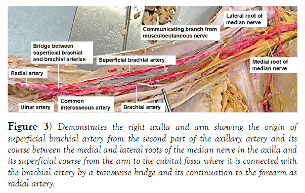
3.The thoracodorsal and lower subscapular nerves were found to branch from the axillary nerve (Figure 3).
Figure 3) Demonstrates the right axilla and arm showing the origin of superficial brachial artery from the second part of the axillary artery and its course between the medial and lateral roots of the median nerve in the axilla and its superficial course from the arm to the cubital fossa where it is connected with the brachial artery by a transverse bridge and its continuation to the forearm as radial artery.
C.Right arm
In the right arm, also three notable vascular, neural and muscular variations were detected.
1. A superficial brachial artery originated from the second part of the axillary artery, passed between the two roots of median nerve and ran superficial to the brachial artery and median nerve to the cubital fossa. It gave no branches in the arm. In the cubital fossa, deep to the bicipital aponeurosis, it joined the brachial artery via an arterial bridge and continued as radial artery to the forearm.
2.The right musculocutaneous nerve gave a communicating branch within the coracobrachialis muscle that joined the median nerve. At the middle of the arm the median nerve also gave a communicating branch that joined the lateral antebrachial cutaneous nerve that branches from the musculocutaneous nerve (Figure 4).
Figure 4) Illustrates the right arm with double communication between median and musculocutaneous nerves. Themusculocutaneous nerve within the coracobrachialis muscle gave a communicating branch to median nerve and at the middle of the arm, the median nerve gave a communicating branch that joined the lateral antebrachial cutaneous branch of musculocutaneous.
3.Moreover, a three-head right biceps brachii muscle was also detected. The short and long heads are formed the usually way but the third (accessory head) originated by fleshy fiber from the ventromedial aspect of the shaft of the humerus between the attachments of coracobrachialis and brachialis muscles (Figure 5)
Discussion
The internal thoracic artery, instead of arising from the first part of the subclavian artery, can be a branch from the thyrocervical trunk, third part of subclavian artery [1] or from the aortic arch [2]. What is found in the current case confirms the variant origin of the internal thoracic artery from the thyrocervical trunk. In a study conducted on 56 cadavers the thoracoacromial artery was foundin all the cases as it arises from the second part of the axillary artery [3]. However, a case of complete absence of this artery with all its branches arising from the second part of the axillary artery was also reported [4], which is in agreement with the finding in this case report (Figure 2), where the right thoracoacromial trunk was absent and all its branches originated from the second part of the axillary artery. The lateral thoracic and subscapular arteries are the other arteries that can have variable origin and branching patterns in which case the lateral thoracic artery can arise from the thoracodorsal artery in the third part of the axillary artery [5] or can arise via a common trunk with the subscapular artery from the second part of the axillary artery [6]. However, in this present case (Figure 2), the two arteries took their usual origins from second and third parts of the axillary artery respectively but they showed variability in their branching patterns. A common lateral thoracic artery branched from the second part of the axillary artery and broke up into proper lateral thoracic and thoracodorsal arteries. The subscapular artery also took it origin from the third part of the axillary artery and divided into muscular branches to teres major and subscapularis muscles and the circumflex scapular artery. The vascular variation in this case report is, therefore, completely different from those previously reported cases.
Though the upper subscapular, thoracodorsal and the lower subscapular nerves frequently arise from the posterior cord of the brachial plexus, a previous study done on 57 cadavers revealed that the upper subscapular nerve (in 50%) and accessory subscapular nerve (in 21.1%) arose from the posterior cord of the brachial plexus, while the lower subscapular nerve (in 54.4%) originated from axillary nerve [6] and in another case all the three subscapular nerves took their origin from the axillary nerve [7]. Whereas, the present case (Figure 2) showed only the thoracodorsal and lower subscapular nerves branched from the axillary nerve.
The existence of superficial brachial artery is long known with the incidence of 12% – 19.7%. In a report that documented three cases of superficial brachial artery, in one of the cases it terminated by bifurcating to radial and ulnar arteries, in the second case it crossed the cubital fossa superficial to the bicipital aponeurosis and continued as radial artery and in the third case it also continued superficially and became ulnar artery [8]. However, the present case (Figure 3) showed that the superficial brachial artery in the cubital fossa undercrossed the bicipital aponeurosis where it communicated with brachial artery by an arterial bridge and then continued as radial artery.
Various forms of communications from the musculocutaneous to median nerve or vise vera were well documented. In a study done on 53 cadavers the most frequent communications are from musculocutaneous to median nerve (17%) while median to musculocutaneous communications constituted on a few (2.8%) [9]. The finding in the present case (Figure 5) is different from this previous report in that there is a unique double communication between the two nerves (musculocutaneous to median and median to lateral antebrachial cutaneous branch of musculocutaneous).
A study on 60 upper limbs revealed an incidence of variation of biceps brachii muscle as high as 15%; 13.3% of which were three headed biceps brachii. In 8.3% of the three headed biceps brachii, the third head took its origin from the anterior surface of the humerus just above the origin of the brachialis muscle [10]. The find in this current report (Figure 5) goes in line with this previous report.
Conclusion
The findings in this report demonstrate the concept that no two parts of the same individual are anatomically exactly alike. The presence of such combinations of variants structures in a very limited area of the body like the root of the neck, axillae and cubital fossa could be causes ofunintended injuries with consequent sequelae during different procedures. Therefore, serious attention should be paid to such major variations during dissection for a better anatomy teaching and during clinical procedures to avoid the unintended damages and injuries of variant structures for a positive clinical outcome.
Acknowledgements
My thanks go to the donor and her families for their invaluable consent and the department of biomedical sciences for its encouragement and support. I also like to express my gratitude to Denelle Kees, Chelsy Swanson and John Opland for their uninterrupted technical assistance during the preparation and dissection of this cadaver.
REFERENCES
- Delmotra P, Goel A, Singla RK. Variant anatomy of internal thoracic artery-Clinical implication. Int J Anat Res. 2019;7:6489-93.
- Desai R, Rubinsztian L, Kacharava A. Anomalous left internal mammary artery arising from an aberrant artery of the aorta in a patient with “pseudo bovine” aortic arch anatomy. JC Cases. 2016;14:11-12.
- Bhattacharya S, Zaman AU. Variations in the branching patterns of thoracoacromial artery among the West Bengal population. Indian J Basic Appl Med Res. 2019;8:314-22.
- Astik R, Dave U. Variations in branching patterns of axillary artery: a study of 40 human cadavers. J Vasc Bra. 2012;11:12-17.
- Bhavya BS, Kadadi SP, Smitha M, et al. A cadaveric study on lateral thoracic artery. Int J Anat Res. 2018;6:4973-6.
- Ballesteros LE, Ramirez LM. Variation of origin of collateral branches emerging from the posterior aspect of the brachial plexus. JBPPNI. 2007;4:14.
- Alibay S. A rare variation in the branching pattern of posterior cord. Int J Anat Var.2016;9:29-31.
- Nkomozepi P, Xhakaza N, Swanepoel E. Superficial brachial artery: A possible cause for idiopathic median nerve entrapment neuropathy. Folia Morphol. 2017;76:527-31.
- Ballesteros LE, Forero PL, Buitrago ER. Communication between the musculocutaneous and median nerves in the arm: Anatomical study and clinical implications. Rev bras ortop. 2015;50:567-72.
- Bharambe VK, Kanaskar NS, Arole V. A study of biceps brachii muscle: anatomical considerations and clinical implications. Sahel Med J. 2015;18:31-7.


