Multiple bilateral anatomical variations of the tendons of the forearm and wrist: a case report based on a cadaver study
Karl Jscobs1,2*, Roelof Jan Oostra1,Raoul Engelbert2,3 and Petra Habets1
1Departments of Anatomy, Embryology and Physiology
2Departments of Anatomy, Embryology and Physiology and Rehabilitation,Amsterdam, Netherlands.
3Academic Medical Centre, University of Amsterdam, Meibergdreef 9, 1105 AZ; Amsterdam School of Health Professions, Education of Physiotherapy, Tafelbergweg 51, 1105 BD
- *Corresponding Author:
- Karl Jacobs
Department of Anatomy Embryology and Physiology Academic Medical Centre University of Amsterdam Meibergdreef 9 1105 A Amsterdam, Netherlands
Tel: +31 626341309
E-mail: k.jacobs@uva.amc.nl
Date of Received: November 14th, 2014
Date of Accepted: September 25th, 2015
Published Online: April 8th, 2016
© Int J Anat Var (IJAV). 2016; 9: 3–7.
[ft_below_content] =>Keywords
variation, extensor pollicis, extensor digiti minimi, abductor pollicis longus,accessory muscle
Introduction
Anatomical variations of muscle and tendons are commonly encountered. These variations may consist of absence of a muscle, supernumerary muscles, deviations from the normal course, or an unusual origin or insertion. Accessory muscles or muscle slips and tendons are anatomical variations representing additional distinct muscles that are encountered along with the normal complement of muscles. Accessory muscles are typically asymptomatic and encountered as incidental findings although they have been implicated as a potential source of clinical symptoms [1].
Historically, the majority of data regarding accessory musculature has been based on serendipitous findings during surgical interventions and anatomical dissections. However, with the upcoming modern cross-section imaging techniques such as ultrasound (US), computed tomography (CT) and magnetic resonance imaging (MRI) anatomical variations are more regularly encountered and can be identified non-invasively [1].
Anatomical variations in the tendons of the wrist have been described for more than a century [2].
Reports of more than one variation in the forearm of one specimen are however quite rare. During routine human cadaver dissection at the Department of Anatomy, Embryology and Physiology of the University of Amsterdam, we found not just a single, but a multiplicity of anatomical variations. In this article we describe these three muscles and tendon variations found on this one cadaver forearm and we will discuss their clinical and educational relevance.
Case Report
The observations described here were done in the upper limbs of a formalin-fixated cadaver obtained through the body donation program of our department. It concerned a male, aged 80 years, with no known medical history. The following combination of variations was found in the left forearm:
1) Concerning the m. abductor pollicis longus (APL)
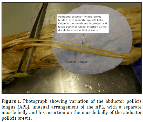
The unusual arrangement of the APL observed in the present study, a muscle with an additional muscular belly which arises from the lateral aspect of the distal portion of the typical APL muscle belly, just proximal to the formation of the APL tendon. The muscle fibers of this additional muscle belly (APL)
2) extended from the belly of the APL. The tendon continued distally and finds his insertion on the muscle belly of the abductor pollicis brevis (Figure 1). This kind of variation has been found and prescribed before by [3]. Fabrizio and Clemente have found a variation in the muscular attachment along the lateral aspect of the distal portion of the abductor pollicis longus muscle belly in the forearm by 15 out of 50 cadavers study.
2) Concerning the m. extensor pollicis longus (EPL)
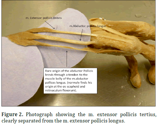
A supernumerary EPL tendon (extensor pollicis tertius) with separate muscle belly was found, originated next to the origin of the EPL on the dorsal surface of the lower third of the ulna. The muscle belly measured 3,5 cm in length en approximately 1 cm in wide. The tendon measured 11 cm and inserted medial to the insertion of the EPL on the basis of the distal phalanx (Figure 2).
The occurrence of an accessory extensor muscle with its own tendon joining the tendon of the extensor pollicis longus and inserting into the thumb alone is extremely rare. Since the early review of LeDouble in 1897, only three other cases have been reported. Even more rare is the presence of an extra extensor muscle and tendon to the thumb. The first reported case in the literature of an extra extensor muscle and tendon to the thumb was written by Abu Hijleh [4]. Our finding is similar as the finding of Abu-Hijleh [4] but the combination with the other anatomical variations found on one single forearm has never been described before.
3) Concerning the m. extensor digit minimi
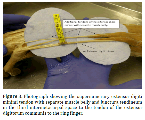
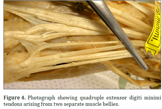
A supernumerary extensor digiti minimi tendon with separate muscle belly and junctura tendineum in the third intermetacarpal space, to the tendon of the extensor digitorum communis was found during routine dissection (Figure 3). The muscle belly of the supernumerary extensor digiti minimi was originated lateral to the origin of the extensor digiti minimi muscle. We also found multiple tendons to the little finger. Altogether it shows 4 tendons (Figure 4) arising from two separate muscle bellies and inserting to the base of the fifth metacarpal bone. This finding is similar as reported by [5] who found the presence of this variation in 4 percent of 50 cases.
The following anatomical variations were found In the right forearm
1) Concerning the m. abductor pollicis longus
We found the presence of a supernumerary tendon of the m. abductor pollicis longus with his origin located alongside the traditional origin on the muscle belly of the m. abductor pollicis longus and his insertion on the base of the first metacarpal, as described by [6].
2) Concerning the m. extensor pollicis longus
We also found a supernumerary muscle belly and tendon of the m. extensor pollicis longus, both tendons inserting as usual to the base of the dorsum of the proximal phalanx [6].
Dissection of the other extremities and the trunk of this specimen revealed no additional variations or malformations.
Discussion
In this article we have described several muscle and tendon variations found in the left and right upper limb of a male cadaver. The first variation we observed was the presence of an unusual arrangement of the abductor pollicis longus muscle, this variation in the muscular attachment was found along the lateral aspect of the distal portion of the abductor pollicis longus muscle belly in the forearm.
The second variation we observed was the presence of a supernumerary extensor pollicis longus tendon, also called the extensor pollicis tertius. Finally, we observed a multiple tendon variation of the extensor digiti minimi muscle. All the described anatomical variations in this article have been reported before as individual variations. However, this is the first reported case in literature that describes the unique finding of a combination of these three anatomical variations in a single forearm.
Common anatomy of the forearm
Knowledge of the most common anatomy of the forearm is necessary for understanding the variations described in this article.
The typical arrangement of the muscles in the region of the thumb and the dorso-lateral aspect of the forearm involves three muscles. There action on the thumb is as follows:
Moving from lateral to medial across the dorsal aspect of the deep compartment in the posterior forearm these muscles are the m. abductor pollicis longus, m. extensor pollicis brevis (EPB), m. extensor pollicis longus [6].
The most common description of the proximal attachment of the m. abductor pollicis longus is the dorsal surface of the middle one-third of the ulna and radius and the interosseus membrane between the ulna and [6].
After the muscle belly traverses the deep compartment of the posterior forearm to emerge superficially, it spirals around the distal dorso-lateral surface of the radius, than it crosses over the tendons of the extensor carpi radialis longus (ECRL) and extensor carpi radialis brevis (ECRB) to finally insert to the base of the first metacarpal [6].
The m. abductor pollicis brevis is a thin, subcutaneous muscle in the proximo-lateral part of the thenar eminence. It arises mainly from the flexor retinaculum, but a few fibers spring from the tuberculum of the scaphoid bone and trapezium, and from the tendon of abductor pollicis longus. Its medial fibers are attached by a thin, flat tendon to the radial side of the base of the proximal phalanx of the thumb, and its lateral fibers join the dorsal digital expansion of the thumb [6].
We found an unusual arrangement of the APL, a muscle with an additional muscular belly.
The origin of extensor pollicis longus arises from the lateral part of the middle third of the posterior surface of the shaft of the ulna below the abductor pollicis longus, and the adjacent interosseous membrane. The tendon passes through a separate compartment of the extensor retinaculum in a narrow, oblique groove on the back of the distal radius. It turns around the “Lister” tubercle, to change the line of pull from that of the forearm to that of the thumb, and is attached to the base of the dorsum of the proximal phalanx [6].
We found a supernumerary EPL tendon (extensor pollicis tertius) with separate muscle belly, originated next to the origin of the EPL on the dorsal surface of the lower third of the ulna inserted medial to the insertion of the EPL on the basis of the distale phalanx.
We also found the presence of an anatomical variation of the tendons of the extensor digiti minimi. The extensor digiti minimi is a slender muscle medial to, and mostly connected with the extensor digitorum. It arises from the common extensor tendon by a thin tendinous and an adjacent intermuscular septum. It frequently has an additional origin from the antebrachial fascia.
Its long tendon slides in a separate compartment of the extensor retinaculum just behind the inferior radio-ulnar joint. Distal to the retinaculum, the tendon typically splits into two, and the lateral slip is joined by a tendon from the extensor digitorum muscle. All three tendons are attached to the dorsal digital expansion of the fifth digit, and there may be a slip to the fourth digit [6].
Anatomical variations found in the hetero-lateral forearm
The hetero-lateral forearm and hand of the same person were also used for dissection, and here we found also multiple tendon and muscle variation. Particularly interesting was the fact that these variations where not the same as we found in left forearm. In the right forearm we found the presence of a supernumerary tendon of the m. abductor pollicis longus but in comparison to the left forearm, its origin located alongside the traditional origin on the muscle belly of the m. abductor pollicis longus and its insertion on the base of the first metacarpal, as described by [6].
We also found a supernumerary muscle belly and tendon of the m. extensor pollicis longus, both tendons inserting as usual to the base of the dorsum of the proximal phalanx [6].
Embryological development of the forearm
When we take in consideration the developmental aspect of the forearm extensor muscle sheet, the presence of such anatomical variations is even more remarkable. The precursor extensor muscle mass differentiates into three bundles. The superficial mass forms the extensor digitorum communis, extensor carpi ulnaris and extensor digiti quinti proprius. The radial mass forms the brachioradialis and the extensor carpi radialis longus and brevis. The deep bundle eventually becomes the abductor pollicis longus, extensor pollicis brevis, extensor pollicis longus and the extensor indicis proprius [4]. This suggest that the found variations described in this case share the same embryologic origin with the deep extensor muscles and that it probably occurs very early in the developing extensor sheet of the forearm.
Clinical relevance
The clinical relevance and knowledge about the existence of this kind of anatomical variations can be found in different topics.
First, there is a direct relation to surgeons in case of tendon transfer operation techniques and also to other specialist for better understanding musculo-skeletal pathologies. In second place, a relation can be made to medical education. When medical students learn more about the possible existence of anatomical variation during their study, they will be more aware that the anatomy of all persons is different and that they need more than only anatomy books to obtain all necessary knowledge of the human anatomy. They will use their knowledge for better understanding musculo-skeletal pathologies and treatment and learn to look at the human body with a broader view of perspective.
A wide array of supernumerary and accessory musculature has been described in literature [7]. Mostly, these muscles are asymptomatic and represent incidental findings.
However, in some cases accessory muscles may produce clinical symptoms related to palpable swelling, or may be the result of mass effect on neurovascular structures, typically in fibro-osseous tunnels [1].
Considering the importance of thumb and wrist mobility in hand function and the complexity of surgical procedures currently being used, knowledge of the existence of such anatomical variations may be useful in surgical identification and planning tendon repairs or transfers [8].
Reports in the literature suggest that variations in the numbers of APL tendons, the tendons distal attachment sites, and the structure of the APL can have clinical relevance [3,9]. Variation in the number of APL tendons and corresponding osseous-fibrous canals are involved in the etiology and subsequent surgical decompression of the Quervains syndrome [9] and an incomplete understanding of the possible variations in the thumb and forearm region can lead to surgical errors.
We can only speculate regarding the mechanical significance and functions of the anatomical variations described in this report. We found an unusual arrangement of the abductor pollicis longus (APL) muscle, and this can be related to tendon sheet pathologies like the De Quervain syndrome [10,11].
Due to the presence of a supernumerary extensor pollicis longus muscle (extensor pollicis tertius), there is a possibility that a greater amount of force is generated in extension movement of the thumb, and the presence of the numerous tendons of the extensor digiti minimi, could be useful by using this tendons for tendon crafts in hand surgery [5].
Conclusion
This is the first reported case in the literature of an unique combination of bilateral anatomical variations in the muscles and tendons of the forearm.
These variations where discovered during routine dissection of a male cadaver. The first variation that we describe is the presence of an unusual arrangement of the abductor pollicis longus muscle. A variation in the muscular attachment was found along the lateral aspect of the distal portion of the abductor pollicis longus muscle belly. The second variation that we describe is the presence of a supernumerary extensor pollicis longus tendon, by some authors called the extensor pollicis tertius. Finally, we describe the presence of a multiple tendon variation of the extensor digiti minimi.
Knowledge of the existence of such anatomical variations may be useful for better understanding musculo-skeletal pathologies, in surgical identification and in planning tendon repairs or transfers.
Medical student should be educated about the existence of these anatomical variations during their study to obtain a more realistic perception of the human anatomy.
Acknowledgements
The help and encouragement of Dr. J.W.M. Jacobs has been at great value for the result of this report.
References
- Sookur PA, Naraghi AM, Bleakney RR, Jalan R, Chan O, White LM. Accessory muscles:anatomy, symptoms, and radiologic evaluation. Radiographics, 2008; 28: 481-499.
- Le Double A. Muscles de la main. Traité des variations du système musculaire de l’Homme.,Tome II. Schleicher frères. 1897; 153-217.
- Fabrizio PA, Clemente FR. A variation in the organization of abductor pollicis longus. Clin Anat. 1996; 9: 371-375.
- Abu-Hijleh MF. Extensor pollicis tertius: an additional extensor muscle to the thumb. Plast Reconstr Surg. 1993; 92: 340-343.
- Zilber S, Oberlin C. Anatomical variations of the extensor tendons to the fingers over the dorsum of the hand: a study of 50 hands and a review of the literature. Plast Reconstr Surg. 2004; 113: 214-221
- Standring S ed. Gray’s Anatomy. The Anatomical Basics of Clinical Practice. 40th Ed., Elsevier Churchill Livingstone, 2008; 851-884.
- Bergman RA, Afifi AK, Miyauchi R. Illustrated encyclopaedia of human anatomic variation. Opus 1: Muscular system: Alphabetical Listing of Muscles. http://www.anatomyatlases.org/ AnatomicVariants/MuscularSystem/Alphabetical/MuscleListing.shtml (accessed January 2013).
- Bluth BE, Wu B, Elena Stark M, Wisco JJ. Variant of the extensor pollicis tertius: a case report on a unique extensor muscle to the thumb. Anat Sci Int. 2011; 86: 160-163.
- Jackson WT, Viegas SF, Coon TM, Stimpson KD, Frogameni AD, Simpson JM. Anatomical variations in the first extensor compartment of the wrist. A clinical and anatomical study. J Bone Joint Surg Am. 1986; 68: 923-926.
- Nayak SR, Hussein M, Krishnamurthy A, Mansur DI, Prabhu LV, D’souza P, Potu BK Chetiar GK. Variation and clinical significance of extensor pollicis brevis: a study in South Indian cadavers. Chang Gung Med J. 2009; 32: 600-604.
- Nayak SR, Krishnamurthy A, Pai MM, Prabhu LV, Ramanathan LA, Ganesh Kumar C, Thomas MM. Multiple variations of the extensor tendons of the forearm. Rom J Morphol Embryol. 2008; 49: 97-100.
Karl Jscobs1,2*, Roelof Jan Oostra1,Raoul Engelbert2,3 and Petra Habets1
1Departments of Anatomy, Embryology and Physiology
2Departments of Anatomy, Embryology and Physiology and Rehabilitation,Amsterdam, Netherlands.
3Academic Medical Centre, University of Amsterdam, Meibergdreef 9, 1105 AZ; Amsterdam School of Health Professions, Education of Physiotherapy, Tafelbergweg 51, 1105 BD
- *Corresponding Author:
- Karl Jacobs
Department of Anatomy Embryology and Physiology Academic Medical Centre University of Amsterdam Meibergdreef 9 1105 A Amsterdam, Netherlands
Tel: +31 626341309
E-mail: k.jacobs@uva.amc.nl
Date of Received: November 14th, 2014
Date of Accepted: September 25th, 2015
Published Online: April 8th, 2016
© Int J Anat Var (IJAV). 2016; 9: 3–7.
Abstract
Anatomic variations of the tendons of the forearm and wrist are frequently encountered during routine cadaver dissection, radiological examination and surgical interventions. A wide array of these muscular and tendinous variations of the forearm and wrist has been described in the anatomic, radiological and surgical literature. During a routine cadaver dissection, we came across multiple anatomical variations in the left and right upper limb of a male cadaver including: an unusual variation of the origin of the abductor pollicis longus muscle; a supernumerary extensor muscle and tendon to the thumb (m. extensor pollicis tertius); and a quadruple tendon variant of the m. extensor digiti minimi. We describe these variations, their possible embryonic origin and their clinical and didactic relevance.
-Keywords
variation, extensor pollicis, extensor digiti minimi, abductor pollicis longus,accessory muscle
Introduction
Anatomical variations of muscle and tendons are commonly encountered. These variations may consist of absence of a muscle, supernumerary muscles, deviations from the normal course, or an unusual origin or insertion. Accessory muscles or muscle slips and tendons are anatomical variations representing additional distinct muscles that are encountered along with the normal complement of muscles. Accessory muscles are typically asymptomatic and encountered as incidental findings although they have been implicated as a potential source of clinical symptoms [1].
Historically, the majority of data regarding accessory musculature has been based on serendipitous findings during surgical interventions and anatomical dissections. However, with the upcoming modern cross-section imaging techniques such as ultrasound (US), computed tomography (CT) and magnetic resonance imaging (MRI) anatomical variations are more regularly encountered and can be identified non-invasively [1].
Anatomical variations in the tendons of the wrist have been described for more than a century [2].
Reports of more than one variation in the forearm of one specimen are however quite rare. During routine human cadaver dissection at the Department of Anatomy, Embryology and Physiology of the University of Amsterdam, we found not just a single, but a multiplicity of anatomical variations. In this article we describe these three muscles and tendon variations found on this one cadaver forearm and we will discuss their clinical and educational relevance.
Case Report
The observations described here were done in the upper limbs of a formalin-fixated cadaver obtained through the body donation program of our department. It concerned a male, aged 80 years, with no known medical history. The following combination of variations was found in the left forearm:
1) Concerning the m. abductor pollicis longus (APL)
The unusual arrangement of the APL observed in the present study, a muscle with an additional muscular belly which arises from the lateral aspect of the distal portion of the typical APL muscle belly, just proximal to the formation of the APL tendon. The muscle fibers of this additional muscle belly (APL)
2) extended from the belly of the APL. The tendon continued distally and finds his insertion on the muscle belly of the abductor pollicis brevis (Figure 1). This kind of variation has been found and prescribed before by [3]. Fabrizio and Clemente have found a variation in the muscular attachment along the lateral aspect of the distal portion of the abductor pollicis longus muscle belly in the forearm by 15 out of 50 cadavers study.
2) Concerning the m. extensor pollicis longus (EPL)
A supernumerary EPL tendon (extensor pollicis tertius) with separate muscle belly was found, originated next to the origin of the EPL on the dorsal surface of the lower third of the ulna. The muscle belly measured 3,5 cm in length en approximately 1 cm in wide. The tendon measured 11 cm and inserted medial to the insertion of the EPL on the basis of the distal phalanx (Figure 2).
The occurrence of an accessory extensor muscle with its own tendon joining the tendon of the extensor pollicis longus and inserting into the thumb alone is extremely rare. Since the early review of LeDouble in 1897, only three other cases have been reported. Even more rare is the presence of an extra extensor muscle and tendon to the thumb. The first reported case in the literature of an extra extensor muscle and tendon to the thumb was written by Abu Hijleh [4]. Our finding is similar as the finding of Abu-Hijleh [4] but the combination with the other anatomical variations found on one single forearm has never been described before.
3) Concerning the m. extensor digit minimi
A supernumerary extensor digiti minimi tendon with separate muscle belly and junctura tendineum in the third intermetacarpal space, to the tendon of the extensor digitorum communis was found during routine dissection (Figure 3). The muscle belly of the supernumerary extensor digiti minimi was originated lateral to the origin of the extensor digiti minimi muscle. We also found multiple tendons to the little finger. Altogether it shows 4 tendons (Figure 4) arising from two separate muscle bellies and inserting to the base of the fifth metacarpal bone. This finding is similar as reported by [5] who found the presence of this variation in 4 percent of 50 cases.
The following anatomical variations were found In the right forearm
1) Concerning the m. abductor pollicis longus
We found the presence of a supernumerary tendon of the m. abductor pollicis longus with his origin located alongside the traditional origin on the muscle belly of the m. abductor pollicis longus and his insertion on the base of the first metacarpal, as described by [6].
2) Concerning the m. extensor pollicis longus
We also found a supernumerary muscle belly and tendon of the m. extensor pollicis longus, both tendons inserting as usual to the base of the dorsum of the proximal phalanx [6].
Dissection of the other extremities and the trunk of this specimen revealed no additional variations or malformations.
Discussion
In this article we have described several muscle and tendon variations found in the left and right upper limb of a male cadaver. The first variation we observed was the presence of an unusual arrangement of the abductor pollicis longus muscle, this variation in the muscular attachment was found along the lateral aspect of the distal portion of the abductor pollicis longus muscle belly in the forearm.
The second variation we observed was the presence of a supernumerary extensor pollicis longus tendon, also called the extensor pollicis tertius. Finally, we observed a multiple tendon variation of the extensor digiti minimi muscle. All the described anatomical variations in this article have been reported before as individual variations. However, this is the first reported case in literature that describes the unique finding of a combination of these three anatomical variations in a single forearm.
Common anatomy of the forearm
Knowledge of the most common anatomy of the forearm is necessary for understanding the variations described in this article.
The typical arrangement of the muscles in the region of the thumb and the dorso-lateral aspect of the forearm involves three muscles. There action on the thumb is as follows:
Moving from lateral to medial across the dorsal aspect of the deep compartment in the posterior forearm these muscles are the m. abductor pollicis longus, m. extensor pollicis brevis (EPB), m. extensor pollicis longus [6].
The most common description of the proximal attachment of the m. abductor pollicis longus is the dorsal surface of the middle one-third of the ulna and radius and the interosseus membrane between the ulna and [6].
After the muscle belly traverses the deep compartment of the posterior forearm to emerge superficially, it spirals around the distal dorso-lateral surface of the radius, than it crosses over the tendons of the extensor carpi radialis longus (ECRL) and extensor carpi radialis brevis (ECRB) to finally insert to the base of the first metacarpal [6].
The m. abductor pollicis brevis is a thin, subcutaneous muscle in the proximo-lateral part of the thenar eminence. It arises mainly from the flexor retinaculum, but a few fibers spring from the tuberculum of the scaphoid bone and trapezium, and from the tendon of abductor pollicis longus. Its medial fibers are attached by a thin, flat tendon to the radial side of the base of the proximal phalanx of the thumb, and its lateral fibers join the dorsal digital expansion of the thumb [6].
We found an unusual arrangement of the APL, a muscle with an additional muscular belly.
The origin of extensor pollicis longus arises from the lateral part of the middle third of the posterior surface of the shaft of the ulna below the abductor pollicis longus, and the adjacent interosseous membrane. The tendon passes through a separate compartment of the extensor retinaculum in a narrow, oblique groove on the back of the distal radius. It turns around the “Lister” tubercle, to change the line of pull from that of the forearm to that of the thumb, and is attached to the base of the dorsum of the proximal phalanx [6].
We found a supernumerary EPL tendon (extensor pollicis tertius) with separate muscle belly, originated next to the origin of the EPL on the dorsal surface of the lower third of the ulna inserted medial to the insertion of the EPL on the basis of the distale phalanx.
We also found the presence of an anatomical variation of the tendons of the extensor digiti minimi. The extensor digiti minimi is a slender muscle medial to, and mostly connected with the extensor digitorum. It arises from the common extensor tendon by a thin tendinous and an adjacent intermuscular septum. It frequently has an additional origin from the antebrachial fascia.
Its long tendon slides in a separate compartment of the extensor retinaculum just behind the inferior radio-ulnar joint. Distal to the retinaculum, the tendon typically splits into two, and the lateral slip is joined by a tendon from the extensor digitorum muscle. All three tendons are attached to the dorsal digital expansion of the fifth digit, and there may be a slip to the fourth digit [6].
Anatomical variations found in the hetero-lateral forearm
The hetero-lateral forearm and hand of the same person were also used for dissection, and here we found also multiple tendon and muscle variation. Particularly interesting was the fact that these variations where not the same as we found in left forearm. In the right forearm we found the presence of a supernumerary tendon of the m. abductor pollicis longus but in comparison to the left forearm, its origin located alongside the traditional origin on the muscle belly of the m. abductor pollicis longus and its insertion on the base of the first metacarpal, as described by [6].
We also found a supernumerary muscle belly and tendon of the m. extensor pollicis longus, both tendons inserting as usual to the base of the dorsum of the proximal phalanx [6].
Embryological development of the forearm
When we take in consideration the developmental aspect of the forearm extensor muscle sheet, the presence of such anatomical variations is even more remarkable. The precursor extensor muscle mass differentiates into three bundles. The superficial mass forms the extensor digitorum communis, extensor carpi ulnaris and extensor digiti quinti proprius. The radial mass forms the brachioradialis and the extensor carpi radialis longus and brevis. The deep bundle eventually becomes the abductor pollicis longus, extensor pollicis brevis, extensor pollicis longus and the extensor indicis proprius [4]. This suggest that the found variations described in this case share the same embryologic origin with the deep extensor muscles and that it probably occurs very early in the developing extensor sheet of the forearm.
Clinical relevance
The clinical relevance and knowledge about the existence of this kind of anatomical variations can be found in different topics.
First, there is a direct relation to surgeons in case of tendon transfer operation techniques and also to other specialist for better understanding musculo-skeletal pathologies. In second place, a relation can be made to medical education. When medical students learn more about the possible existence of anatomical variation during their study, they will be more aware that the anatomy of all persons is different and that they need more than only anatomy books to obtain all necessary knowledge of the human anatomy. They will use their knowledge for better understanding musculo-skeletal pathologies and treatment and learn to look at the human body with a broader view of perspective.
A wide array of supernumerary and accessory musculature has been described in literature [7]. Mostly, these muscles are asymptomatic and represent incidental findings.
However, in some cases accessory muscles may produce clinical symptoms related to palpable swelling, or may be the result of mass effect on neurovascular structures, typically in fibro-osseous tunnels [1].
Considering the importance of thumb and wrist mobility in hand function and the complexity of surgical procedures currently being used, knowledge of the existence of such anatomical variations may be useful in surgical identification and planning tendon repairs or transfers [8].
Reports in the literature suggest that variations in the numbers of APL tendons, the tendons distal attachment sites, and the structure of the APL can have clinical relevance [3,9]. Variation in the number of APL tendons and corresponding osseous-fibrous canals are involved in the etiology and subsequent surgical decompression of the Quervains syndrome [9] and an incomplete understanding of the possible variations in the thumb and forearm region can lead to surgical errors.
We can only speculate regarding the mechanical significance and functions of the anatomical variations described in this report. We found an unusual arrangement of the abductor pollicis longus (APL) muscle, and this can be related to tendon sheet pathologies like the De Quervain syndrome [10,11].
Due to the presence of a supernumerary extensor pollicis longus muscle (extensor pollicis tertius), there is a possibility that a greater amount of force is generated in extension movement of the thumb, and the presence of the numerous tendons of the extensor digiti minimi, could be useful by using this tendons for tendon crafts in hand surgery [5].
Conclusion
This is the first reported case in the literature of an unique combination of bilateral anatomical variations in the muscles and tendons of the forearm.
These variations where discovered during routine dissection of a male cadaver. The first variation that we describe is the presence of an unusual arrangement of the abductor pollicis longus muscle. A variation in the muscular attachment was found along the lateral aspect of the distal portion of the abductor pollicis longus muscle belly. The second variation that we describe is the presence of a supernumerary extensor pollicis longus tendon, by some authors called the extensor pollicis tertius. Finally, we describe the presence of a multiple tendon variation of the extensor digiti minimi.
Knowledge of the existence of such anatomical variations may be useful for better understanding musculo-skeletal pathologies, in surgical identification and in planning tendon repairs or transfers.
Medical student should be educated about the existence of these anatomical variations during their study to obtain a more realistic perception of the human anatomy.
Acknowledgements
The help and encouragement of Dr. J.W.M. Jacobs has been at great value for the result of this report.
References
- Sookur PA, Naraghi AM, Bleakney RR, Jalan R, Chan O, White LM. Accessory muscles:anatomy, symptoms, and radiologic evaluation. Radiographics, 2008; 28: 481-499.
- Le Double A. Muscles de la main. Traité des variations du système musculaire de l’Homme.,Tome II. Schleicher frères. 1897; 153-217.
- Fabrizio PA, Clemente FR. A variation in the organization of abductor pollicis longus. Clin Anat. 1996; 9: 371-375.
- Abu-Hijleh MF. Extensor pollicis tertius: an additional extensor muscle to the thumb. Plast Reconstr Surg. 1993; 92: 340-343.
- Zilber S, Oberlin C. Anatomical variations of the extensor tendons to the fingers over the dorsum of the hand: a study of 50 hands and a review of the literature. Plast Reconstr Surg. 2004; 113: 214-221
- Standring S ed. Gray’s Anatomy. The Anatomical Basics of Clinical Practice. 40th Ed., Elsevier Churchill Livingstone, 2008; 851-884.
- Bergman RA, Afifi AK, Miyauchi R. Illustrated encyclopaedia of human anatomic variation. Opus 1: Muscular system: Alphabetical Listing of Muscles. http://www.anatomyatlases.org/ AnatomicVariants/MuscularSystem/Alphabetical/MuscleListing.shtml (accessed January 2013).
- Bluth BE, Wu B, Elena Stark M, Wisco JJ. Variant of the extensor pollicis tertius: a case report on a unique extensor muscle to the thumb. Anat Sci Int. 2011; 86: 160-163.
- Jackson WT, Viegas SF, Coon TM, Stimpson KD, Frogameni AD, Simpson JM. Anatomical variations in the first extensor compartment of the wrist. A clinical and anatomical study. J Bone Joint Surg Am. 1986; 68: 923-926.
- Nayak SR, Hussein M, Krishnamurthy A, Mansur DI, Prabhu LV, D’souza P, Potu BK Chetiar GK. Variation and clinical significance of extensor pollicis brevis: a study in South Indian cadavers. Chang Gung Med J. 2009; 32: 600-604.
- Nayak SR, Krishnamurthy A, Pai MM, Prabhu LV, Ramanathan LA, Ganesh Kumar C, Thomas MM. Multiple variations of the extensor tendons of the forearm. Rom J Morphol Embryol. 2008; 49: 97-100.


