Multiple congenital vertebral anomalies identified with multidetector CT
Aysegul Altunkas, Basar Sarikaya*
Gaziosmanpasa University, Faculty of Medicine, Department of Radiology, Tokat, Turkey.
- *Corresponding Author:
- Basar Sarikaya, MD
Assistant Professor of Radiology, Gaziosmanpasa University, Faculty of Medicine, Department of Radiology, 60100 Tokat, Turkey.
Tel: +90 356 2129500
Fax: +90 356 2129417
E-mail: basarsarikayamd@yahoo.com
Date of Received: 14-04-2011
Date of Accepted: 13-05-2011
Published Online: 15-11-2011
© IJAV. 2009; 2: 2–3.
[ft_below_content] =>Keywords
computed tomography, cervical vertebrae, craniovertebral junction
Introduction
Although some of them may remain asymptomatic, congenital vertebral anomalies lead to spinal deformity and scoliosis. However they are relatively rarely seen in the cervical vertebrae [1]. We present a 10-year-old patient with several coexistent vertebral anomalies affecting the craniovertebral junction and upper vertebrae, which we believe to be a didactic example to the imaging anatomy of these anomalies.
Case Report
Ten year-old male patient, presenting with short stature, underwent cervical vertebra CT imaging for detailed visualization of suspicious findings on the plain films. CT was performed in an 8-channel multidetector CT suite (GE Medical Systems, Milwaukee, WI, US). Raw data was transferred to a commercially available workstation (AW 4.2; GE Medical Systems, Milwaukee, WI, US) for 2D and 3D reformatting.
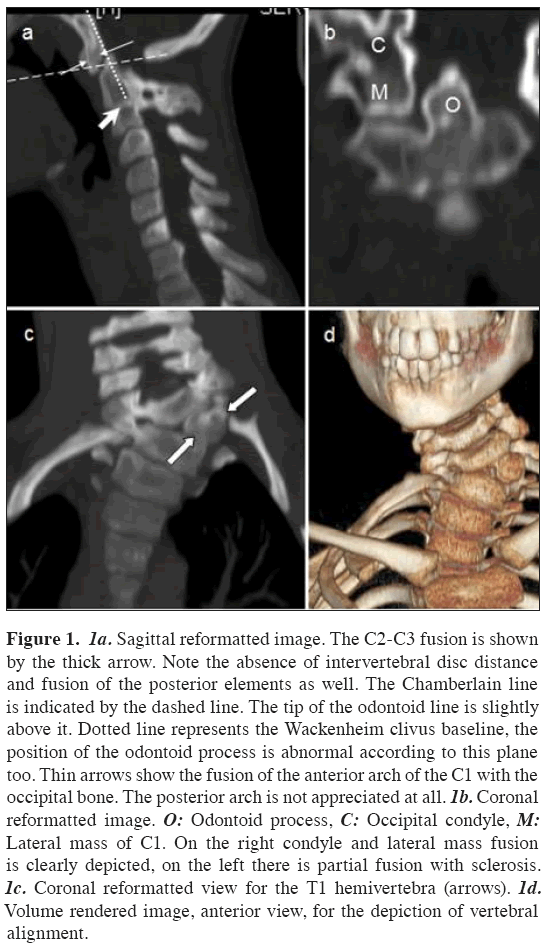
Upon reviewing of reformatted images, numerous anomalies were identified involving the cervical and upper thoracic vertebrae which are partial atlantooccipital assimilation with C2-C3 block vertebrae and T1 hemivertebra in the order of cranial to caudal (Figure 1).
Figure 1: 1a. Sagittal reformatted image. The C2-C3 fusion is shown by the thick arrow. Note the absence of intervertebral disc distance and fusion of the posterior elements as well. The Chamberlain line is indicated by the dashed line. The tip of the odontoid line is slightly above it. Dotted line represents the Wackenheim clivus baseline, the position of the odontoid process is abnormal according to this plane too. Thin arrows show the fusion of the anterior arch of the C1 with the occipital bone. The posterior arch is not appreciated at all. 1b. Coronal reformatted image. O: Odontoid process, C: Occipital condyle, M: Lateral mass of C1. On the right condyle and lateral mass fusion is clearly depicted, on the left there is partial fusion with sclerosis. 1c. Coronal reformatted view for the T1 hemivertebra (arrows). 1d. Volume rendered image, anterior view, for the depiction of vertebral alignment.
Cardiovascular and urogenital systems were examined to rule out any anomaly that may accompany the vertebral anomalies and none was found.
Discussion
Although they may seem non-interrelated, this case is unique in harboring several coexisting anomalies that deserve to be individually discussed. Atlanto-occipital assimilation is one of the most common congenital anomalies of the craniovertebral junction, and it usually is asymptomatic. Also known as occipitalization, it is congenital synostosis of the atlas to the occiput caused by a failure of segmentation and separation of the most caudal occipital sclerotome during the first few weeks of fetal life [1]. There is male predominance of up to 5:1, with a population incidence as high as 0.75-3% [1,2].
It may coexist with basilar invagination that is described as the protrusion of odontoid process into foramen magnum [1]. Chamberlain first described the condition in 1939. There are two types of basilar invagination: Primary, a congenital abnormality often associated with other vertebral defects such as atlanto-occipital fusion, hypoplasia of the atlas, bifid posterior arch of the atlas, Klippel-Feil syndrome, and Goldenhar syndrome; secondary, a developmental condition usually attributed to softening of the osseous structures at the base of the skull, with the deformity developing later in life. The secondary form is more commonly termed as “basilar impression”. This is occasionally seen in conditions such as osteomalacia, rickets, Paget’s disease, osteogenesis imperfecta, renal osteodystrophy, and rheumatoid arthritis [3]. Basilar invagination is difficult to diagnose, though radiological diagnosis can be established with the help of three constructed lines on the lateral cephalogram which are McGregor’s line, Chamberlain’s line and Wackenheim clivus baseline [1,3,4]. The tip of odontoid process should violate Chamberlain’s line over 3 mm to be classified as a basilar invagination (Figure 1a).
The term block vertebrae is applied to congenital synostosis of vertebrae in which bony continuity results from failure of normal segmentation of the vertebral somites at the preosseous stage during embryonic development (3-8 weeks of gestation) [5]. Fusion is also widely used for these segmentation errors, although it refers to intervening union between previously well-developed vertebrae. Congenital block vertebrae can occur as an isolated malformation or in association with other conditions, and may involve two or more contiguous vertebrae. The vertebral body, the posterior elements, or both structures may be affected. The intervertebral disc may be completely absent or may appear as a rudimentary irregularly calcified structure. Block vertebrae can be seen at any level in the spine. In the cervical spine, C5-6 fusion is the most common site, followed by C2-3 [6].
Hemivertebra is a rare congenital abnormality of the spine where only one side of the vertebral body develops, leading to deformation of the spine, such as scoliosis or kyphosis. It results from failure of a vertebra to form on one side, resulting in a laterally based wedge vertebra with half a vertebral body, a single pedicle, and hemilamina. The cause of hemivertebra is unknown. The distribution pattern of the anomaly does not implicate a specific environmental or genetic factor. Hemivertebra should be differentiated from the other vertebral abnormalities (wedge vertebra, butterfly vertebra, bloc vertebra, bar vertebra or any combination) that cause congenital scoliosis, open neural-tube defects, and diastematomyelia [7,8]. Because hemivertebra might be associated with other anomalies that might place a great burden on the future outcome [8,9], the diagnosis of such a skeletal spinal deformity calls for a comprehensive anatomic scan and a detailed echocardiographic examination. Anomalies of the central nervous system and gastrointestinal tract have also been reported. Hemivertebra may be part of a syndrome including Jarcho-Levin, Klippel-Feil, and Vacterl syndrome [8].
Conclusion
Multiplanner imaging with multidetector CT provides excellent insights into craniovertebral junction and vertebral anomalies. Rapid scanning time is an advantage in especially pediatric patients, commonly giving the opportunity to complete the examination without sedation.
References
- Guebert GM, Rowe LJ, Yochum TR, Thompson JR, Masia CJ. Congenital anomalies and normal skeletal variants. In: Yochum TR, Rowe LJ, eds. Essentials of Skeletal Radiology. 3rd Ed., Baltimore, Lippincott Williams & Wilkins. 2005; 257–403.
- Bharucha EP, Dastur HM. Craniovertebral anomalies (a report on 40 cases). Brain. 1964; 87: 469–480.
- Hensinger RN. Congenital anomalies of the cervical spine. In: Rothman RH, Simeone FA, eds. The Spine. Vol. 1. 4th Ed., Philadelphia, WB Saunders Company. 1999; 232–245.
- David KM, Crockard A. Congenital malformations of the base of the skull, atlas, and the dens. In: Clark CR, ed. The Cervical Spine. 4th Ed., Philadelphia, Lippincott Williams & Wilkins. 2005; 420–425.
- Moore K, Persaud T. The Developing Human. 5th Ed., Philadelphia, WB Saunders. 1993; 358–364.
- Yildiz A, Apaydin FD, Ozer C, Egilmez H, Duce MN, Yalcinoglu O. Kranyovertebral bölge ve servikal vertebra anomalileri. Tanısal ve Girisimsel Radyoloji (Diagn. Interv. Radiol.) 2002; 8: 38–42. (Turkish)
- Weisz B, Achiron R, Schindler A, Eisenberg VH, Lipitz S, Zalel Y. Prenatal sonographic diagnosis of hemivertebra. J. Ultrasound Med. 2004; 23: 853–857.
- Basu PS, Elsebaie H, Noordeen MH. Congenital spinal deformity: a comprehensive assessment at presentation. Spine. 2002; 27: 2255–2259.
- McMaster MJ, David CV. Hemivertebra as a cause of scoliosis. A study of 104 patients. J. Bone Joint Surg. Br. 1986; 68: 588–595.
Aysegul Altunkas, Basar Sarikaya*
Gaziosmanpasa University, Faculty of Medicine, Department of Radiology, Tokat, Turkey.
- *Corresponding Author:
- Basar Sarikaya, MD
Assistant Professor of Radiology, Gaziosmanpasa University, Faculty of Medicine, Department of Radiology, 60100 Tokat, Turkey.
Tel: +90 356 2129500
Fax: +90 356 2129417
E-mail: basarsarikayamd@yahoo.com
Date of Received: 14-04-2011
Date of Accepted: 13-05-2011
Published Online: 15-11-2011
© IJAV. 2009; 2: 2–3.
-Keywords
computed tomography, cervical vertebrae, craniovertebral junction
Introduction
Although some of them may remain asymptomatic, congenital vertebral anomalies lead to spinal deformity and scoliosis. However they are relatively rarely seen in the cervical vertebrae [1]. We present a 10-year-old patient with several coexistent vertebral anomalies affecting the craniovertebral junction and upper vertebrae, which we believe to be a didactic example to the imaging anatomy of these anomalies.
Case Report
Ten year-old male patient, presenting with short stature, underwent cervical vertebra CT imaging for detailed visualization of suspicious findings on the plain films. CT was performed in an 8-channel multidetector CT suite (GE Medical Systems, Milwaukee, WI, US). Raw data was transferred to a commercially available workstation (AW 4.2; GE Medical Systems, Milwaukee, WI, US) for 2D and 3D reformatting.
Upon reviewing of reformatted images, numerous anomalies were identified involving the cervical and upper thoracic vertebrae which are partial atlantooccipital assimilation with C2-C3 block vertebrae and T1 hemivertebra in the order of cranial to caudal (Figure 1).
Figure 1: 1a. Sagittal reformatted image. The C2-C3 fusion is shown by the thick arrow. Note the absence of intervertebral disc distance and fusion of the posterior elements as well. The Chamberlain line is indicated by the dashed line. The tip of the odontoid line is slightly above it. Dotted line represents the Wackenheim clivus baseline, the position of the odontoid process is abnormal according to this plane too. Thin arrows show the fusion of the anterior arch of the C1 with the occipital bone. The posterior arch is not appreciated at all. 1b. Coronal reformatted image. O: Odontoid process, C: Occipital condyle, M: Lateral mass of C1. On the right condyle and lateral mass fusion is clearly depicted, on the left there is partial fusion with sclerosis. 1c. Coronal reformatted view for the T1 hemivertebra (arrows). 1d. Volume rendered image, anterior view, for the depiction of vertebral alignment.
Cardiovascular and urogenital systems were examined to rule out any anomaly that may accompany the vertebral anomalies and none was found.
Discussion
Although they may seem non-interrelated, this case is unique in harboring several coexisting anomalies that deserve to be individually discussed. Atlanto-occipital assimilation is one of the most common congenital anomalies of the craniovertebral junction, and it usually is asymptomatic. Also known as occipitalization, it is congenital synostosis of the atlas to the occiput caused by a failure of segmentation and separation of the most caudal occipital sclerotome during the first few weeks of fetal life [1]. There is male predominance of up to 5:1, with a population incidence as high as 0.75-3% [1,2].
It may coexist with basilar invagination that is described as the protrusion of odontoid process into foramen magnum [1]. Chamberlain first described the condition in 1939. There are two types of basilar invagination: Primary, a congenital abnormality often associated with other vertebral defects such as atlanto-occipital fusion, hypoplasia of the atlas, bifid posterior arch of the atlas, Klippel-Feil syndrome, and Goldenhar syndrome; secondary, a developmental condition usually attributed to softening of the osseous structures at the base of the skull, with the deformity developing later in life. The secondary form is more commonly termed as “basilar impression”. This is occasionally seen in conditions such as osteomalacia, rickets, Paget’s disease, osteogenesis imperfecta, renal osteodystrophy, and rheumatoid arthritis [3]. Basilar invagination is difficult to diagnose, though radiological diagnosis can be established with the help of three constructed lines on the lateral cephalogram which are McGregor’s line, Chamberlain’s line and Wackenheim clivus baseline [1,3,4]. The tip of odontoid process should violate Chamberlain’s line over 3 mm to be classified as a basilar invagination (Figure 1a).
The term block vertebrae is applied to congenital synostosis of vertebrae in which bony continuity results from failure of normal segmentation of the vertebral somites at the preosseous stage during embryonic development (3-8 weeks of gestation) [5]. Fusion is also widely used for these segmentation errors, although it refers to intervening union between previously well-developed vertebrae. Congenital block vertebrae can occur as an isolated malformation or in association with other conditions, and may involve two or more contiguous vertebrae. The vertebral body, the posterior elements, or both structures may be affected. The intervertebral disc may be completely absent or may appear as a rudimentary irregularly calcified structure. Block vertebrae can be seen at any level in the spine. In the cervical spine, C5-6 fusion is the most common site, followed by C2-3 [6].
Hemivertebra is a rare congenital abnormality of the spine where only one side of the vertebral body develops, leading to deformation of the spine, such as scoliosis or kyphosis. It results from failure of a vertebra to form on one side, resulting in a laterally based wedge vertebra with half a vertebral body, a single pedicle, and hemilamina. The cause of hemivertebra is unknown. The distribution pattern of the anomaly does not implicate a specific environmental or genetic factor. Hemivertebra should be differentiated from the other vertebral abnormalities (wedge vertebra, butterfly vertebra, bloc vertebra, bar vertebra or any combination) that cause congenital scoliosis, open neural-tube defects, and diastematomyelia [7,8]. Because hemivertebra might be associated with other anomalies that might place a great burden on the future outcome [8,9], the diagnosis of such a skeletal spinal deformity calls for a comprehensive anatomic scan and a detailed echocardiographic examination. Anomalies of the central nervous system and gastrointestinal tract have also been reported. Hemivertebra may be part of a syndrome including Jarcho-Levin, Klippel-Feil, and Vacterl syndrome [8].
Conclusion
Multiplanner imaging with multidetector CT provides excellent insights into craniovertebral junction and vertebral anomalies. Rapid scanning time is an advantage in especially pediatric patients, commonly giving the opportunity to complete the examination without sedation.
References
- Guebert GM, Rowe LJ, Yochum TR, Thompson JR, Masia CJ. Congenital anomalies and normal skeletal variants. In: Yochum TR, Rowe LJ, eds. Essentials of Skeletal Radiology. 3rd Ed., Baltimore, Lippincott Williams & Wilkins. 2005; 257–403.
- Bharucha EP, Dastur HM. Craniovertebral anomalies (a report on 40 cases). Brain. 1964; 87: 469–480.
- Hensinger RN. Congenital anomalies of the cervical spine. In: Rothman RH, Simeone FA, eds. The Spine. Vol. 1. 4th Ed., Philadelphia, WB Saunders Company. 1999; 232–245.
- David KM, Crockard A. Congenital malformations of the base of the skull, atlas, and the dens. In: Clark CR, ed. The Cervical Spine. 4th Ed., Philadelphia, Lippincott Williams & Wilkins. 2005; 420–425.
- Moore K, Persaud T. The Developing Human. 5th Ed., Philadelphia, WB Saunders. 1993; 358–364.
- Yildiz A, Apaydin FD, Ozer C, Egilmez H, Duce MN, Yalcinoglu O. Kranyovertebral bölge ve servikal vertebra anomalileri. Tanısal ve Girisimsel Radyoloji (Diagn. Interv. Radiol.) 2002; 8: 38–42. (Turkish)
- Weisz B, Achiron R, Schindler A, Eisenberg VH, Lipitz S, Zalel Y. Prenatal sonographic diagnosis of hemivertebra. J. Ultrasound Med. 2004; 23: 853–857.
- Basu PS, Elsebaie H, Noordeen MH. Congenital spinal deformity: a comprehensive assessment at presentation. Spine. 2002; 27: 2255–2259.
- McMaster MJ, David CV. Hemivertebra as a cause of scoliosis. A study of 104 patients. J. Bone Joint Surg. Br. 1986; 68: 588–595.


