Multiple forearm muscle variants including a rare hypothenar variant
2 College of Naturopathic Medicine, University of Bridgeport, Bridgeport, CT, USA
Received: 23-Jul-2012 Accepted Date: Sep 20, 2012; Published: 29-Aug-2013
Citation: Kelliher KR, Terfera D, Pracella J, et al. Multiple forearm muscle variants including a rare hypothenar variant. Int J Anat Var. 2013;6:124-127.
This open-access article is distributed under the terms of the Creative Commons Attribution Non-Commercial License (CC BY-NC) (http://creativecommons.org/licenses/by-nc/4.0/), which permits reuse, distribution and reproduction of the article, provided that the original work is properly cited and the reuse is restricted to noncommercial purposes. For commercial reuse, contact reprints@pulsus.com
Abstract
During a routine dissection of the upper extremities we observed multiple muscle variants in the forearm of a male cadaver. Variants of the palmaris longus, flexor pollicis longus and a rare hypothenar muscle were observed bilaterally. The palmaris longus presented as a different variant on either side, a reverse palmaris longus and a bifid palmaris longus. The accessory head of the flexor policis longus was observed in both forearms and a rarely seen accessory hypothenar muscle was found bilaterally.
Keywords
hypothenar, palmaris longus, pisiform, bilateral, variant
Introduction
Variant muscles in the forearm are not particularly rare however they are clinically relevant. They lead to compression syndromes, can be confused as soft tissue pathologies or become burdensome during surgical procedures. While there are many reports of individual muscle variations occurring unilaterally or bilaterally there are very few reports showing multiple variants occurring simultaneously. Here during a routine forearm and hand dissection we observed, in a single cadaver, multiple forearm muscle variations bilaterally.
Case Report
During a routine classroom dissection on the upper extremities it was discovered that a 78-year-old male cadaver had multiple variants present in both left and right forearms.
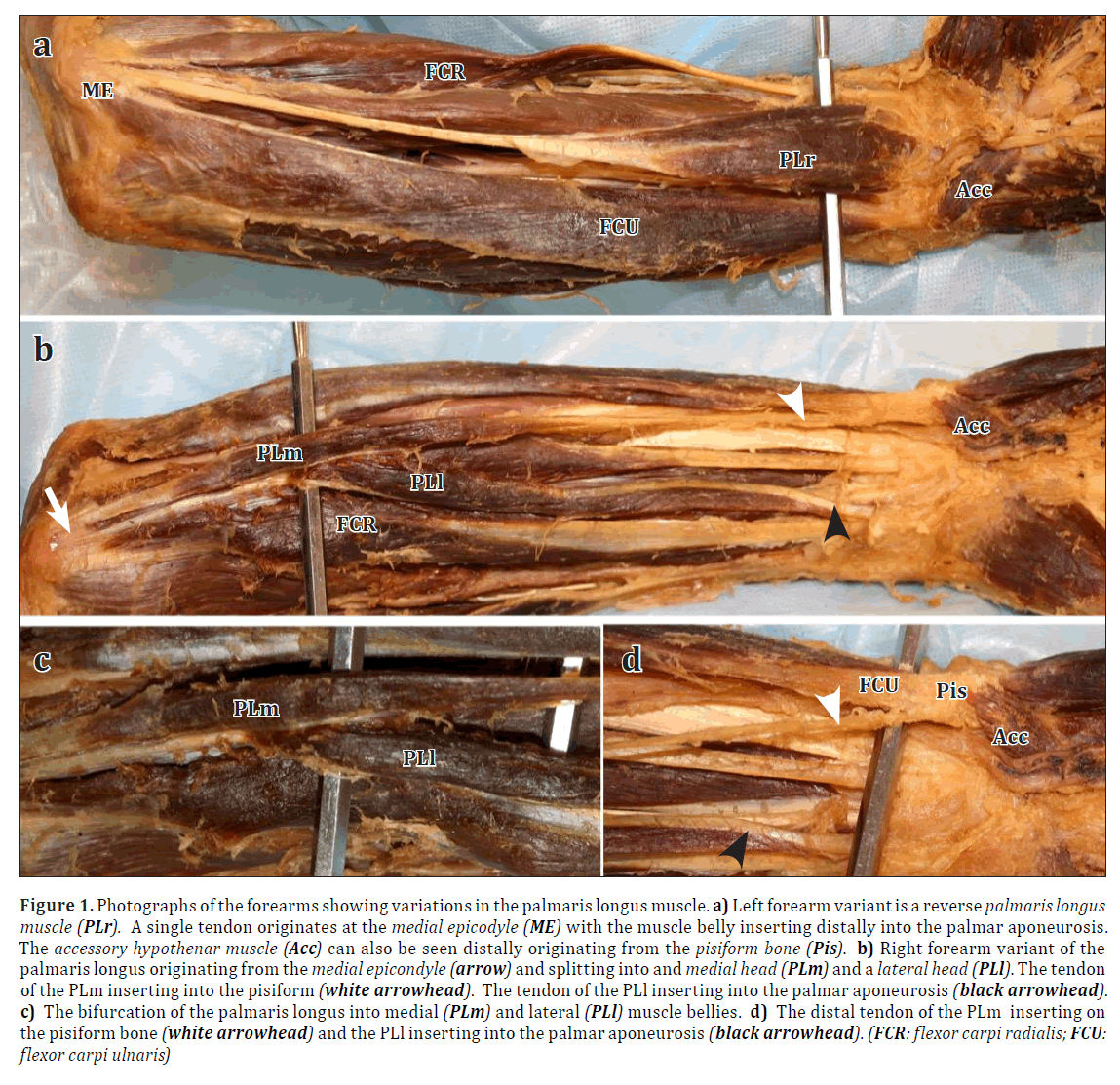
The palmaris longus muscle (PL) varied bilaterally, however each side had different variations. The left forearm had a reversed PL (Figure 1a). The muscle tendon of the PL began at the medial epicondyle and extended distally 16 cm before broadening to a muscle belly. The muscle belly measured 11.5 cm by 2.5 cm and extended into the forearm and inserted into the palmar aponeurosis. In the right forearm the PL was present as a bifid variant. The muscle originated on the medial epicondyle and continued for 8 cm before splitting into a lateral and medial head. The lateral head continued as a narrow strip for 20 cm. The tendon of this laterally located head measured 5.5 cm and inserted distally into the palmar aponeurosis (Figures 1b, c). The medial head continued from the bifurcation for another 11 cm. The tendon of the medial head measured 12 cm and inserted onto the pisiform bone distally (Figure 1d). We observed branches of the median nerve passing into both of these muscle bellies.
Figure 1. Photographs of the forearms showing variations in the palmaris longus muscle. a) Left forearm variant is a reverse palmaris longus muscle (PLr). A single tendon originates at the medial epicodyle (ME) with the muscle belly inserting distally into the palmar aponeurosis. The accessory hypothenar muscle (Acc) can also be seen distally originating from the pisiform bone (Pis). b) Right forearm variant of the palmaris longus originating from the medial epicondyle (arrow) and splitting into and medial head (PLm) and a lateral head (PLl). The tendon of the PLm inserting into the pisiform (white arrowhead). The tendon of the PLl inserting into the palmar aponeurosis (black arrowhead). c) The bifurcation of the palmaris longus into medial (PLm) and lateral (PLl) muscle bellies. d) The distal tendon of the PLm inserting on the pisiform bone (white arrowhead) and the PLl inserting into the palmar aponeurosis (black arrowhead). (FCR: flexor carpi radialis; FCU: flexor carpi ulnaris)
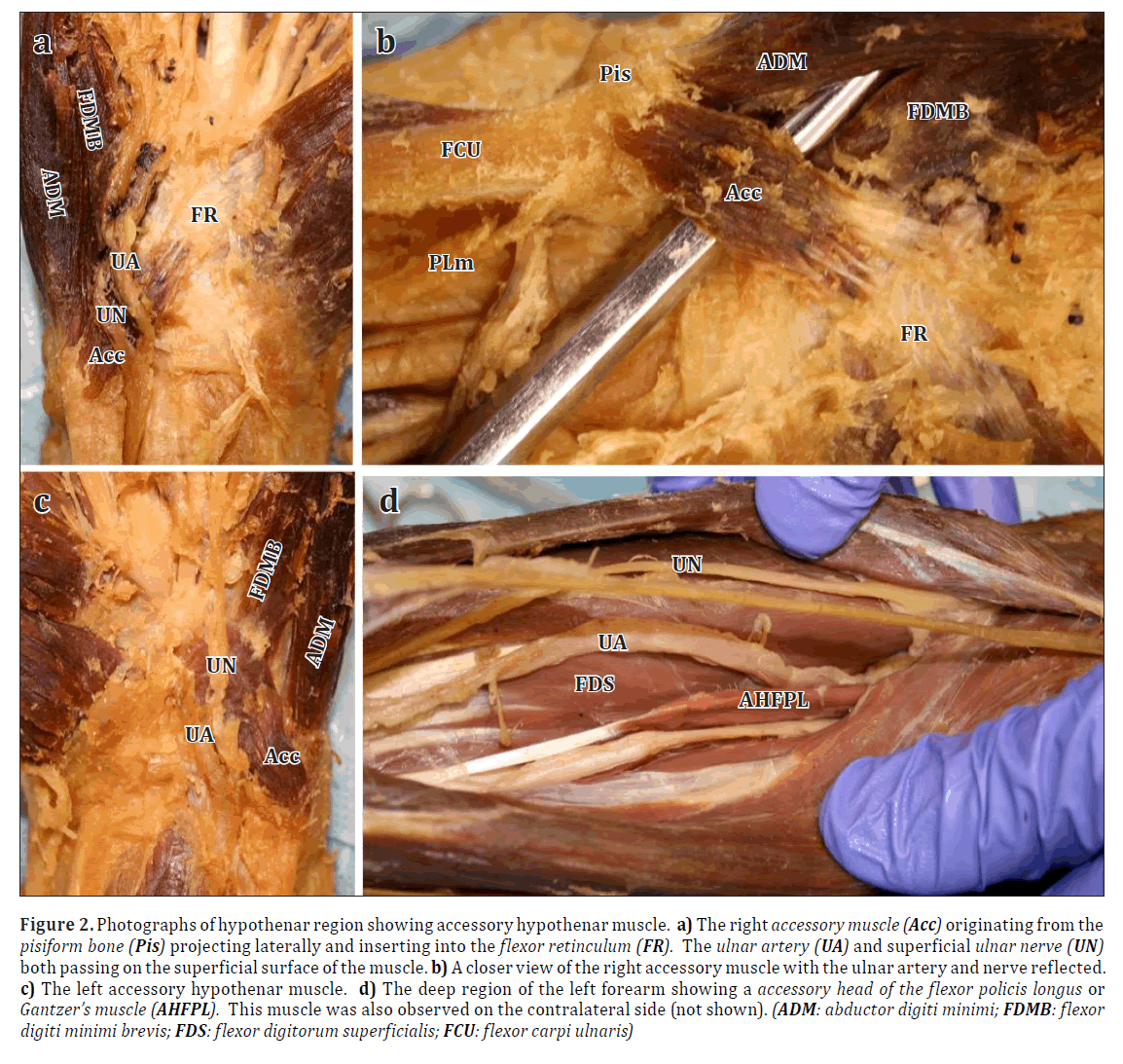
A rarely seen accessory muscle was present bilaterally. The 3.6 cm by 1.3 cm muscle originated on the pisiform and inserted medially into the flexor retinaculum (Figure 2a, b, c). The ulnar nerve split into its superficial and deep branches just proximal to the variant muscle with the superficial branch passing superficial and deep branch passing deep to the muscle belly. The ulnar artery also passed superficial to the muscle.
Figure 2. Photographs of hypothenar region showing accessory hypothenar muscle. a) The right accessory muscle (Acc) originating from the pisiform bone (Pis) projecting laterally and inserting into the flexor retinculum (FR). The ulnar artery (UA) and superficial ulnar nerve (UN) both passing on the superficial surface of the muscle. b) A closer view of the right accessory muscle with the ulnar artery and nerve reflected. c) The left accessory hypothenar muscle. d) The deep region of the left forearm showing a accessory head of the flexor policis longus or Gantzer’s muscle (AHFPL). This muscle was also observed on the contralateral side (not shown). (ADM: abductor digiti minimi; FDMB: flexor digiti minimi brevis; FDS: flexor digitorum superficialis; FCU: flexor carpi ulnaris)
The third variant also present bilaterally in the forearm was an accessory head of the flexor pollicis longus or Gantzer’s muscle (Figure 2d).
Discussion
The palmaris longus muscle is the most variable muscle in the forearm if not the entire musculoskeletal system being absent in 13% of individuals and variant in another 9% [1]. It typically presents as a fleshy belly proximally, originating from the medial epicondyle and inserting distally as a tendon into the palmar aponeurosis. Variants have been identified that present a proximal tendon and distal muscle belly (reverse palmaris longus), a proximal and distal belly connected by a central tendon (digastric), a muscle belly throughout its length (non-tendinous) or as two bellies originating from the medial epicondyle with two distal insertions (bifid) [2]. In our case we observed two different variations on the same individual, a reverse palmaris longus in the left forearm and a bifid palmaris longus in the right forearm. In our case the medial head of the palmaris longus inserted into the pisiform bone. Alternate insertion of the palmaris longus, in particular the bifid variety, into the pisiform is not uncommon [3]. Pugh et al. observed a similar bifid muscle to the one we observed that originated from the common flexor tendon and inserted onto the pisiform bone [4]. They identified that muscle as an accessory flexor carpi ulnaris. They reported that the muscle was innervated by both the median and the ulnar nerves. However, in our case we only observed branches of the median nerve innervating both bellies and observed no branches of the ulnar nerve innervating it. This suggests that the variant we observed is a variant of the palmaris longus and not the flexor carpi ulnaris.
Accessory muscles in the wrist are a common occurrence with variations of the abductor minimi muscle being one of the most prevalent [5,6]. These variations also occur bilaterally with a high frequency [5]. In our case, the variant muscle originated on the pisiform, however it inserted medially into the flexor retinaculum. Murata et al. described an unusual muscle similar to ours originating from the pisiform; however, the muscle remained superficial to the ulnar nerve [7]. Two other reports have described a similar presentation to what we observed. In a 29-year-old cadaver, the hypothenar muscle was found passing between the superficial and deep branches of the ulnar nerve [8]. Pugh et al. described a muscular slip arising from the pisiform bone and inserting on to the hamate [4]. They also observed the muscle passing between the superficial and deep branches of the ulnar nerve. Interestingly, in the cases we reviewed this accessory hypothenar muscle occurs bilaterally [4,8].
There is a wide body of literature identifying and examining individual forearm muscle variations. We have identified on a single cadaver multiple variants, some common (Gantzer’s muscle) and some rare (hypothenar muscle) that appear bilaterally. The cataloging of these multiple variants will lead to a better understanding of the developmental origins of human muscle variation.
REFERENCES
- Reimann AF, Daseler EH, Anson BJ, Beaton LE. The palmaris longus muscle and tendon: a study of 1600 extremities. Anat Rec. 1944: 89: 495–505.
- Tountas CP, Bergman RA. Anatomic variations of the upper extremity. New York, Churchill Livingstone. 1993: 141, 147–170.
- Bergman RA, Thompson SA, Afifi AK. Catalog of Human Anatomical Variation. Baltimore, Urban & Schwarzenberg. 1984; 31–32.
- Pugh AM, Silvestri BA, Ward MA, Whited WM, Capehart AA. Unusual bilateral muscular variation in the medial forearm: separate humeral and ulnar bellies of flexor carpi ulnaris and anomalous muscle addition. Anatomy. 2010: 4: 67–71.
- Dodds GA 3rd, Hale D, Jackson WT. Incidence of anatomic variants in Guyon’s canal. J Hand Surg Am. 1990: 15: 352–355.
- Zeiss J, Guilliam-Haidet L. MR demonstration of anomalous muscles about the volar aspect of the wrist and forearm. Clin Imaging. 1996: 20: 219–221.
- Murata K, Tamai M, Gupta A. Anatomic study of variations of hypothenar muscles and arborization patterns of the ulnar nerve in the hand. J Hand Surg Am. 2004: 29: 500–509.
- Depukat P, Mizia E, Walocha J. An anomalous bilateral muscle in Guyon’s canal found during cadaver study. Folia Morphol (Warsz). 2010: 69: 65–67.


