Multiple renal vascular variations: A case series
Received: 26-Jul-2018 Accepted Date: Aug 18, 2018; Published: 28-Aug-2018
Citation: Potaliya P, Sharma S, Kataria D, et al. Multiple renal vascular variations: A case Series. Int J Anat Var. 2018;11(3):90-92.
This open-access article is distributed under the terms of the Creative Commons Attribution Non-Commercial License (CC BY-NC) (http://creativecommons.org/licenses/by-nc/4.0/), which permits reuse, distribution and reproduction of the article, provided that the original work is properly cited and the reuse is restricted to noncommercial purposes. For commercial reuse, contact reprints@pulsus.com
Abstract
Renal vascular abnormalities represent a heterogeneous group of diseases and etiologies. It is important to differentiate these vascular anomalies from other renal enhancing lesions prior to any attempt to perform any surgical intervention. This study aims to find out anatomical variations in renal vasculature of resected specimens. This study was conducted on 60 resected kidney specimens in Department of Anatomy, All India Institute of Medical Sciences, Jodhpur (Rajasthan) during routine Anatomy practical sessions of MBBS students. We found variations in renal artery where the main renal artery divided into pre-hilar branches (3.33%) and there was presence of additional renal artery (6.67%) and polar artery (1.67%). Additional renal veins were also found (3.33%) along with variation in relationship of hilar structures (16.67%). The knowledge of these variations can be useful in various surgical interventions including renal transplants
Keywords
Renal; Vascular variation; Kidney; Vasculature; Hilum
Introduction
Renal vessels are known with a wide range of variations that have also been evidenced during multiple surgeries in recent years. These variations can lead to significant surgical complications or even life-threatening events if unrecognized. Classically renal vasculature is formed by one artery and one vein only but unfortunately it is evident in only less than 25% of cases [1].
Renal artery
Normally each kidney is supplied by a single renal artery, which arises as a lateral branch of abdominal aorta, between the levels of L1 and L2. Each renal artery divides into an anterior and a posterior division. The branches from the anterior division supplies the apical, upper, middle and lower segments while posterior division supplies the posterior segment of the kidney [1].
Morphological variations of renal artery are its variable number and unusual branches called as aberrant, supernumerary, supplementary, accessory, among other terms [2]. According to Sampaio and Passos these arteries should be called multiple, and they should be named according to the territory supplied by them as- hilar, superior polar and inferior polar [3].
Renal vein
The renal veins lie anterior to the renal arteries and open into the inferior vena cava almost at right angles at the level of L2 vertebra. The left renal vein is three times longer than the right (7.5 cm and 2.5 cm) and for this reason the left kidney is the preferred side for live donor nephrectomy. The renal vein may be double, lying anterior and posterior to the aorta before joining inferior vena cava. This is known as ‘renal collar’ [4]. Morphological variation found in renal veins is its variable number known as additional renal vein. Venous blood of the kidney is drained by an additional vein into the inferior vena cava with the similar course and termination as its normal vein, named as accessory or supernumerary renal vein [5].
Renal hilum
Kidney being bean shaped, has thick and rounded superior pole and thin and pointed inferior pole. Renal hilum is deep vertical slit situated in its medial border which lies about 5 cm from the midline opposite the lower border of L1 vertebra. At the hilum, usually the renal vein is the anterior most structure with the renal artery posterior to it and the pelvis of kidney lying further posteriorly (Figure 1) [6].
So, our study demarcates various anatomical variations associated with renal vasculature. Knowledge of these variations accompanied by pathologies may be important as it can affect diagnostic and treatment procedures and it may avoid unnecessary complications associated with different types of renal interventions like renal transplantation, urological and retroperitoneal surgeries.
Methodology
This study was conducted on thirty embalmed cadavers in the Department of Anatomy at All India Institute of Medical Sciences, Jodhpur (Rajasthan) during routine abdominal dissection conducted for medical undergraduates. Cadavers were explored and the morphological variations of renal arteries, renal veins and hilar structures were noted.
Results
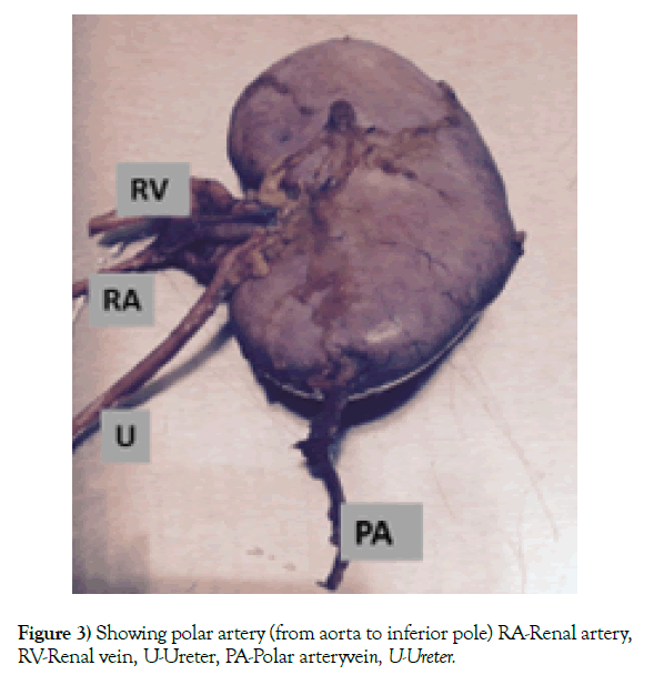
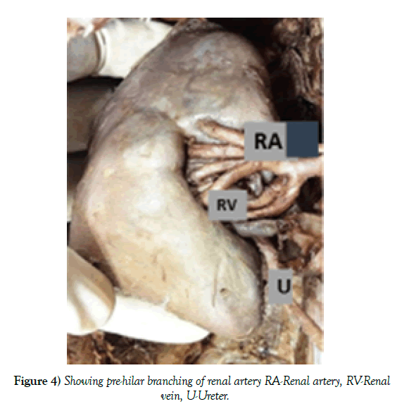
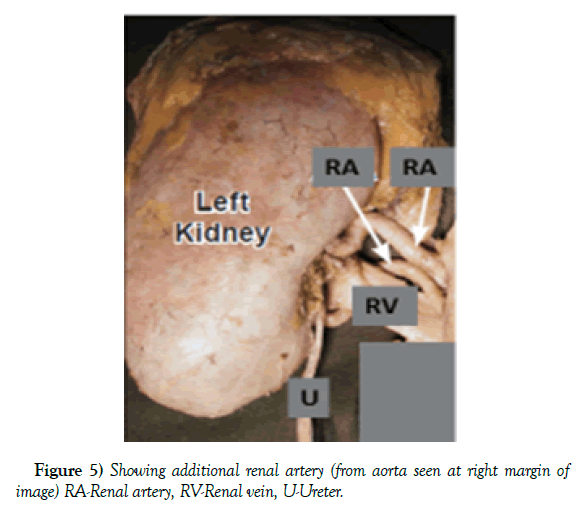
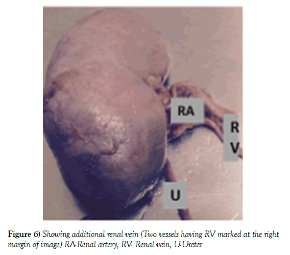
Out of 60 kidney specimens, (Table 1) variation in renal hilum was found in 16.67% kidneys (10 out of 60 specimens) (Figure 2). Usually the order in which structures are present in the hilum is vein, artery and pelvis (anterior to posterior). But in our study, the order of these structures in 16.67% kidneys was observed to be artery, vein and pelvis (anterior to posterior). Variations found in renal artery were polar artery, pre-hilar branching of renal artery and additional renal artery in 1,2 and 4 kidneys respectively i.e. 1.67%, 3.33% and 6.67% respectively (Figures 3-5). Additional renal arteries arose from the aorta. They followed the main renal artery to the hilum of the kidney. Accessory renal artery entered the kidney directly into inferior pole. These are known as polar arteries. More than one segmental arteries arose from main renal artery known as pre-hilar branches. Additional renal vein was seen in 2 (3.33%) kidneys (Figure 6). Emergence of more than one renal vein from hilum of kidney going to inferior vena cava.
| (Total number of resected kidney specimens = 60) | ||
|---|---|---|
| Variation | Number of Kidneys (n) | Percentage (%) |
| Relationship at hilum (artery-vein-pelvis) | 10 | 16.67 |
| Pre-hilar branching of renal artery | 2 | 3.33 |
| Polar artery | 1 | 1.67 |
| Additional renal artery | 4 | 6.67 |
| Additional renal vein | 2 | 3.33 |
Table 1: Showing spectrum of renal vascular variations
Discussion
Renal artery
Vertebrate urogenital system has history of evolution. Many conditions which are considered anomalous in human are normally present in some lower animals. Multiple renal arteries were found originating from aorta in frog, lizard, domestic fowl etc. Hence, multiple renal arteries in human may be explained phylogenetically [7]. Keibel and Mall explained accessory renal artery on the basis of ontogeny. As the kidneys ascend from the pelvis, they receive their blood supply from the vascular structures close to them. Initially renal arteries are the branches of common iliac arteries. Later, while the kidneys ascend they receive new branches from the aorta, and the inferior branches disappear. In the ninth week of the intrauterine life the kidneys come in contact with the suprarenal glands and the ascent ceases. The kidneys receive their most cranial branches from the aorta. These are the permanent renal arteries. Failure of degeneration of initial branches leads to formation of accessory renal artery [8].
Many researchers have reported variations in renal artery. Bordei et al. and Krishnasamy et al. reported 54 cases of double renal arteries supplying one kidney originating from aorta [9,10] Saldarriage et al. observed bilateral additional artery in 7.7% of the cases. He also reported that additional arteries had entered through the hilum in 12% of cases and through the inferior pole of the kidney in 1.8% of cases [11]. In our present study additional renal artery was found in 4 out of 60 kidneys (6.67%) (Figure 5) and polar artery was found in 1 out of 60 kidneys (1.67%) (Figure 3). The additional renal arteries can be of importance to the clinicians. Additional renal artery play role in causing hydronephrosis. Incidence of ureteropelvic junction obstruction (Shoja et al.) is very high in case of inferior polar artery [12]. Before reaching the hilum, early division of renal artery was found in 75% cases by Sarfraz et al. 81.67% cases by Daescu et al. [13,14]. According to an another study pre-hilar branching was found in 33.3% and 28.5% for right and left kidneys respectively. 2 In our present study pre-hilar branching of renal artery was found in 2 out of 60 kidneys (3.33%) (Figure 4).
Renal vein
Bilaterally symmetrical cardinal venous system becomes unilateral rightsided inferior vena cava at 8th week of intrauterine life. Two renal veins are present, one ventrally and another dorsally [15]. Then confluence of these two veins occur and hence a single vessel is formed. If in case; confluence of veins doesn’t occur, then additional renal vein is formed [16].
Various studies have shown renal vein variation. Variations have been found on both the sides as well as separately. Accessory renal vein was observed in 36 % by Chugh KS et al. and in 42 % by Brodie P et al. [17,9]. Gupta et al. in his study found accessory renal vein on the right side in 33% and on the left side in 3.3% of cases [18]. Krishnasamy et al. and Satyapal et al. noted multiple renal vein in 0.4% of the cases [10,19]. While Dhar P et al. observed additional renal vein in 3% on left side and 12% on right side [20]. Girdhar S et al. reported a rare presentation of triple renal vein [21]. In our present study additional renal vein was found in 2 out of 60 kidneys (3.33%) (Figure 6).
Renal hilum
Das S et al. noted variation in relationship of hilar structures with renal artery lying anterior to vein and pelvis [22]. Trivedi S et al. in their study concluded that in three-fourth of the cases arrangement of hilar structures does not confirm to the normal description (i.e. vein, artery and pelvis) [23]. According to our present study, relationship at hilum of artery, vein and ureter (anterior to posterior) was observed in 10 out of 60 cases (16.67%) (Figure 2).
Conclusion
It is of paramount importance for surgeons to have thorough knowledge of renal vasculature development and to readily identify their anomalies in order to prevent adverse bleeding effects. With the increasing demand for kidney transplantation, living donor grafts have become major source for maintaining donor pool and successful allograft with multiple arteries has been observed. Knowledge of variation in renal vasculature may be useful to the clinicians and radiologists performing invasive techniques and to surgeons performing nephrectomy and renal transplantation. Variations in the renal vasculature often remain unnoticed but are very significant from the surgical point of view.
REFERENCES
- Maheswararao SU, Vanju VVL, Shahajeer B, et al. A cadaveric study of renal hilar structures and their variations in Andhra Pradesh population of South India. J Evid Based Med Healthc. 2016;3:3760-3.
- Budhiraja V, Rastogi R, Jain V, et al. Anatomical variations of renal artery and its clinical correlations: A cadaveric study from central India. J Morphol Sci. 2013;30:228-33.
- Sampaio FJ, Passos MA. Renal arteries: Anatomic study for surgical and radiological practice. Surg Radiol Anat. 1992;14:113-7.
- Mukundan M, Vijaynath V, Ravi V. Bilateral variation of renal vein: A case report. Int J Anat Res. 2016;4:3153-5.
- Kumar N, Swamy RS, Patil J, et al. Accessory right renal vein draining into inferior vena cava. OA Case Reports. 2014;3:48.
- Kumar N, Aithal AP, Guru A, et al. Bilateral vascular variations at the renal hilum: A case report. Case Rep Vasc Med. 2012.
- Gupta A, Gupta R, Singhla RK. The accessory renal arteries: A comparative study in vertebrates with its clinical implications. J Clin Diagn Res. 2011;5:970-3.
- Keibel F, Mall FP. Manual of human embryology, J. B. Lippincott, Philadelphia. 1912;2:820-5.
- Bordei P, Sapte E, Iliescu D. Double renal artery originating from aorta. Surg Radiol Anat. 2004;26:474-9.
- Krishnasamy N, Rao M, Somayaji SN, et al. An unusual case of unilateral additional right renal artery and vein. Int J Anat Var. 2010;3:9-11.
- Saldarrriage AF, Perez LE, Ballesteros LE. A direct anatomical study of additional renal arteries in a Colombian Mestizo population. Folia Morphol. 2008;67:129-34.
- Shoja MM, Tubbs RS, Shakeri AB, et al. Origins of the gonadal artery: Embryologic implications. Clin Anat. 2007;20:428-32.
- Sarfraz R, Tahir M, Sami W. Presegmental arterial pattern of human kidneys in local population. Ann Pak Ins Med Sci. 2008;4:212-5.
- Daescu E, Zahoi De, Otoc A, et al. morphological variability of the renal artery branching pattern: a brief review and an anatomical study. Rom J Morphol Embryol. 2012;53:287- 91.
- Tatar I, Tore HG, Celik HH. Retroaortic and circumaortic left renal veins with their finding and review of the literature. Anatomy. 2008;2:72-6.
- Mankhause WS, Khalique A. The adrenal and renal mass and their connection with Azygos and lumber vein. J Anat. 1986;146:105-15.
- Chugh KS, Malik N, Gosh AK, et al. Pattern of renal arteries in normal subjects: A study of 170 renal donor angiograms. Indian J Nephrol. 1993;3:9-11.
- Gupta A, Gupta R, Singhla RK. Congenital variations of renal veins: Embryological background and clinical implications. J Clin Diagn Res. 2011;6:1140-3.
- Satyapal KS, Kalideen JM, Singu B, et al. Why we use the donor left kidney in live related transplantation. S Afr J Surg. 2003;41:24-6.
- Dhar P, Lal K. Main and accessory renal arteries-A morphological study. Ital J Anat Embryol. 2005;110:101-10.
- Girdhar S, Sharma V, Gupta OP, et al. Triple right renal veins and bilateral multiple renal arteries-A case report. J Anat Sciences. 2015;23:10-13.
- Das S, Paul S. Variation of renal hilar structures: A cadaveric case. Eur J Anat. 2006;10:41-3.
- Trivedi S, Athavale S, Kotgiriwar S. Normal and variant anatomy of renal hilar structures and its clinical significance. Int J Morphol. 2011; 29:1379-83.


