Multiple variations in vertebrobasilar system: A cadaveric study
Received: 07-Nov-2017 Accepted Date: Nov 28, 2017; Published: 20-Dec-2017
Citation: Sarah SS, Jyoti C, Anita R, et al. Multiple variations in vertebrobasilar system: A cadaveric study. Int J Anat Var. 2017;10(4): 112-114.
This open-access article is distributed under the terms of the Creative Commons Attribution Non-Commercial License (CC BY-NC) (http://creativecommons.org/licenses/by-nc/4.0/), which permits reuse, distribution and reproduction of the article, provided that the original work is properly cited and the reuse is restricted to noncommercial purposes. For commercial reuse, contact reprints@pulsus.com
Abstract
The intracranial parts of the vertebral arteries (VAs), basilar artery (BA) and their branches together form the vertebrobasilar system (VBS). Numerous anomalies have been seen in VBS, which are usually benign. Many authors have reported unilateral hypoplasia of VAs, posterior and anterior inferior cerebellar arteries and variation in origin of branches of VA and BA. These are thought to develop due to some errors during development. Aplasia of VA is a common variant but seldom reported. During routine dissection a rare variant of VA along with multiple variations in other arteries of vertebrobasilar system was observed in one cadaveric brain. It was observed that there was aplasia of left VA and left PICA took origin from the right VA, crossed midline and supplied the contralateral side of the cerebellar hemisphere. Double origin of right anterior inferior cerebellar artery (AICA) and bilateral fetal PCA (posterior cerebral artery) was also observed.
Keywords
Variations; Vertebrobasilar system; Aplasia; Vertebral artery; PICA
Introduction
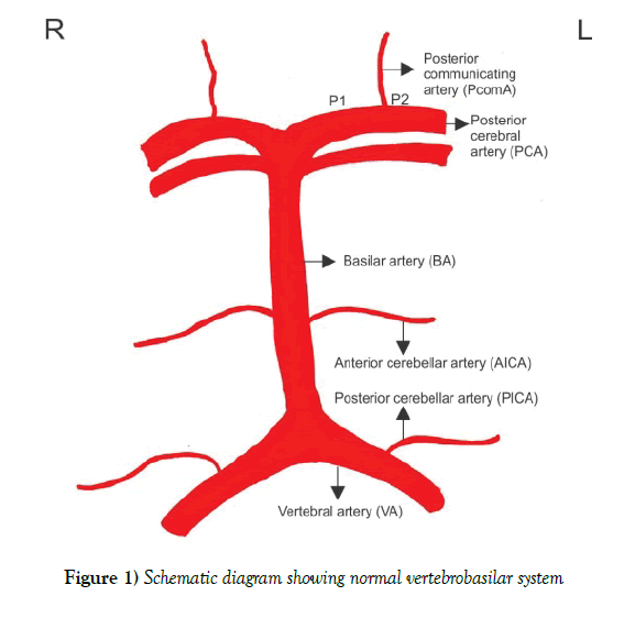
Vertebral artery (VA) arises from postero-superior aspect of subclavian artery, passes through foramina of all cervical transverse processes except seventh, curves medially behind the lateral mass of atlas vertebra and then enters cranium via foramen magnum. At lower pontine border it joins its fellow to form basilar artery. Posterior inferior cerebellar artery (PICA), the largest branch of vertebral artery, arises near the lower end of olive. Basilar artery (BA), a median vessel, formed by the union of vertebral arteries, extends from lower to upper pontine border in cisterna pontis. It lodges in a shallow groove on the ventral pontine surface and at the upper pontine border it terminates into two posterior cerebral arteries. The Anterior inferior cerebellar artery (AICA) arises from lower part of basilar artery. Superior cerebellar artery (SCA) arises near the termination of basilar artery. Its terminal branch Posterior cerebral artery (PCA) is larger than Superior cerebellar artery and is separated from it near its origin by the occulomotor nerve. As it passes laterally parallel with Superior cerebellar artery it receives Posterior communicating artery (PCoA) [1]. The segment of PCA proximal to this communication is known as P1 and distal one is P2 of PCA (Figure 1).
During development of circulation of brain internal carotid artery is the first artery to form from third aortic arch and it provides all the blood required by the primitive brain. As the occipital region, brain stem and cerebellum enlarge; supply from internal carotid artery becomes insufficient, triggering the development of posterior circulation, first with BA, later with VAs [2]. At 4-5 mm embryonic stage the hind brain is supplied by two parallel longitudinal vascular channels. These channels obtain their blood supply from carotid-vertebrobasilar anastomosis given by the trigeminal artery (TA), otic artery (OA), hypoglossal artery (HA) and proatlantal artery (ProA). These two parallel longitudinal vascular channels joint each other to form basilar artery. At 7-12 mm stage, the definitive VA forms through a series of longitudinal anastomosis beginning with ProA and linking downwards the first six cervical intersegmental arteries. Eventually, the proximal connections of the intersegmental arteries to the dorsal aorta regresses, at the exception of the seventh intersegmental artery, which remains prominent and becomes the adult subclavian artery which involves the origin of VAs [3].
The VA is classically divided into 4 segments. The first segment (V1) starts from its origin on the subclavian artery to the C6 transverse process, the second (V2) from C6 to C2 transverse process, third (V3) from C2 to the foramen magnum, and the fourth (V4) form the foramen magnum to vertebrobasilar junction [4]. The V1 is formed by the dorsal division of the 7th cervical intersegmental artery. The V2 is formed from the post-costal anastomoses between the first to sixth cervical intersegmental arteries. The V3 is derived from the spinal branch of the first cervical intersegmental artery and the V4 from ProA and first cervical intersegmental artery [5].
Hypoplasia or atresia of VA is not very rare congenital anomaly. 1.8% incidence of right VA hypoplasia or atresia and 3.1% of left VA hypoplasia or atresia have been report [6]. The presence of such anomaly causes haemodynamic changes in VBS resulting into headaches, hypertension, vertigo, pulsatile tinnitus and posterior circulation stroke [4,7]. Aplasia of VA is usually seen to be associated with persistence of primitive carotid-vertebrobasilar anastomosing channels like Hypoglossal artery, Trigeminal artery or Pro-atlantal artery, therefore, their presence should always be looked for. Presence of these primitive arteries might alter the surgical procedures in this region [7,8].
In the present study we report a case of unilateral aplasia of V4 segment of VA of left side along with variant origin of left PICA, double origin of right AICA and bilateral fetal PCA.
Case Report
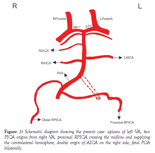
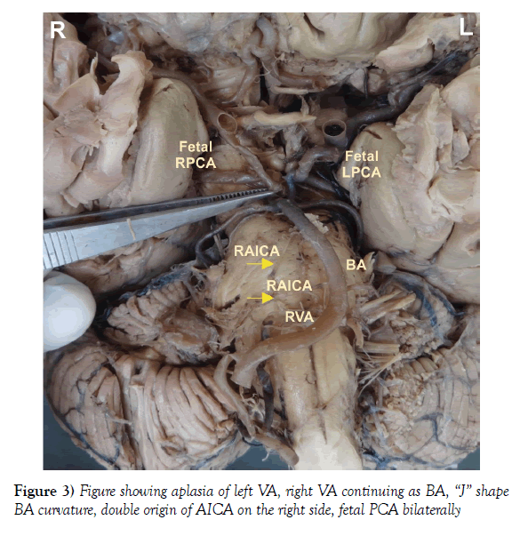
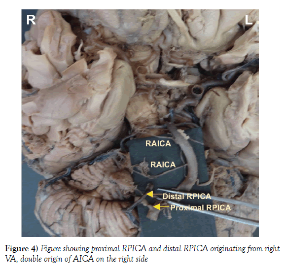
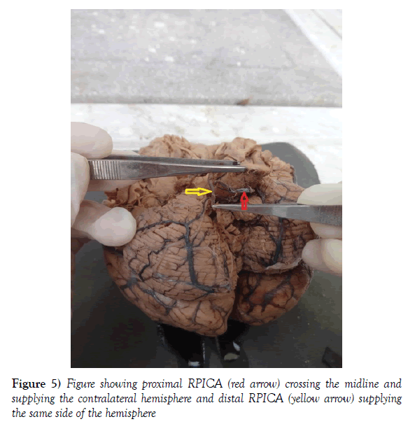
During routine dissection, in the Department of Anatomy, King George’s Medical University, while studying the arteries on the external surface of brain, absence of intracranial (V4 segment) part of left VA and a very rare variant origin of left posterior inferior cerebellar artery (PICA) was observed. Basilar artery commenced at pontomedullary junction as a continuation of RVA, as intracranial segment of LVA was absent. At the commencement of BA, it was deflected towards the left side. The diameter of right vertebral artery as well as of BA was 5 mm. Careful dissection of the area of interest revealed that from right vertebral artery two posterior inferior cerebellar arteries were arising. The distal one supplied the right cerebellar hemisphere and its course was normal, whereas the proximal one crossed midline and supplied the left cerebellar hemisphere. We also found two right anterior inferior cerebellar arteries (AICA) arising from the basilar artery. The diameters of P1 segments of both right and left posterior cerebral artery (PCA) was 2 mm and that of P2 segments of both side was 4 mm. The diameter of posterior communicating artery (PcomA) of both right and left side was 4 mm. Fetal PCA was found bilaterally as the diameter of PcomA was larger than P1 segments on both sides (Figures 2-5).
Discussion
In the present case aplasia of V4 segment of left VA was associated with multiple variations involving multiple vessels of VBS. Survey of literature also revealed that cases of hypoplasia or aplasia of VA were associated with anomalies of other vessels of VBS [7,9]. Incidence of left VA hypoplasia or aplasia has been observed to be more than right VA (3.1% vs. 1.8%) [6] and we also found aplasia of left VA. Segmental agenesis of VA is usually found to be associated with persistant trigeminal, hypoglossal or pro-atlantal arteries but we did not find any persistant primitive artery. Vasuki et al., also observed absence of RVA with no persistant primitive arteries [7].
It is commonly observed that in cases of segmental aplasia of VA (V4 segment), the artery of the affected side continues its course as posterior inferior cerebellar artery and contralateral artery continues as BA [4]. In the present case report the RVA was continuing as BA but the left PICA was not the continuation of V3 segment of left VA but it was originating from right VA. Double trunk of PICA is a very rare anomaly and in such cases one trunk is larger than the other and supplies the ipsilateral side of cerebellar hemisphere [10], but in the present case the second trunk of PICA was crossing to the opposite side and was supplying the contralateral side. Cases of bihemispheric PICA have also been reported [11]. The bihemispheric PICA arises from dominant VA and contraletral PICA is usually hypoplastic or absent. In these cases there is only one PICA which is supplying both cerebellar hemisphere but in our case two PICAs were arising from dominant VA and the proximal one crossed to the contralateral side to supply blood.
We also found two right anterior inferior cerebellar arteries (AICA) arising from the basilar artery. The left AICA was normal in its origin and course. Pekcevik (2015) reported a case of right AICA which had two origins, however it was different from our case as in that one AICA arose from vertebral artery and the other from basilar artery. The left AICA was absent [12].
Bilateral fetal PCA was also observed in the present case. We hypothesize that development of bilateral fetal PCA was body’s compensatory mechanism because in the present case VBS was receiving its blood only from single VA, this might have resulted into inadequate vertebrobasilar circulation. To overcome this insufficiency fetal PCA developed, so that PCA received blood mainly from ICA. If on one hand it was a boon on other it could be a bane as it allows thrombotic material from atherosclerotic internal carotid artery to pass into the PCA, where it may cause an ischemic event [13].
Unilateral hypoplasia or aplasia of VA can cause significant haemodynamic changes in the contralateral VA which may contribute to BA curvature. Hong et al. (2009) found VA significantly larger on the left side than on the right side and the curvature of BA was on the opposite side of VA dominant. Their findings were consistent to our present case as we found the curvature of BA was opposite the right VA. They concluded that the asymmetrical flow patterns of VAs around the vertebrobasilar junction might be an important mechanical force in the origin of BA curvature as well as a causative factor of peri-vertebrobasilar junctional infarcts [14]. Headache, hypertension and posterior circulation stroke may be the presenting feature in the absence or hypoplasia of vertebral artery. Aneurysms, atherosclerosis and thrombosis are more common in persistent primitive arteries [7].
Conclusion
Knowledge of variations of vertebral artery (VAs) supplying the brain can be a useful guideline to the surgeons and interventional radiologist for careful pre-operative planning and thus help them in avoiding potentially life threatening complications. The knowledge of the anatomical and morphological variations of vertebral artery may also be important in interpreting the anatomic features of the vasculature in MRI, CT and MR angiographies and may be significant in surgical procedures of head and neck region. Unilateral VA aplasia was found to be associated with multiple other anomalies of arteries of VBS like, double trunk of PICA and AICA, bilateral fetal PCA, therefore it is highly recommended that if one comes across with any variation of a single vessel, the whole VBS should be thoroughly observed.
REFERENCES
- Bannister LH, Late Peter L Williams, Martin M Berry, et al. Gray’s Anatomy. Churchill Livingstone. 1995.
- Padget DH. The development of the cranial arteries in the human embryo. Contrib Embryol. 1948;32:205-61.
- Kathuria S, Gregg L, Chen J, et al. Normal cerebral arterial development and variations. Semin Ultrasound CT MR. 2011; 32:242-51.
- Tuncer MC, Akgul YH, Karabulut O. MR angiography ımaging of absence vertebral artery causing of pulsatile tinnitus: a case report. Int J Morphol. 2010;28:357-63.
- Singh I. Human Embryology. 2014.
- Blickenstaff KL, Weaver FA, Yellin AE, et al. Trends in the management of traumatic vertebral artery injuries. Am J Surg. 1989;158:101-6.
- Vasuki AKM, Devi MN, Kandavadivelu MJ, et al. Absent Intracranial Part of right Vertebral Artery– A Case Report. Inno J Med Health Sci.2015;5:6-8.
- Woodcock RJ, Cloft HJ, Dion JE. Bilateral type 1 proatlantal arteries with absence of vertebral arteries. Am J Neuroradiol. 2001;22:418-20.
- Tuccar E, Yazar F, Kirici Y, et al. A complex variation of the vertebrobasilar system. Neuroanat. 2002;1:12-3.
- Sharifi M, Ungier E, Ciszek B. Double posterior inferior cerebellar artery. Surg Radiol Anat. 2010;32:87.
- Cullen SP, Ozanne A, Alvarez H, et al. The bihemispheric posterior inferior cerebellar artery. Neuroradiol. 2005; 47:809-12.
- Pekcevik Y. Double origin and early bifurcation of the anterior inferior cerebellar artery diagnosed by CT angiography. Surg Radiol Anat. 2015.
- De Silva KRD, Silva TRN, Gunasekera WSL, et al. Variation in the origin of the posterior cerebral artery in adult Sri Lankans. Neurol India. 2009;57:46-9.
- Hong JM, Chung CS, Bang OY, et al. Vertebral artery dominance contributes to basilar artery curvature and peri-vertebrobasilar junctional infarcts. J Neurol Neurosurg Psychiatry. 2009;80:1087-92.


