Multiple variations of the abdominal vessels in a single cadaver
Gulnur Ozguner*, Osman Sulak, Esra Koyuncu
Department of Anatomy, Faculty of Medicine, Suleyman Demirel University, Isparta, Turkey.
- *Corresponding Author:
- Dr. Gulnur Ozguner
Assistant Professor, Department of Anatomy, Suleyman Demirel University, Faculty of Medicine, Isparta, 32260, Turkey.
Tel: +90 (246) 2113301
E-mail: ozgunerg@hotmail.com
Date of Received: April 13th, 2012
Date of Accepted: October 25th, 2012
Published Online: February 21st, 2013
© Int J Anat Var (IJAV). 2013; 6: 52–55.
[ft_below_content] =>Keywords
abdominal vessels, accessory hepatic artery, bilateral accessory renal arteries, retroaortic–additional renal vein, testicular artery
Introduction
The abdominal aorta begins at 12th thoracic vertebra, and it descends anterior to the lumbar vertebrae, and bifurcates at the 4th lumbal vertebral body. It supplies all the abdominal organs, abdominal walls, gonads, and lower extremity [1].
In the usual vasculature, celiac trunk is divided into three branches: common hepatic artery (CHA), splenic artery (SA), and left gastric artery (LGA). The CHA gives hepatic artery proper (HAP) and gastroduodenal artery. HAP divides into left hepatic branch and right hepatic branch. The renal arteries arise from the side of the aorta, immediately below the superior mesenteric artery. The testicular arteries arise from the front of the aorta, a little below the renal arteries. The left renal vein is longer than the right, and passes in front of the aorta. It receives the left testicular and left inferior phrenic veins [1].
Assessment of the hepatic arterial anatomy is one of significant interest in liver transplantation. Moreover, hepatic vasculature is extremely important in chemotherapy treatment of primary or secondary liver tumors. Injuries to accessory or aberrant hepatic arteries can lead to liver ischemia or necrosis [2,3].
Good knowledge of the renal vascular variations is essential in case of renal transplantation, renal surgeries, gonadal surgeries, uroradiology, gonadal color Doppler imaging, and in surgeries of aneurysms of abdominal aorta [4].
Therefore, it is very important to know normal anatomy, and their variations of the abdominal vessels.
Case Report
During the routine educational dissection of abdominal organs and branches of abdominal aorta at our department, a 45-year-old male cadaver was dissected. We observed the following variations in the abdominal vasculature.
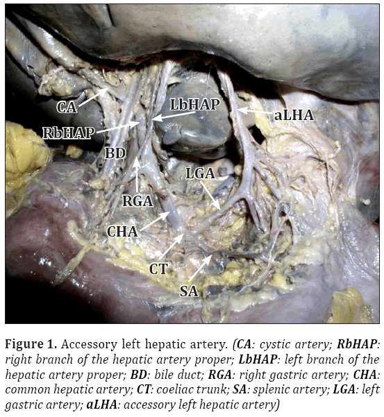
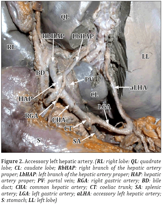
An accessory left hepatic artery was (aLHA) originated from the left gastric artery and was supplied the left lobe of the liver. The celiac trunk was originated from the abdominal aorta and divided into three branches as CHA, SA, LGA. The right and the left branches of the HAP were arising from the CHAs. The right branch gave the cystic artery. The left branch of the HAP gave the right gastric artery and then divided into two branches before entering the porta hepatis. The right branch of the HAP supplied the right lobe, and the left branch of the HAP supplied the caudate and quadrate lobes, and left accessory hepatic artery arising from the LGA supplied the left lobe of the liver (Figures 1, 2). The diameters of these arteries were: SA: 4.5 mm, LGA: 4 mm, CHA: 5 mm, right branch of HAP: 4 mm, left branch of HAP: 3 mm, and aLHA: 4 mm.
Figure 1: Accessory left hepatic artery. (CA: cystic artery; RbHAP: right branch of the hepatic artery proper; LbHAP: left branch of the hepatic artery proper; BD: bile duct; RGA: right gastric artery; CHA: common hepatic artery; CT: coeliac trunk; SA: splenic artery; LGA: left gastric artery; aLHA: accessory left hepatic artery)
Figure 2: Accessory left hepatic artery. (RL: right lobe: QL: quadrate lobe; CL: caudate lobe; RbHAP: right branch of the hepatic artery proper; LbHAP: left branch of the hepatic artery proper; HAP: hepatic artery proper; PV: portal vein; RGA: right gastric artery; BD: bile duct; CHA: common hepatic artery; CT: coeliac trunk; SA: splenic artery; LGA: left gastric artery; aLHA: accessory left hepatic artery; S: stomach; LL: left lobe)
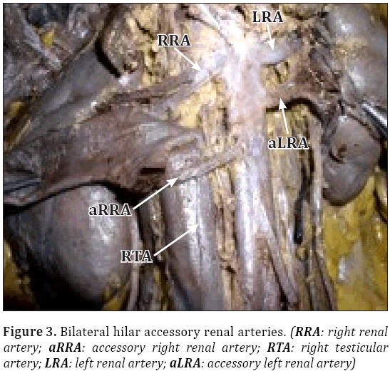
Bilateral hilar accessory renal arteries were originated from the abdominal aorta. The right and left main renal arteries arose from the lateral surface of the aorta with a symmetric origin, just below the origin of superior mesenteric artery (Figure 3). The diameters of the right and the left main renal arteries were 7 mm each, and lengths of the right and the left main renal arteries were 40 mm and 42 mm, respectively. On the right side, an accessory renal artery was originated from the aorta,14 mm above the origin of inferior mesenteric artery, and on the left side, an accessory renal artery arose from the aorta, 32 mm above the origin of inferior mesenteric artery. The distances between the accessory renal artery and main renal artery was 25 mm on the right side, and was 10 mm on the left side. The diameters of the right and the left accessory renal arteries were 3.8 mm and 6 mm, respectively. The lengths of the right and the left accessory renal arteries were 44 mm and 42 mm, respectively.
Right testicular artery was originated from the right accessory renal artery (Figure 3). The left testicular artery was originated from the 10 mm above the bifurcation of abdominal aorta.
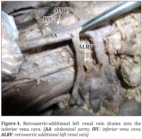
Retroaortic-additional left renal vein was drained into the inferior vena cava (Figure 4).
Discussion
According to Michel’s [5] classification of hepatic arteries, the left accessory hepatic artery is originated from the left gastric artery in 8 percent of the cases, while replaced left hepatic artery is originated from the left gastric artery in 10 percent of the cases. In our case, an accessory hepatic artery originates from the LGA, and enters into the left lobe of the liver. The CHA divides into right and left HAP. The right HAP supplies the right lobe, and the left HAP supplies the caudate and quadrate lobes of the liver. It is important to identify this variant for left hepatectomy because this artery must be identified and ligated [6]. In the fetal life, the right hepatic artery originates from the superior mesenteric artery, and the left hepatic artery originates from the left gastric artery, thus partial or complete persistence of the fetal pattern could result in arterial variations of the liver [7].
The presence of multiple renal arteries increases the complexity of renal transplantation; also renal vascular variations are associated with a significantly higher failure rate than kidneys with a single artery [8].
In previous studies the frequency of bilateral accessory renal arteries was 5% [9], 7.7% [8], 12% [10]. In our case, bilateral hilar accessory renal arteries originate from the abdominal aorta, and the right accessory renal artery gives the right testicular artery. The variations in the renal arteries are mainly due to the various developmental positions of kidneys. The kidney develops in the pelvic cavity and than ascends to lumbal region. During this course, it is supplied by the branches of common iliac artery and the lower level of the abdominal aorta. Normally, the caudal branches disappear. The presence of accessory renal artery might be due to the failure in the disappearance of these arteries [11].
The anatomy of the gonadal arteries is very important because of the development of new operative techniques for operations such as varicocele and undescended testis [12]. It was reported that, the anatomic variations of gonadal arteries might be important due to the influence on the blood flow from the kidney to gonadal glands [13]. Cicekcibasi et al. reported that 5.5% of gonadal arteries originate from the renal artery. Moreover, they noted that these variations are more commonly in the male fetuses than the female, and are more frequently on the right side than on the left [14]. In our case, the right testicular artery was originated from the right accessory renal artery, and the left testicular artery was originated from the aorta, 10 mm above the bifurcation.
In the literature, the prevalence of retroaortic left renal vein is 0.5-1.66% and additional renal vein is 0.4% [15,16]. During the fifth to seventh week of embryological period, subcardinal veins develop. The anastomosis between the subcardinal veins form the left renal vein. Unusual persistence or regression of the anastomoses at this level result in the formation of left renal variations, such as retroaortic left renal vein [15]. In our case, the retroaortic accessory left renal vein was crossed behind the abdominal aorta, and was drained directly to the inferior vena cava. The knowledge of such variations is quite useful in planning any upper abdominal surgery.
Awareness of the arterial and venous variations in the abdomen, such as shown in our case, is very important during surgical and radiological procedures pertaining to the abdominal organs.
References
- Standring S, ed. Gray’s Anatomy: The Anatomical Basis of Clinical Practice. 39th Ed., Edinburgh, Elsevier Churchill Livingstone. 2005; 1271.
- Saba L, Mallarini G. Multidetector row CT angiography in the evaluation of the hepatic artery and its anatomical variants. Clin Radiol. 2008; 63: 312–321.
- Molmenti EP, Pinto PA, Klein J, Klein AS. Normal and variant arterial supply of the liver and gallbladder. Pediatr Transplant. 2003; 7: 80–82.
- Jetti R, Jevoor P, Vollala VR, Potu BK, Ravishankar M, Virupaxi R. Multiple variations of the urogenital vascular system in a single cadaver: a case report. Cases J. 2008; 1: 344.
- Michels NA. Newer anatomy of the liver and its variant blood supply and collateral circulation. Am J Surg. 1966; 112: 337–347.
- Iezzi R, Cotroneo AR, Giancristofaro D, Santoro M, Storto ML. Multidetector-row CT angiographic imaging of the celiac trunk: anatomy and normal variants. Surg Radiol Anat. 2008; 30: 303–310.
- Polguj M, Gabryniak T, Topol M. The right accessory hepatic artery; a case report and review of the literature. Surg Radiol Anat. 2010; 32:175–179.
- Cicekcibasi AE, Ziylan T, Salbacak A, Seker M, Buyukmumcu M, Tuncer I. An investigation of the origin, location and variations of the renal arteries in human fetuses and their clinical relevance. Ann Anat. 2005; 187: 421–427.
- Ozkan U, Oguzkurt L, Tercan F, Kizilkilic O, Koc Z, Koca N. Renal artery origins and variations: angiographic evaluation of 855 consecutive patients. Diagn Iterv Radiol. 2006; 12: 183–186.
- Urban BA, Ratner LE, Fishman EK. Three dimensional volume-rendered CT angiography of the renal arteries and veins: normal anatomy, variants, and clinical application. Radiographics. 2001; 21: 373–386.
- Moore KL, Persaud TVN. The Developing Human: Clinically Oriented Embryology. 7th Ed., Philadelphia, WB Saunders. 2002; 293.
- Brohi RA, Sargon MF, Yener N. High origin and suprarenal branch of a testicular artery. Surg Radiol Anat. 2001; 23: 207–208.
- Onderoglu S, Yuksel M, Arik Z. Unusual branching and course of the testicular artery. Ann Anat. 1993; 175: 541–544.
- Cicekcibasi AE, Salbacak A, Seker M, Ziylan T, Buyukmumcu M, Uysal II. The origin of gonadal arteries in human fetuses: Anatomical variations. Ann Anat. 2002; 184: 275–279.
- Dilli A, Ayaz UY, Karabacak OR, Tatar IG, Hekimoglu B. Study of the left renal variations by means of magnetic resonance imaging. Surg Radiol Anat. 2012; 34: 267–270.
- Satyapal KS, Kalideen JM, Haffejee AA, Singh B, Robbs JV. Left renal vein variations. Surg Radiol Anat. 1999; 21: 77–81.
Gulnur Ozguner*, Osman Sulak, Esra Koyuncu
Department of Anatomy, Faculty of Medicine, Suleyman Demirel University, Isparta, Turkey.
- *Corresponding Author:
- Dr. Gulnur Ozguner
Assistant Professor, Department of Anatomy, Suleyman Demirel University, Faculty of Medicine, Isparta, 32260, Turkey.
Tel: +90 (246) 2113301
E-mail: ozgunerg@hotmail.com
Date of Received: April 13th, 2012
Date of Accepted: October 25th, 2012
Published Online: February 21st, 2013
© Int J Anat Var (IJAV). 2013; 6: 52–55.
Abstract
Variant branching pattern of the abdominal vessels was observed in a single cadaver. A left accessory hepatic artery is originated from the left gastric artery and is supplied the left lobe of the liver. Bilateral hilar accessory renal arteries are originated from the abdominal aorta. Right testicular artery is originated from the right accessory renal artery. Retroaortic-additional left renal vein is drained into the inferior vena cava.
-Keywords
abdominal vessels, accessory hepatic artery, bilateral accessory renal arteries, retroaortic–additional renal vein, testicular artery
Introduction
The abdominal aorta begins at 12th thoracic vertebra, and it descends anterior to the lumbar vertebrae, and bifurcates at the 4th lumbal vertebral body. It supplies all the abdominal organs, abdominal walls, gonads, and lower extremity [1].
In the usual vasculature, celiac trunk is divided into three branches: common hepatic artery (CHA), splenic artery (SA), and left gastric artery (LGA). The CHA gives hepatic artery proper (HAP) and gastroduodenal artery. HAP divides into left hepatic branch and right hepatic branch. The renal arteries arise from the side of the aorta, immediately below the superior mesenteric artery. The testicular arteries arise from the front of the aorta, a little below the renal arteries. The left renal vein is longer than the right, and passes in front of the aorta. It receives the left testicular and left inferior phrenic veins [1].
Assessment of the hepatic arterial anatomy is one of significant interest in liver transplantation. Moreover, hepatic vasculature is extremely important in chemotherapy treatment of primary or secondary liver tumors. Injuries to accessory or aberrant hepatic arteries can lead to liver ischemia or necrosis [2,3].
Good knowledge of the renal vascular variations is essential in case of renal transplantation, renal surgeries, gonadal surgeries, uroradiology, gonadal color Doppler imaging, and in surgeries of aneurysms of abdominal aorta [4].
Therefore, it is very important to know normal anatomy, and their variations of the abdominal vessels.
Case Report
During the routine educational dissection of abdominal organs and branches of abdominal aorta at our department, a 45-year-old male cadaver was dissected. We observed the following variations in the abdominal vasculature.
An accessory left hepatic artery was (aLHA) originated from the left gastric artery and was supplied the left lobe of the liver. The celiac trunk was originated from the abdominal aorta and divided into three branches as CHA, SA, LGA. The right and the left branches of the HAP were arising from the CHAs. The right branch gave the cystic artery. The left branch of the HAP gave the right gastric artery and then divided into two branches before entering the porta hepatis. The right branch of the HAP supplied the right lobe, and the left branch of the HAP supplied the caudate and quadrate lobes, and left accessory hepatic artery arising from the LGA supplied the left lobe of the liver (Figures 1, 2). The diameters of these arteries were: SA: 4.5 mm, LGA: 4 mm, CHA: 5 mm, right branch of HAP: 4 mm, left branch of HAP: 3 mm, and aLHA: 4 mm.
Figure 1: Accessory left hepatic artery. (CA: cystic artery; RbHAP: right branch of the hepatic artery proper; LbHAP: left branch of the hepatic artery proper; BD: bile duct; RGA: right gastric artery; CHA: common hepatic artery; CT: coeliac trunk; SA: splenic artery; LGA: left gastric artery; aLHA: accessory left hepatic artery)
Figure 2: Accessory left hepatic artery. (RL: right lobe: QL: quadrate lobe; CL: caudate lobe; RbHAP: right branch of the hepatic artery proper; LbHAP: left branch of the hepatic artery proper; HAP: hepatic artery proper; PV: portal vein; RGA: right gastric artery; BD: bile duct; CHA: common hepatic artery; CT: coeliac trunk; SA: splenic artery; LGA: left gastric artery; aLHA: accessory left hepatic artery; S: stomach; LL: left lobe)
Bilateral hilar accessory renal arteries were originated from the abdominal aorta. The right and left main renal arteries arose from the lateral surface of the aorta with a symmetric origin, just below the origin of superior mesenteric artery (Figure 3). The diameters of the right and the left main renal arteries were 7 mm each, and lengths of the right and the left main renal arteries were 40 mm and 42 mm, respectively. On the right side, an accessory renal artery was originated from the aorta,14 mm above the origin of inferior mesenteric artery, and on the left side, an accessory renal artery arose from the aorta, 32 mm above the origin of inferior mesenteric artery. The distances between the accessory renal artery and main renal artery was 25 mm on the right side, and was 10 mm on the left side. The diameters of the right and the left accessory renal arteries were 3.8 mm and 6 mm, respectively. The lengths of the right and the left accessory renal arteries were 44 mm and 42 mm, respectively.
Right testicular artery was originated from the right accessory renal artery (Figure 3). The left testicular artery was originated from the 10 mm above the bifurcation of abdominal aorta.
Retroaortic-additional left renal vein was drained into the inferior vena cava (Figure 4).
Discussion
According to Michel’s [5] classification of hepatic arteries, the left accessory hepatic artery is originated from the left gastric artery in 8 percent of the cases, while replaced left hepatic artery is originated from the left gastric artery in 10 percent of the cases. In our case, an accessory hepatic artery originates from the LGA, and enters into the left lobe of the liver. The CHA divides into right and left HAP. The right HAP supplies the right lobe, and the left HAP supplies the caudate and quadrate lobes of the liver. It is important to identify this variant for left hepatectomy because this artery must be identified and ligated [6]. In the fetal life, the right hepatic artery originates from the superior mesenteric artery, and the left hepatic artery originates from the left gastric artery, thus partial or complete persistence of the fetal pattern could result in arterial variations of the liver [7].
The presence of multiple renal arteries increases the complexity of renal transplantation; also renal vascular variations are associated with a significantly higher failure rate than kidneys with a single artery [8].
In previous studies the frequency of bilateral accessory renal arteries was 5% [9], 7.7% [8], 12% [10]. In our case, bilateral hilar accessory renal arteries originate from the abdominal aorta, and the right accessory renal artery gives the right testicular artery. The variations in the renal arteries are mainly due to the various developmental positions of kidneys. The kidney develops in the pelvic cavity and than ascends to lumbal region. During this course, it is supplied by the branches of common iliac artery and the lower level of the abdominal aorta. Normally, the caudal branches disappear. The presence of accessory renal artery might be due to the failure in the disappearance of these arteries [11].
The anatomy of the gonadal arteries is very important because of the development of new operative techniques for operations such as varicocele and undescended testis [12]. It was reported that, the anatomic variations of gonadal arteries might be important due to the influence on the blood flow from the kidney to gonadal glands [13]. Cicekcibasi et al. reported that 5.5% of gonadal arteries originate from the renal artery. Moreover, they noted that these variations are more commonly in the male fetuses than the female, and are more frequently on the right side than on the left [14]. In our case, the right testicular artery was originated from the right accessory renal artery, and the left testicular artery was originated from the aorta, 10 mm above the bifurcation.
In the literature, the prevalence of retroaortic left renal vein is 0.5-1.66% and additional renal vein is 0.4% [15,16]. During the fifth to seventh week of embryological period, subcardinal veins develop. The anastomosis between the subcardinal veins form the left renal vein. Unusual persistence or regression of the anastomoses at this level result in the formation of left renal variations, such as retroaortic left renal vein [15]. In our case, the retroaortic accessory left renal vein was crossed behind the abdominal aorta, and was drained directly to the inferior vena cava. The knowledge of such variations is quite useful in planning any upper abdominal surgery.
Awareness of the arterial and venous variations in the abdomen, such as shown in our case, is very important during surgical and radiological procedures pertaining to the abdominal organs.
References
- Standring S, ed. Gray’s Anatomy: The Anatomical Basis of Clinical Practice. 39th Ed., Edinburgh, Elsevier Churchill Livingstone. 2005; 1271.
- Saba L, Mallarini G. Multidetector row CT angiography in the evaluation of the hepatic artery and its anatomical variants. Clin Radiol. 2008; 63: 312–321.
- Molmenti EP, Pinto PA, Klein J, Klein AS. Normal and variant arterial supply of the liver and gallbladder. Pediatr Transplant. 2003; 7: 80–82.
- Jetti R, Jevoor P, Vollala VR, Potu BK, Ravishankar M, Virupaxi R. Multiple variations of the urogenital vascular system in a single cadaver: a case report. Cases J. 2008; 1: 344.
- Michels NA. Newer anatomy of the liver and its variant blood supply and collateral circulation. Am J Surg. 1966; 112: 337–347.
- Iezzi R, Cotroneo AR, Giancristofaro D, Santoro M, Storto ML. Multidetector-row CT angiographic imaging of the celiac trunk: anatomy and normal variants. Surg Radiol Anat. 2008; 30: 303–310.
- Polguj M, Gabryniak T, Topol M. The right accessory hepatic artery; a case report and review of the literature. Surg Radiol Anat. 2010; 32:175–179.
- Cicekcibasi AE, Ziylan T, Salbacak A, Seker M, Buyukmumcu M, Tuncer I. An investigation of the origin, location and variations of the renal arteries in human fetuses and their clinical relevance. Ann Anat. 2005; 187: 421–427.
- Ozkan U, Oguzkurt L, Tercan F, Kizilkilic O, Koc Z, Koca N. Renal artery origins and variations: angiographic evaluation of 855 consecutive patients. Diagn Iterv Radiol. 2006; 12: 183–186.
- Urban BA, Ratner LE, Fishman EK. Three dimensional volume-rendered CT angiography of the renal arteries and veins: normal anatomy, variants, and clinical application. Radiographics. 2001; 21: 373–386.
- Moore KL, Persaud TVN. The Developing Human: Clinically Oriented Embryology. 7th Ed., Philadelphia, WB Saunders. 2002; 293.
- Brohi RA, Sargon MF, Yener N. High origin and suprarenal branch of a testicular artery. Surg Radiol Anat. 2001; 23: 207–208.
- Onderoglu S, Yuksel M, Arik Z. Unusual branching and course of the testicular artery. Ann Anat. 1993; 175: 541–544.
- Cicekcibasi AE, Salbacak A, Seker M, Ziylan T, Buyukmumcu M, Uysal II. The origin of gonadal arteries in human fetuses: Anatomical variations. Ann Anat. 2002; 184: 275–279.
- Dilli A, Ayaz UY, Karabacak OR, Tatar IG, Hekimoglu B. Study of the left renal variations by means of magnetic resonance imaging. Surg Radiol Anat. 2012; 34: 267–270.
- Satyapal KS, Kalideen JM, Haffejee AA, Singh B, Robbs JV. Left renal vein variations. Surg Radiol Anat. 1999; 21: 77–81.


