Multiple variations of the branches of abdominal aortaor internal
Mehmet Fatih Inceikli1, Ilker Mustafa Kafa2*, Murat Uysal2, Sinan Bakirci2 and Ibrahim Hakan Oygucu2
1Department of Radiology, Uludag University, Faculty of Medicine, Bursa, Turkey
2Department of Radiology and Anatomy, Uludag University, Faculty of Medicine, Bursa, Turkey
- *Corresponding Author:
- Ilker Mustafa Kafa, MD
Department of Anatomy, Uludag University, Faculty of Medicine, Gorukle, 16059, Bursa, Turkey
Tel: +90 (224) 295 23 16
E-mail: imkafa@uludag.edu.tr
Date of Received: November 20th, 2009
Date of Accepted: June 9th, 2010
Published Online: June 23rd, 2010
© IJAV. 2010; 3: 88–90.
[ft_below_content] =>Keywords
abdominal aorta, aorta abdominalis, variation, artery
Introduction
The abdominal aorta begins and descends after aortic hiatus at the level of the twelfth thoracic vertebrae, courses downward with the inferior vena cava and terminates at its bifurcation at the level of the fourth lumbar vertebra. Branches of abdominal aorta may be described as ventral, lateral, dorsal and terminal, corresponding to their origins. The ventral braches are the celiac trunk, superior and inferior mesenteric arteries; the dorsal branches are the lumbar and median sacral arteries; the lateral branches are the inferior phrenic, middle suprarenal, renal, ovarian or testicular arteries and the terminal branches are the common iliac arteries [1].
The primitive aorta appears firstly in the embryo and gives segmental branches to the digestive tube (ventral splanchnic arteries), to the mesonephric ridge (lateral splanchnic arteries) and to the body wall (somatic arteries); many intersegmental branches supply the somites and their derivatives during the development [2]. Inferior phrenic arteries may arise separately from the aorta, just above celiac trunk and help to supply the diaphragm. Classically, each kidney is supplied by a single renal artery; however, anatomic variations are common as two or more renal arteries can supply a kidney. The testicular arteries usually arise from the anterior aspect of the abdominal aorta just inferior to the renal arteries at the level of the second lumbar vertebra; however, they may also originate from the renal artery, middle suprarenal artery, one of the lumbar arteries, common aortaor internal iliac artery. Each passes obliquely downward and laterally on the psoas major muscle [3]. The median sacral artery is a small posterior branch of abdominal aorta and leaves from the aorta at level of its bifurcation; it is originating infrequently from the common stem with the fourth lumbar vertebral arteries.
Arterial variations in the abdominal region are important during laparoscopic and various other surgical procedures.
Case Report
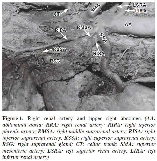
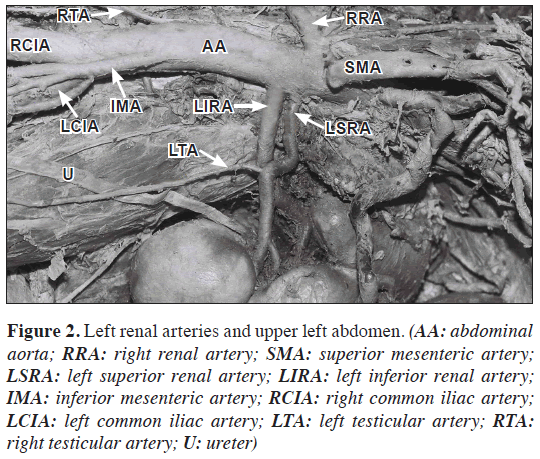
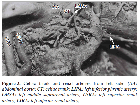
During routine dissections, multiple variations of the abdominal aorta were observed in the abdominal retroperitoneal region of 54-year-old Caucasian male cadaver. We found double renal arteries on the left side. The upper renal artery was arising from aorta and advancing about 2.8 cm laterally, then progressing downwards about 1.3 cm straightly and showed approximately 80º of kinking then crossed the lower one and entered to the inferior pole of hilum of the kidney (Figures 1,2,3). The left testicular artery was originating after this kinking of renal artery (Figure 2).
Figure 1: Right renal artery and upper right abdomen. (AA: abdominal aorta; RRA: right renal artery; RIPA: right inferior phrenic artery; RMSA: right middle suprarenal artery; RISA: right inferior suprarenal artery; RSSA: right superior suprarenal artery; RSG: right suprarenal gland; CT: celiac trunk; SMA: superior mesenteric artery; LSRA: left superior renal artery; LIRA: left inferior renal artery)
Figure 2: Left renal arteries and upper left abdomen. (AA: abdominal aorta; RRA: right renal artery; SMA: superior mesenteric artery; LSRA: left superior renal artery; LIRA: left inferior renal artery; IMA: inferior mesenteric artery; RCIA: right common iliac artery; LCIA: left common iliac artery; LTA: left testicular artery; RTA: right testicular artery; U: ureter)
The left inferior phrenic artery was originating from the aorta, 1.1 cm below to the celiac trunk. However the left middle suprarenal artery was branching from celiac trunk (Figure 3).
The right renal and testicular arteries were arising from the right side of abdominal aorta, as usual (Figures 1,2). However, the right inferior phrenic artery was originating from the right renal artery (Figure 1). Then, right inferior phrenic artery ascended upwards and branched out to the diaphragm. The right middle and superior suprarenal arteries branched from the right inferior phrenic artery. Right inferior suprarenal artery was branching from right renal artery (Figure 1).
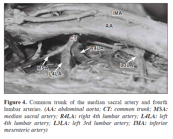
The median sacral artery and fourth lumbar arteries have been branched from a common trunk from the posterior surface of the abdominal aorta (Figure 4).
The abdominal aorta itself was spanning normally between the levels of T12-L4 vertebrae.
Discussion
To our knowledge, Pick and Anson [4] evaluated the largest series regarding the origin of the right inferior phrenic artery. They reported the origin of this artery as follows: 47% from aorta, 40% from celiac trunk, 10.5% from right renal artery, 2% from left gastric artery and 0.5% from hepatic artery. In another study, 26 of 180 patients had right phrenic arteriogram; the origin of artery was from right renal artery in two cases (7.6%) [5]. Knowledge of all possible variations and particularly the origins of the left and right inferior phrenic arteries may be useful in the discussion and treatment of hepatic, suprarenal or diaphragmatic lesions [6]. The renal artery is usually singular on each side. However, it is not rare for multiple renal arteries to be found on one side. The incidence of anomalous renal pedicles may be due to the embryological development of the kidneys which do not rotate medially when they ascend from the pelvis to the abdomen. Supplementary renal arteries with an aortic origin are frequently appeared as vascular variations and represent the persistence of the embryonic vessels within the renal ascent according to Langman and Sadler [7]. The incidence of left double hilar renal arteries was found as 4.98% in a largest study [8]. The double hilar renal arteries also found as 11.1% (15.5% right side – 6.6 % left side) in another study [9]. The variations in the renal vascular anatomy have increased clinical importance. Doppler ultrasound and magnetic resonance angiography of the renal arteries can be used in combination to achieve adequate screening of patients prior to conventional angiography [10]. Lumbar artery anatomy also has an importance for spinal cord surgeons and interventional radiologists. The fourth lumbar arteries on either side may provide the middle sacral or both arteries may arise as a common stem with the middle sacral artery [11].
Every surgeon and radiologist must need precise knowledge for the single or multiple variations of the abdominal aorta in diagnostic and/or surgical investigations. Ligation or damage of these arteries without knowing the possible variations in laparotomy, nephrectomy, renal transplantation, arterial reconstruction and laparoscopy or in other surgical applications may cause unpredictable complications, such as segmental or total visceral ischemia and failure. The variations of the branches of abdominal aorta were investigated in various studies in literature. Although these variations might encounter in different cadavers, we cannot found in literature that including all of these variations in same cadaver.
References
- Williams LP, Warwick R, Dyson M, Bannister LH. Gray’s Anatomy. 37th Ed., Edinburgh, Churchill Livingstone. 1989; 766.
- Moore KL, Persaude TVN. The Developing Human, Clinically Oriented Embryology. 5th Ed., London, W.B. Saunders. 1993; 309.
- Moore KL, Dalley AF. Clinically Oriented Anatomy. 4th Ed., Philadelphia, Lippincott Williams & Wilkins. 1999; 292.
- Pick J, Anson B. The inferior phrenic artery. Origin and suprarenal branches. Anat Rec. 1940; 78: 413–427.
- Hiwatashi A, Yoshida K. The origin of right inferior phrenic artery on multidetector row helical CT. Clin Imaging. 2003; 27: 298–303.
- Loukas M, Hullett J, Wagner T. Clinical anatomy of the ınferior phrenic artery. Clin Anat. 2005; 18: 357–365.
- Langman J, Sadler TW. Embryologie médicale. Pradel, Paris. 1996; 301.
- Khamanarong K, Prachaney P, Utraravichien A, Tong-Un T, Sripaoraya K. Anatomy of renal arterial supply. Clin Anat. 2004; 17: 334–336.
- Cicekcibasi AE, Ziylan T, Salbacak A, Seker M, Buyukmumcu M, Tuncer I. An investigation of the origin, location and variations of the renal arteries in human fetuses and their clinical revelance. Ann Anat. 2005; 187: 421–427.
- Soulez G, Oliva VL, Turpin S, Lambert R, Nicolet V, Therasse E. Imaging of renovascular hypertension: Respective values of renal scintigraphy, renal Doppler US, and MR angiography. Radiographics. 2000; 20: 1355–1368.
- Bergman RA, Afifi K, Ryosuke Miyauchi MS. Illustrated Encyclopedia of Human Anatomic Variation. Opus II: Cardiovascular System, Arteries, Abdomen, Lumbar arteries. http://www.anatomyatlases.org/AnatomicVariants/Cardiovascular/Text/Arteries/Lumbar.shtml (accessed June 2010)
Mehmet Fatih Inceikli1, Ilker Mustafa Kafa2*, Murat Uysal2, Sinan Bakirci2 and Ibrahim Hakan Oygucu2
1Department of Radiology, Uludag University, Faculty of Medicine, Bursa, Turkey
2Department of Radiology and Anatomy, Uludag University, Faculty of Medicine, Bursa, Turkey
- *Corresponding Author:
- Ilker Mustafa Kafa, MD
Department of Anatomy, Uludag University, Faculty of Medicine, Gorukle, 16059, Bursa, Turkey
Tel: +90 (224) 295 23 16
E-mail: imkafa@uludag.edu.tr
Date of Received: November 20th, 2009
Date of Accepted: June 9th, 2010
Published Online: June 23rd, 2010
© IJAV. 2010; 3: 88–90.
Abstract
Multiple variations were found in a 54-year-old male cadaver during routine dissections of abdominal retroperitoneal region. Right inferior phrenic artery was arising from right renal artery, while the right middle and superior suprarenal artery branched from the right inferior phrenic artery. Left inferior phrenic artery was originating from the abdominal aorta below celiac trunk. The left middle suprarenal artery appeared as the branch of celiac trunk. Double left renal artery was arising separately from the left side of the aorta. The upper left renal artery showed approximately 80º of kinking which then crossed the lower one and entered to the inferior pole of hilum of kidney. Left testicular artery was originating from the upper left renal artery after this kinking. The left and right fourth lumbar arteries and median sacral artery have branched from a common trunk posterior to the abdominal aorta. In spite of these abundant variations in the branches, abdominal aorta itself did not show any variation, spanning normally between the levels of T12–L4 vertebrae. The variations of abdominal aorta may have clinical importance, especially in surgical and radiologic investigations.
-Keywords
abdominal aorta, aorta abdominalis, variation, artery
Introduction
The abdominal aorta begins and descends after aortic hiatus at the level of the twelfth thoracic vertebrae, courses downward with the inferior vena cava and terminates at its bifurcation at the level of the fourth lumbar vertebra. Branches of abdominal aorta may be described as ventral, lateral, dorsal and terminal, corresponding to their origins. The ventral braches are the celiac trunk, superior and inferior mesenteric arteries; the dorsal branches are the lumbar and median sacral arteries; the lateral branches are the inferior phrenic, middle suprarenal, renal, ovarian or testicular arteries and the terminal branches are the common iliac arteries [1].
The primitive aorta appears firstly in the embryo and gives segmental branches to the digestive tube (ventral splanchnic arteries), to the mesonephric ridge (lateral splanchnic arteries) and to the body wall (somatic arteries); many intersegmental branches supply the somites and their derivatives during the development [2]. Inferior phrenic arteries may arise separately from the aorta, just above celiac trunk and help to supply the diaphragm. Classically, each kidney is supplied by a single renal artery; however, anatomic variations are common as two or more renal arteries can supply a kidney. The testicular arteries usually arise from the anterior aspect of the abdominal aorta just inferior to the renal arteries at the level of the second lumbar vertebra; however, they may also originate from the renal artery, middle suprarenal artery, one of the lumbar arteries, common aortaor internal iliac artery. Each passes obliquely downward and laterally on the psoas major muscle [3]. The median sacral artery is a small posterior branch of abdominal aorta and leaves from the aorta at level of its bifurcation; it is originating infrequently from the common stem with the fourth lumbar vertebral arteries.
Arterial variations in the abdominal region are important during laparoscopic and various other surgical procedures.
Case Report
During routine dissections, multiple variations of the abdominal aorta were observed in the abdominal retroperitoneal region of 54-year-old Caucasian male cadaver. We found double renal arteries on the left side. The upper renal artery was arising from aorta and advancing about 2.8 cm laterally, then progressing downwards about 1.3 cm straightly and showed approximately 80º of kinking then crossed the lower one and entered to the inferior pole of hilum of the kidney (Figures 1,2,3). The left testicular artery was originating after this kinking of renal artery (Figure 2).
Figure 1: Right renal artery and upper right abdomen. (AA: abdominal aorta; RRA: right renal artery; RIPA: right inferior phrenic artery; RMSA: right middle suprarenal artery; RISA: right inferior suprarenal artery; RSSA: right superior suprarenal artery; RSG: right suprarenal gland; CT: celiac trunk; SMA: superior mesenteric artery; LSRA: left superior renal artery; LIRA: left inferior renal artery)
Figure 2: Left renal arteries and upper left abdomen. (AA: abdominal aorta; RRA: right renal artery; SMA: superior mesenteric artery; LSRA: left superior renal artery; LIRA: left inferior renal artery; IMA: inferior mesenteric artery; RCIA: right common iliac artery; LCIA: left common iliac artery; LTA: left testicular artery; RTA: right testicular artery; U: ureter)
The left inferior phrenic artery was originating from the aorta, 1.1 cm below to the celiac trunk. However the left middle suprarenal artery was branching from celiac trunk (Figure 3).
The right renal and testicular arteries were arising from the right side of abdominal aorta, as usual (Figures 1,2). However, the right inferior phrenic artery was originating from the right renal artery (Figure 1). Then, right inferior phrenic artery ascended upwards and branched out to the diaphragm. The right middle and superior suprarenal arteries branched from the right inferior phrenic artery. Right inferior suprarenal artery was branching from right renal artery (Figure 1).
The median sacral artery and fourth lumbar arteries have been branched from a common trunk from the posterior surface of the abdominal aorta (Figure 4).
The abdominal aorta itself was spanning normally between the levels of T12-L4 vertebrae.
Discussion
To our knowledge, Pick and Anson [4] evaluated the largest series regarding the origin of the right inferior phrenic artery. They reported the origin of this artery as follows: 47% from aorta, 40% from celiac trunk, 10.5% from right renal artery, 2% from left gastric artery and 0.5% from hepatic artery. In another study, 26 of 180 patients had right phrenic arteriogram; the origin of artery was from right renal artery in two cases (7.6%) [5]. Knowledge of all possible variations and particularly the origins of the left and right inferior phrenic arteries may be useful in the discussion and treatment of hepatic, suprarenal or diaphragmatic lesions [6]. The renal artery is usually singular on each side. However, it is not rare for multiple renal arteries to be found on one side. The incidence of anomalous renal pedicles may be due to the embryological development of the kidneys which do not rotate medially when they ascend from the pelvis to the abdomen. Supplementary renal arteries with an aortic origin are frequently appeared as vascular variations and represent the persistence of the embryonic vessels within the renal ascent according to Langman and Sadler [7]. The incidence of left double hilar renal arteries was found as 4.98% in a largest study [8]. The double hilar renal arteries also found as 11.1% (15.5% right side – 6.6 % left side) in another study [9]. The variations in the renal vascular anatomy have increased clinical importance. Doppler ultrasound and magnetic resonance angiography of the renal arteries can be used in combination to achieve adequate screening of patients prior to conventional angiography [10]. Lumbar artery anatomy also has an importance for spinal cord surgeons and interventional radiologists. The fourth lumbar arteries on either side may provide the middle sacral or both arteries may arise as a common stem with the middle sacral artery [11].
Every surgeon and radiologist must need precise knowledge for the single or multiple variations of the abdominal aorta in diagnostic and/or surgical investigations. Ligation or damage of these arteries without knowing the possible variations in laparotomy, nephrectomy, renal transplantation, arterial reconstruction and laparoscopy or in other surgical applications may cause unpredictable complications, such as segmental or total visceral ischemia and failure. The variations of the branches of abdominal aorta were investigated in various studies in literature. Although these variations might encounter in different cadavers, we cannot found in literature that including all of these variations in same cadaver.
References
- Williams LP, Warwick R, Dyson M, Bannister LH. Gray’s Anatomy. 37th Ed., Edinburgh, Churchill Livingstone. 1989; 766.
- Moore KL, Persaude TVN. The Developing Human, Clinically Oriented Embryology. 5th Ed., London, W.B. Saunders. 1993; 309.
- Moore KL, Dalley AF. Clinically Oriented Anatomy. 4th Ed., Philadelphia, Lippincott Williams & Wilkins. 1999; 292.
- Pick J, Anson B. The inferior phrenic artery. Origin and suprarenal branches. Anat Rec. 1940; 78: 413–427.
- Hiwatashi A, Yoshida K. The origin of right inferior phrenic artery on multidetector row helical CT. Clin Imaging. 2003; 27: 298–303.
- Loukas M, Hullett J, Wagner T. Clinical anatomy of the ınferior phrenic artery. Clin Anat. 2005; 18: 357–365.
- Langman J, Sadler TW. Embryologie médicale. Pradel, Paris. 1996; 301.
- Khamanarong K, Prachaney P, Utraravichien A, Tong-Un T, Sripaoraya K. Anatomy of renal arterial supply. Clin Anat. 2004; 17: 334–336.
- Cicekcibasi AE, Ziylan T, Salbacak A, Seker M, Buyukmumcu M, Tuncer I. An investigation of the origin, location and variations of the renal arteries in human fetuses and their clinical revelance. Ann Anat. 2005; 187: 421–427.
- Soulez G, Oliva VL, Turpin S, Lambert R, Nicolet V, Therasse E. Imaging of renovascular hypertension: Respective values of renal scintigraphy, renal Doppler US, and MR angiography. Radiographics. 2000; 20: 1355–1368.
- Bergman RA, Afifi K, Ryosuke Miyauchi MS. Illustrated Encyclopedia of Human Anatomic Variation. Opus II: Cardiovascular System, Arteries, Abdomen, Lumbar arteries. http://www.anatomyatlases.org/AnatomicVariants/Cardiovascular/Text/Arteries/Lumbar.shtml (accessed June 2010)


