Notched liver associated with subhepatic caecum and appendix – a case report
Satheesha Nayak B*, Prasad AM, Narendra Pamidi and Bincy M George
Department of Anatomy, Melaka Manipal Medical College (Manipal Campus), International Centre for Health Sciences, Manipal University, Madhav Nagar, Manipal, Karnataka, India
- *Corresponding Author:
- Dr. Satheesha Nayak B.
Professor and Head, Department of Anatomy, MMMC Int. Centre for Health Sci. Manipal University, Madhav Nagar, Manipal, Udupi District, Karnataka, 576 104, India
Tel: +91 820 2922519
E-mail: nayaksathish@yahoo.com
Date of Received: May 26th, 2011
Date of Accepted: March 28th, 2012
Published Online: October 12th, 2012
© Int J Anat Var (IJAV). 2012; 5: 48–50.
[ft_below_content] =>Keywords
liver, variation, caecum, appendix, subhepatic
Introduction
The liver is the largest gland of the human body. It is situated under the right dome of the diaphragm. Anatomically, the liver is divided into right and left lobes by the line of reflection of falciform ligament anteriorly, the fissure for ligamentum venosum posteriorly and the fissure for ligamentum teres inferiorly. The anatomical right lobe is larger than that of the left lobe. The anatomical right lobe is further divided into smaller lobes called quadrate and caudate lobes.
The caecum is the initial part of the large intestine. Usually, it is situated in the right iliac fossa, just above the lateral part of the inguinal ligament. It receives the opening of ilium on its left wall and opening of the vermiform appendix on its posteromedial wall. The vermiform appendix is a diverticulum of caecum which is variable in its position in relation to the caecum. It is suspended by a fold of peritoneum called mesoappendix and hence it is mobile. It is supplied by the appendicular artery, which is a branch of ileocolic artery.
In the current case, we encountered two large unusual notches on the right lobe of the liver, which is a very rare variation. The caecum and appendix were also in subhepatic position.
Case Report
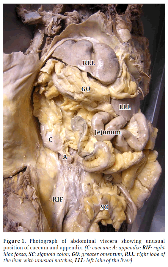
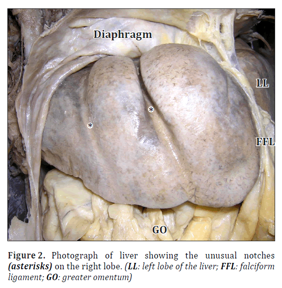
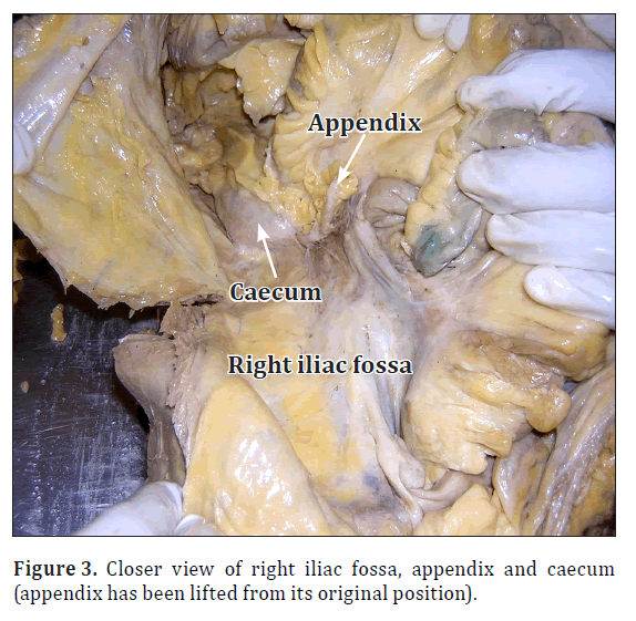
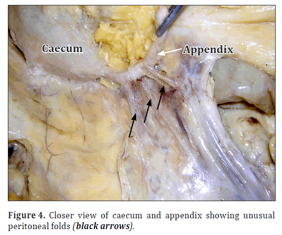
During regular dissection classes for first year medical undergraduates, variations in the right lobe of the liver and the position of caecum were noted in a female cadaver aged approximately 60-65 years. The liver was very large and occupied epigastrium, right and left hypochondriums completely. The right lobe of the liver had two deep notches on the antero-superior surface (Figures 1, 2). The left lobe of the liver was larger than that of the right lobe. The caecum was sub-hepatic in position. It was conical in shape and the vermiform appendix was attached to the tip of the conical caecum (Figures 1, 3, 4). The right iliac fossa was empty. The vermiform appendix had a mesoappendix and in addition, there were three abnormal peritoneal folds radiating from it to the right iliac fossa (Figure 4). The epiploic foramen was covered by the right colic flexure and the greater omentum.
Discussion
Variations in the lobes of the liver are very rare. Presence of unusual lobes or fissures may lead to wrong radiographic diagnosis. Fitzgerald et al., [1] have reported the presence of an additional lobe. The preoperative imaging of the unusual lobe had led to the misdiagnosis as a lesser omental lymphadenopathy. Llorente and Dardik [2] have reported the presence of a large symptomatic accessory liver lobe in a 70-year-old female. The accessory lobes of liver may herniate into the thorax through the diaphragm and cause serious problems [3]. In an extensive study conducted by Joshi et al. on variations of the liver, notching along the inferior border of the caudate lobe was found in 18% of livers, a vertical fissure was found in 30% of livers, and prominent papillary process was found in 32% of cases [4]. Accessory fissures may be present rarely on the antero-superior surface of the liver [5,6]. The accessory fissures are the potential source of errors in diagnosis in imaging techniques [7]. Collection of any fluid in accessory fissures may be mistaken for a cyst, liver abscess or intrahepatic hematoma. Studies are lacking with regards to the overall shape and size of the liver. In the current case, the right lobe of the liver had two large notches and the left lobe was larger than the right lobe. The liver occupied both the hypochondria and epigastrium completely. The presence of the unusual notches on antero-superior surface of the right lobe may lead to radiological misdiagnosis.
Vermiform appendix and caecum develop from the caecal bud of postarterial segment of midgut loop. In early stages of development they are of same diameter but later the appendix narrows down due to the faster growth of the proximal part of the caecal bud. At the birth, the appendix is attached to the tip of the conical caecum. Rarely this condition persists in the adulthood. The caecum and appendix herniate to the umbilical cord as a part of physiological umbilical hernia, and return back to the abdomen. When they return, at first they occupy the subhepatic position. Later they descend to the right iliac fossa. Agenesis of the caecum and vermiform appendix is very rare. The estimated incidence is 1/100,000 laparotomies performed for suspected appendicitis and even in such patients the symptoms of the acute appendicitis may be found. Zetina-Mejía et al., [8] have reported a case of a 48-year-old male patient with congenital absence of caecum and appendix with symptoms of acute appendicitis. A case of undescended caecum and primary torsion of vermiform appendix has also been reported [9]. In a study conducted by Delic et al., [10], conical caecum was found in 56% cases and square type of caecum was found in 44% of cases. It was constant in its position in right iliac fossa in 100% of the investigated cases. The vermiform appendix was attached to the tip of the caecum in 58% if cases, attached to the medial wall in 32% of cases and attached to lateral wall in 10% of cases.
In the current case, the subhepatic position of caecum and appendix might congest the subhepatic region and minimize the intestinal movements. The appendicular movements may be restricted by the three peritoneal bands extending from the appendix to the abdominal wall. The knowledge of this type of variations may be useful for the radiologists and surgeons.
References
- Fitzgerald R, Hale M, Williams CR. Case report: Accessory lobe of the liver mimicking lesser omental lymphadenopathy. Br J Radiol. 1993; 66: 839–841.
- Llorente J, Dardik H. Symptomatic accessory lobe of the liver associated with absence of the left lobe. Arch Surg. 1971; 102: 221–223.
- Feist JH, Lasser EC. Identification of uncommon liver lobulations. J Am Med Assoc. 1959; 169: 1859–1862.
- Joshi SD, Joshi SS, Athavale SA. Some interesting observations on the surface features of the liver and their clinical implications. Singapore Med J. 2009; 50: 715–719.
- Macchi V, Feltrin G, Parenti A, De Caro R. Diaphragmatic sulci and portal fissures. J Anat. 2003; 202: 303–308.
- Macchi V, Porzionato A, Parenti A, Macchi C, Newell R, De Caro R. Main accessory sulcus of the liver. Clin Anat. 2005; 18: 39–45.
- Auh YH, Rubenstein WA, Zirinsky K, Kneeland JB, Pardes JC, Engel IA, Whalen JP, Kazam E. Accessory fissures of the liver: CT and sonographic appearance. AJR Am J Roentgenol. 1984; 143: 565–572.
- Zetina-Mejía CA, Alvarez-Cosío JE, Quillo-Olvera J. Congenital absence of the cecal appendix. Case report. Cir Cir. 2009; 77: 407–410.
- Montes-Tapia F, Quiroga-Garza A, Abrego-Moya V. Primary torsion of the vermiform appendix and undescended caecum treated by video-assisted transumbilical appendectomy. J Laparoendosc Adv Surg Tech A. 2009; 19: 839–841.
- Delic J, Savkovic A, Isakovic E. Variations in the position and point of origin of the vermiform appendix. Med Arh. 2002; 56: 5–8. (Croatian)
Satheesha Nayak B*, Prasad AM, Narendra Pamidi and Bincy M George
Department of Anatomy, Melaka Manipal Medical College (Manipal Campus), International Centre for Health Sciences, Manipal University, Madhav Nagar, Manipal, Karnataka, India
- *Corresponding Author:
- Dr. Satheesha Nayak B.
Professor and Head, Department of Anatomy, MMMC Int. Centre for Health Sci. Manipal University, Madhav Nagar, Manipal, Udupi District, Karnataka, 576 104, India
Tel: +91 820 2922519
E-mail: nayaksathish@yahoo.com
Date of Received: May 26th, 2011
Date of Accepted: March 28th, 2012
Published Online: October 12th, 2012
© Int J Anat Var (IJAV). 2012; 5: 48–50.
Abstract
Developmental variations of the liver, caecum and vermiform appendix are relatively rare. We found a large notched liver and an infantile subhepatic caecum in an adult female cadaver. The right lobe of the liver had two deep notches on the antero-superior surface. The right iliac fossa was empty. The caecum was conical and the appendix was attached to the tip of the conical caecum. Knowledge of these variations may be of use to the radiologists and surgeons.
-Keywords
liver, variation, caecum, appendix, subhepatic
Introduction
The liver is the largest gland of the human body. It is situated under the right dome of the diaphragm. Anatomically, the liver is divided into right and left lobes by the line of reflection of falciform ligament anteriorly, the fissure for ligamentum venosum posteriorly and the fissure for ligamentum teres inferiorly. The anatomical right lobe is larger than that of the left lobe. The anatomical right lobe is further divided into smaller lobes called quadrate and caudate lobes.
The caecum is the initial part of the large intestine. Usually, it is situated in the right iliac fossa, just above the lateral part of the inguinal ligament. It receives the opening of ilium on its left wall and opening of the vermiform appendix on its posteromedial wall. The vermiform appendix is a diverticulum of caecum which is variable in its position in relation to the caecum. It is suspended by a fold of peritoneum called mesoappendix and hence it is mobile. It is supplied by the appendicular artery, which is a branch of ileocolic artery.
In the current case, we encountered two large unusual notches on the right lobe of the liver, which is a very rare variation. The caecum and appendix were also in subhepatic position.
Case Report
During regular dissection classes for first year medical undergraduates, variations in the right lobe of the liver and the position of caecum were noted in a female cadaver aged approximately 60-65 years. The liver was very large and occupied epigastrium, right and left hypochondriums completely. The right lobe of the liver had two deep notches on the antero-superior surface (Figures 1, 2). The left lobe of the liver was larger than that of the right lobe. The caecum was sub-hepatic in position. It was conical in shape and the vermiform appendix was attached to the tip of the conical caecum (Figures 1, 3, 4). The right iliac fossa was empty. The vermiform appendix had a mesoappendix and in addition, there were three abnormal peritoneal folds radiating from it to the right iliac fossa (Figure 4). The epiploic foramen was covered by the right colic flexure and the greater omentum.
Discussion
Variations in the lobes of the liver are very rare. Presence of unusual lobes or fissures may lead to wrong radiographic diagnosis. Fitzgerald et al., [1] have reported the presence of an additional lobe. The preoperative imaging of the unusual lobe had led to the misdiagnosis as a lesser omental lymphadenopathy. Llorente and Dardik [2] have reported the presence of a large symptomatic accessory liver lobe in a 70-year-old female. The accessory lobes of liver may herniate into the thorax through the diaphragm and cause serious problems [3]. In an extensive study conducted by Joshi et al. on variations of the liver, notching along the inferior border of the caudate lobe was found in 18% of livers, a vertical fissure was found in 30% of livers, and prominent papillary process was found in 32% of cases [4]. Accessory fissures may be present rarely on the antero-superior surface of the liver [5,6]. The accessory fissures are the potential source of errors in diagnosis in imaging techniques [7]. Collection of any fluid in accessory fissures may be mistaken for a cyst, liver abscess or intrahepatic hematoma. Studies are lacking with regards to the overall shape and size of the liver. In the current case, the right lobe of the liver had two large notches and the left lobe was larger than the right lobe. The liver occupied both the hypochondria and epigastrium completely. The presence of the unusual notches on antero-superior surface of the right lobe may lead to radiological misdiagnosis.
Vermiform appendix and caecum develop from the caecal bud of postarterial segment of midgut loop. In early stages of development they are of same diameter but later the appendix narrows down due to the faster growth of the proximal part of the caecal bud. At the birth, the appendix is attached to the tip of the conical caecum. Rarely this condition persists in the adulthood. The caecum and appendix herniate to the umbilical cord as a part of physiological umbilical hernia, and return back to the abdomen. When they return, at first they occupy the subhepatic position. Later they descend to the right iliac fossa. Agenesis of the caecum and vermiform appendix is very rare. The estimated incidence is 1/100,000 laparotomies performed for suspected appendicitis and even in such patients the symptoms of the acute appendicitis may be found. Zetina-Mejía et al., [8] have reported a case of a 48-year-old male patient with congenital absence of caecum and appendix with symptoms of acute appendicitis. A case of undescended caecum and primary torsion of vermiform appendix has also been reported [9]. In a study conducted by Delic et al., [10], conical caecum was found in 56% cases and square type of caecum was found in 44% of cases. It was constant in its position in right iliac fossa in 100% of the investigated cases. The vermiform appendix was attached to the tip of the caecum in 58% if cases, attached to the medial wall in 32% of cases and attached to lateral wall in 10% of cases.
In the current case, the subhepatic position of caecum and appendix might congest the subhepatic region and minimize the intestinal movements. The appendicular movements may be restricted by the three peritoneal bands extending from the appendix to the abdominal wall. The knowledge of this type of variations may be useful for the radiologists and surgeons.
References
- Fitzgerald R, Hale M, Williams CR. Case report: Accessory lobe of the liver mimicking lesser omental lymphadenopathy. Br J Radiol. 1993; 66: 839–841.
- Llorente J, Dardik H. Symptomatic accessory lobe of the liver associated with absence of the left lobe. Arch Surg. 1971; 102: 221–223.
- Feist JH, Lasser EC. Identification of uncommon liver lobulations. J Am Med Assoc. 1959; 169: 1859–1862.
- Joshi SD, Joshi SS, Athavale SA. Some interesting observations on the surface features of the liver and their clinical implications. Singapore Med J. 2009; 50: 715–719.
- Macchi V, Feltrin G, Parenti A, De Caro R. Diaphragmatic sulci and portal fissures. J Anat. 2003; 202: 303–308.
- Macchi V, Porzionato A, Parenti A, Macchi C, Newell R, De Caro R. Main accessory sulcus of the liver. Clin Anat. 2005; 18: 39–45.
- Auh YH, Rubenstein WA, Zirinsky K, Kneeland JB, Pardes JC, Engel IA, Whalen JP, Kazam E. Accessory fissures of the liver: CT and sonographic appearance. AJR Am J Roentgenol. 1984; 143: 565–572.
- Zetina-Mejía CA, Alvarez-Cosío JE, Quillo-Olvera J. Congenital absence of the cecal appendix. Case report. Cir Cir. 2009; 77: 407–410.
- Montes-Tapia F, Quiroga-Garza A, Abrego-Moya V. Primary torsion of the vermiform appendix and undescended caecum treated by video-assisted transumbilical appendectomy. J Laparoendosc Adv Surg Tech A. 2009; 19: 839–841.
- Delic J, Savkovic A, Isakovic E. Variations in the position and point of origin of the vermiform appendix. Med Arh. 2002; 56: 5–8. (Croatian)


