Obturator venous ring encompassing the variant origin inferior vesical artery*
Soner Albay1,*, Neslihan Yuzbasioglu2, Marios Loukas3
1Department of Anatomy, Suleyman Demirel University Faculty of Medicine, Isparta, Turkey.
2Department of Anatomy, Istanbul Medipol University Faculty of Medicine, Istanbul, Turkey.
3St. George’s University Faculty of Medicine Department of Anatomical Sciences, Grenada, West Indies.
- *Corresponding Author:
- Soner ALBAY, MD
Assistant Professor, Department of Anatomy, Suleyman Demirel University, Faculty of Medicine, 32260, Isparta, Turkey.
Tel: +90 (246) 211 36 80
E-mail: soneralbay@yahoo.com
Date of Received: October 12th, 2012
Date of Accepted: March 31st, 2013
Published Online: April 14th, 2013
© Int J Anat Var (IJAV). 2013; 6: 56–58.
[ft_below_content] =>Keywords
inferior vesical artery, obturator vein, pelvic vessels, variation
Introduction
Pelvic vessels are generally accepted to show more variations [1]. Internal iliac artery (IIA) and vein variations are not rare [2]. Normally, obturator artery (OA) is a branch of the anterior division of IIA [3]. But some variations have been reported in literature. For example OA may have dual origin, occurring with a frequency of 1% or it may arise from an arterial trunk which is originated from external iliac artery, and an accessory obturator artery may be present as reported by Sañudo et al [4]. The origin of the OA from the posterior division of the IIA has also been reported [5]. Inferior vesical artery (IVA) has the same origin and it supplies the prostate. Venous variations are most commonly due to errors during embryogenesis [6].
The present case provides an opportunity for embryologic discussion and may be useful information to surgeons who operate the pelvis as well as radiologists who interpret imaging of this region.
Case Report
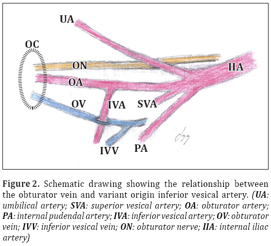
The pelvic vessels were dissected in a 25 year-old male cadaver whose cause of death was a traffic accident. On the right side, we observed a venous ring formed by the obturator vein (OV) (Figures 1, 2). The IVA originated from the OA instead of the IIA and passed through this venous ring. We observed only one OV that divided into two veins for encompassing the IVA and then they merged to form the single vein again. The other branches of right IIA and internal iliac vein (IIV) were as usual. No venous ring was observed on the left side of the cadaver, but the IVA on the left side was also originated from the OA instead of the IIA. The remaining branches of the left IIV showed no variation. There was no accessory obturator vein.
Figure 1: Figure showing the pelvic vessels on the right side and obturator venous ring encompassing the variant origin inferior vesical artery. (UA: umbilical artery; SVA: superior vesical artery; OA: obturator artery; PA: internal pudendal artery; IVA: inferior vesical artery; OV: obturator vein; IVV: inferior vesical vein; ON: obturator nerve; B: bladder; U: ureter; EIV: external iliac vein; IIA: internal iliac artery)
Figure 2: Schematic drawing showing the relationship between the obturator vein and variant origin inferior vesical artery. (UA: umbilical artery; SVA: superior vesical artery; OA: obturator artery; PA: internal pudendal artery; IVA: inferior vesical artery; OV: obturator vein; IVV: inferior vesical vein; ON: obturator nerve; IIA: internal iliac artery)
Discussion
Among the most important considerations in the study of the vascular system are its significant variations. Although many of these cause no disturbance in the functions of the body, they may be of importance to the surgeon. One group of variations represents persistent fetal forms of circulation. Another group represents individual variations, some of which may be explained as developmentally or typical anastomoses [2]. The anatomical variations of the pelvic vessels constitute congenital morphologic differences observed in the human body [7].
Recently, Jusoh et al. have studied 34 pelvic halves to determine origin and branching pattern of the OA. They observed only two specimens (5.8%) in which OA gave off inferior vesical branch [5]. IVA arising from OA seems not rare. But, arising from the posterior division of IIA, both of these obturator arteries were variant. Different from Jusohet al.’s study, in the present case the OA originated from the anterior division of IIA as expected.
Variations in venous pathways are generally more common than arterial variations. The internal and the external iliac veins are frequently manipulated during interventions in the pelvis such as the retroperitoneal lymphadenectomy, anastomosis during a kidney transplant, hypogastric neurectomy and hysterectomy [7]. For the IIV and its tributaries, a systematic look for any possible variants of the retroperitoneal veins is needed. Particularly in lymphadenectomy, the complexity of the variations of the IIV may lead to modification on the technical aspects of the surgical procedure. In hysterectomy, surgical interference on these veins may affect venous drainage and precipitate edema on one or both legs. In orthopedic surgery, the IIV is most at risk during operations involving screw placement [7]. Patients with unusual venous anatomy may also have unusual patterns of lymphatic drainage and lymph node metastases [6].
In conclusion, venous variants, although rare, may be dangerous if not recognized [6]. To our knowledge, the variations described herein have not been previously published. It has been also emphasized that the detailed anatomy of obturator canal and the variations of obturator vessels are important for obturator nerve block [8].
Awareness of these pelvic venous variations and anomalies is necessary to reduce surgical risk and to determine strategy in interventional radiology. Advances in imaging techniques enable these unusual presentations to be defined preoperatively [9].
References
- Putz R, Pabst R. Sobotta Atlas of Human Anatomy. Vol. 2. 20th Ed., Munich, Urban and Schwarzenberg. 1994; 214–217.
- Bergman RA, Afifi AK, Miyauchi R. Illustrated Encyclopedia of Human Anatomic Variation: Opus II: Cardiovascular System: Arteries: Pelvis, Internal Iliac Artery. http://www.anatomyatlases.org/AnatomicVariants/Cardiovascular/Text/Arteries/IliacInternal.shtml (accessed June 2010).
- Standring S. Gray’s Anatomy. The Anatomical Basis of Clinical Practice. 40th Ed., Edinburgh, Churchill Livingstone. 2008; 1086–1089.
- Sanudo JR, Roig M, Rodriguez A, Ferreira B, Domenech JM. Rare origin of the obturator, inferior epigastric and medial circumflex femoral arteries from a common trunk. J Anat. 1993; 183: 161–163.
- Jusoh AR, Abd Rahman N, Abd Latiff A, Othman F, Das S, Abd Ghafar N, Haji Suhaimi F, Hussan F, Maatoq Sulaiman I. The anomalous origin and branches of the obturator artery with its clinical implicaitons. Rom J Morphol Embryol. 2010; 51: 163–166.
- Surucu HS, Erbil KM, Tastan C, Yener N. Anomalous veins of retroperitoneum: clinical considerations. Surg Radiol Anat. 2001; 23: 443–445.
- Cardinot TM, Aragao AHBM, Babinski MA, Favorito LA. Rare variation in course and affluence of internal iliac vein due to its anatomical and surgical significance. Surg Radiol Anat. 2006; 28: 422–425.
- Kendir S, Akkaya T, Comert A, Sayin M, Tatlisumak E, Elhan A, Tekdemir I. The location of the obturator nerve: a three dimensional description of the obturator canal. Surg Radiol Anat. 2008; 30: 495–501.
- Morita S, Higuchi M, Saito N, Mitsuhashi N. Pelvic venous variations in patients with congenital inferior vena cava anomalies: classification with computed tomography. Acta Radiol. 2007; 48: 974–979.
Soner Albay1,*, Neslihan Yuzbasioglu2, Marios Loukas3
1Department of Anatomy, Suleyman Demirel University Faculty of Medicine, Isparta, Turkey.
2Department of Anatomy, Istanbul Medipol University Faculty of Medicine, Istanbul, Turkey.
3St. George’s University Faculty of Medicine Department of Anatomical Sciences, Grenada, West Indies.
- *Corresponding Author:
- Soner ALBAY, MD
Assistant Professor, Department of Anatomy, Suleyman Demirel University, Faculty of Medicine, 32260, Isparta, Turkey.
Tel: +90 (246) 211 36 80
E-mail: soneralbay@yahoo.com
Date of Received: October 12th, 2012
Date of Accepted: March 31st, 2013
Published Online: April 14th, 2013
© Int J Anat Var (IJAV). 2013; 6: 56–58.
Abstract
During routine dissection of a 25-year-old male cadaver, we observed a venous ring formed by the right obturator vein. The inferior vesical artery also originated from obturator artery bilaterally, and the right inferior vesical artery traveled through the venous ring described above. Knowledge of such variation may be important during surgery of the pelvis and interpretation of pelvic imaging.
-Keywords
inferior vesical artery, obturator vein, pelvic vessels, variation
Introduction
Pelvic vessels are generally accepted to show more variations [1]. Internal iliac artery (IIA) and vein variations are not rare [2]. Normally, obturator artery (OA) is a branch of the anterior division of IIA [3]. But some variations have been reported in literature. For example OA may have dual origin, occurring with a frequency of 1% or it may arise from an arterial trunk which is originated from external iliac artery, and an accessory obturator artery may be present as reported by Sañudo et al [4]. The origin of the OA from the posterior division of the IIA has also been reported [5]. Inferior vesical artery (IVA) has the same origin and it supplies the prostate. Venous variations are most commonly due to errors during embryogenesis [6].
The present case provides an opportunity for embryologic discussion and may be useful information to surgeons who operate the pelvis as well as radiologists who interpret imaging of this region.
Case Report
The pelvic vessels were dissected in a 25 year-old male cadaver whose cause of death was a traffic accident. On the right side, we observed a venous ring formed by the obturator vein (OV) (Figures 1, 2). The IVA originated from the OA instead of the IIA and passed through this venous ring. We observed only one OV that divided into two veins for encompassing the IVA and then they merged to form the single vein again. The other branches of right IIA and internal iliac vein (IIV) were as usual. No venous ring was observed on the left side of the cadaver, but the IVA on the left side was also originated from the OA instead of the IIA. The remaining branches of the left IIV showed no variation. There was no accessory obturator vein.
Figure 1: Figure showing the pelvic vessels on the right side and obturator venous ring encompassing the variant origin inferior vesical artery. (UA: umbilical artery; SVA: superior vesical artery; OA: obturator artery; PA: internal pudendal artery; IVA: inferior vesical artery; OV: obturator vein; IVV: inferior vesical vein; ON: obturator nerve; B: bladder; U: ureter; EIV: external iliac vein; IIA: internal iliac artery)
Figure 2: Schematic drawing showing the relationship between the obturator vein and variant origin inferior vesical artery. (UA: umbilical artery; SVA: superior vesical artery; OA: obturator artery; PA: internal pudendal artery; IVA: inferior vesical artery; OV: obturator vein; IVV: inferior vesical vein; ON: obturator nerve; IIA: internal iliac artery)
Discussion
Among the most important considerations in the study of the vascular system are its significant variations. Although many of these cause no disturbance in the functions of the body, they may be of importance to the surgeon. One group of variations represents persistent fetal forms of circulation. Another group represents individual variations, some of which may be explained as developmentally or typical anastomoses [2]. The anatomical variations of the pelvic vessels constitute congenital morphologic differences observed in the human body [7].
Recently, Jusoh et al. have studied 34 pelvic halves to determine origin and branching pattern of the OA. They observed only two specimens (5.8%) in which OA gave off inferior vesical branch [5]. IVA arising from OA seems not rare. But, arising from the posterior division of IIA, both of these obturator arteries were variant. Different from Jusohet al.’s study, in the present case the OA originated from the anterior division of IIA as expected.
Variations in venous pathways are generally more common than arterial variations. The internal and the external iliac veins are frequently manipulated during interventions in the pelvis such as the retroperitoneal lymphadenectomy, anastomosis during a kidney transplant, hypogastric neurectomy and hysterectomy [7]. For the IIV and its tributaries, a systematic look for any possible variants of the retroperitoneal veins is needed. Particularly in lymphadenectomy, the complexity of the variations of the IIV may lead to modification on the technical aspects of the surgical procedure. In hysterectomy, surgical interference on these veins may affect venous drainage and precipitate edema on one or both legs. In orthopedic surgery, the IIV is most at risk during operations involving screw placement [7]. Patients with unusual venous anatomy may also have unusual patterns of lymphatic drainage and lymph node metastases [6].
In conclusion, venous variants, although rare, may be dangerous if not recognized [6]. To our knowledge, the variations described herein have not been previously published. It has been also emphasized that the detailed anatomy of obturator canal and the variations of obturator vessels are important for obturator nerve block [8].
Awareness of these pelvic venous variations and anomalies is necessary to reduce surgical risk and to determine strategy in interventional radiology. Advances in imaging techniques enable these unusual presentations to be defined preoperatively [9].
References
- Putz R, Pabst R. Sobotta Atlas of Human Anatomy. Vol. 2. 20th Ed., Munich, Urban and Schwarzenberg. 1994; 214–217.
- Bergman RA, Afifi AK, Miyauchi R. Illustrated Encyclopedia of Human Anatomic Variation: Opus II: Cardiovascular System: Arteries: Pelvis, Internal Iliac Artery. http://www.anatomyatlases.org/AnatomicVariants/Cardiovascular/Text/Arteries/IliacInternal.shtml (accessed June 2010).
- Standring S. Gray’s Anatomy. The Anatomical Basis of Clinical Practice. 40th Ed., Edinburgh, Churchill Livingstone. 2008; 1086–1089.
- Sanudo JR, Roig M, Rodriguez A, Ferreira B, Domenech JM. Rare origin of the obturator, inferior epigastric and medial circumflex femoral arteries from a common trunk. J Anat. 1993; 183: 161–163.
- Jusoh AR, Abd Rahman N, Abd Latiff A, Othman F, Das S, Abd Ghafar N, Haji Suhaimi F, Hussan F, Maatoq Sulaiman I. The anomalous origin and branches of the obturator artery with its clinical implicaitons. Rom J Morphol Embryol. 2010; 51: 163–166.
- Surucu HS, Erbil KM, Tastan C, Yener N. Anomalous veins of retroperitoneum: clinical considerations. Surg Radiol Anat. 2001; 23: 443–445.
- Cardinot TM, Aragao AHBM, Babinski MA, Favorito LA. Rare variation in course and affluence of internal iliac vein due to its anatomical and surgical significance. Surg Radiol Anat. 2006; 28: 422–425.
- Kendir S, Akkaya T, Comert A, Sayin M, Tatlisumak E, Elhan A, Tekdemir I. The location of the obturator nerve: a three dimensional description of the obturator canal. Surg Radiol Anat. 2008; 30: 495–501.
- Morita S, Higuchi M, Saito N, Mitsuhashi N. Pelvic venous variations in patients with congenital inferior vena cava anomalies: classification with computed tomography. Acta Radiol. 2007; 48: 974–979.


