Occipitalization of the atlas: its occurrence and embryological basis
Vineeta Saini1, Royana Singh2*, Manimay Bandopadhyay3, Sunil Kumar Tripathi1 and Satya Narayan Shamal2
1Department of Forensic Medicine, Banaras Hindu University, Varanasi 221005, U.P, India
2Department of Forensic Medicine and Anatomy, Institute of Medical Sciences, Banaras Hindu University, Varanasi 221005, U.P, India
3Department of Anatomy, Calcutta National Medical College, Kolkata, India
- *Corresponding Author:
- Royana Singh, MBBS, MD, DNB
Department of Anatomy, Institute of Medical Sciences, Banaras Hindu University, Varanasi, 221005, Uttar Pradesh, India
Tel: +91 0542-6703451
E-mail: singhroyana@rediffmail.com
Date of Received: January 20th, 2009
Date of Accepted: June 8th, 2009
Published Online: June 30th, 2009
© IJAV. 2009; 2: 65–68.
[ft_below_content] =>Keywords
occipitalization, atlas, occipital bone, sclerotome
Introduction
Occipitalization is a congenital synostosis of the atlas to the occiput, which is a result of failure of segmentation and separation of the most caudal occipital sclerotome and the first cervical sclerotome during the first few weeks of fetal life [1]. There may be varying degrees of bony fusion between atlas and occiput; complete and partial assimilation have been described [2,3]. In a majority of cases, assimilation occurs between the anterior arch of the atlas and the anterior rim of the foramen magnum and is associated with other skeletal malformations such as basilar invagination, occipital vertebra, spina bifida of atlas, or fusion of the second and third cervical vertebrae (Klippel-Feil syndrome) [4]. The incidence of atlanto-occipital fusion ranges from 0.14 to 0.75% of the population, with both sexes being equally affected [1]. Occipitalization of atlas is associated with abnormalities as a result of narrowing of the foramen magnum, compressing the spinal cord or the brain stem [5,6]. However, this anatomical variation may often go unnoticed but, incidentally, reveals its presence as a radiological, operative or autopsy finding. We present two cases of atlanto-occipital fusion laying emphasis on its embryological origin and trying to evaluate the prevalence in Northern India.
Case Report
A total of 126 human adult skulls of Northern Indian origin were examined. Since the sex of the human skulls collected from the Department of Forensic were known, the skulls obtained from the Department of Anatomy were scrutinized and grouped, according to their sex [7]. Each of the skull was examined. The variations present in the atlas vertebra and the occipital bone of the skull, as a consequence to the assimilation were noted. The area of the foramen magnum = π × 1/4 × W × H, where W is the maximum width of the foramen magnum and H is its maximum antero-posterior diameter in the median plane, was calculated. An estimation of the reduced area was performed with the square counting technique using millimeter graph paper. The percentage reduction in the effective area was then calculated. The different cranial indices were compared with that of other normal skull in order to designate any change, which may have resulted due to the assimilation.
Case 1
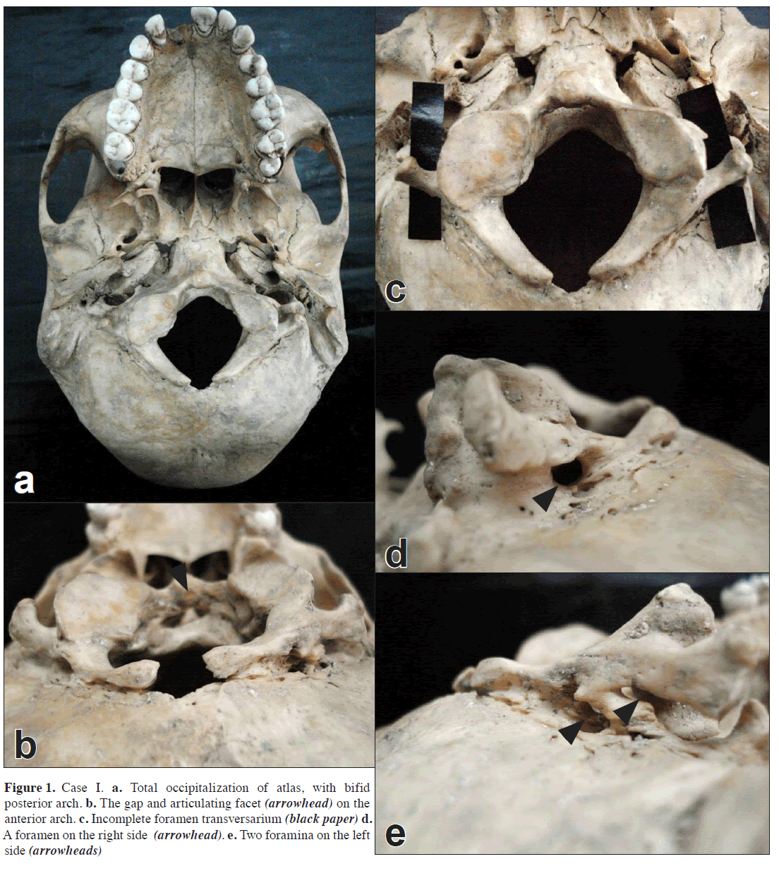
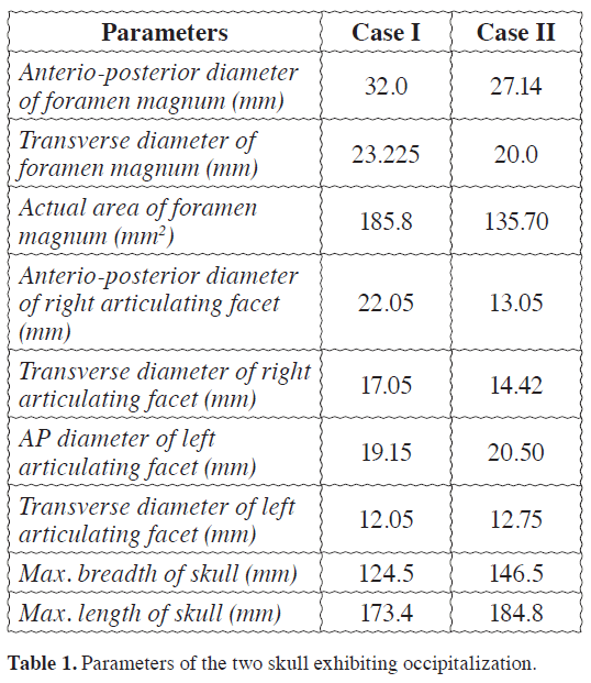
A female skull from Northern India region exhibited complete fusion of the atlas vertebra with the occipital bone (Figure 1a). The anterior arch of the atlas was completely fused except for a slit like opening, above the anterior tubercle. The slit extended more on the left than the right side (Figure 1b). The area posterior to the anterior tubercle had a small articulating facet (Figure 1b). The posterior arch was bifid with serrated margins, and completely fused to the posterior rim of the foramen magnum. The lateral masses were completely fused to different extents, i.e., more on the right than the left side, such that the transverse plane was inclined on the right side. Both the right and the left transverse process were free from the occipital bone and were club shaped (Figure 1a,b). The left transverse process exhibited an incomplete foramen transversarium with a protruding anterior costal lamella (Figure 1d). The right transverse process had no foramen transversarium (Figure 1d). The right articulating facet had a larger transverse diameter as well as longitudinal length. It possessed a concavity with two constrictions and the longitudinal axis turned inwards. The left articulating facet was more or less rhomboid shaped and more flat (Figure 1c). The assimilated atlas bore two foramina on the left side between the fused posterior arch and the occipital bone (Figure 1e). The right side possessed only one foramen. The hypoglossal canals on both sides were normal. The inferior surface of the posterior arch had a definite sulcus on both the sides, more deep on the right side. No foramen existed for the first cervical nerve on the right. The skull was bradycephalic (Table 1).
Case II
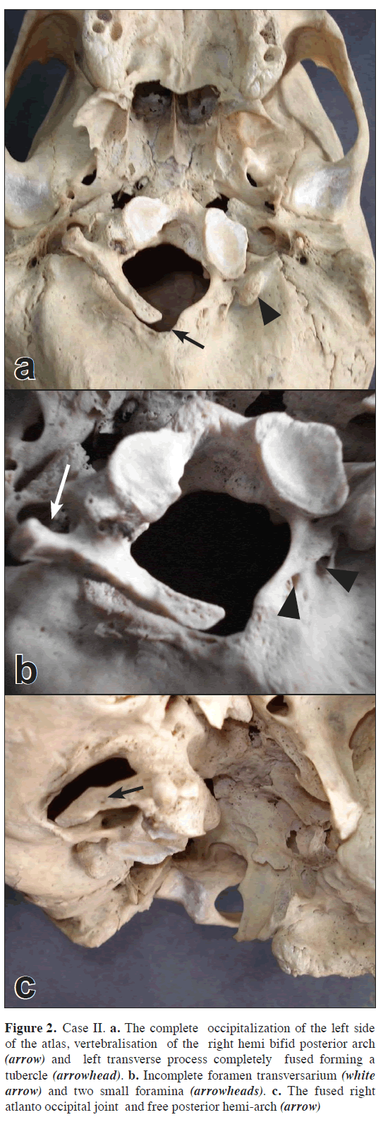
The other case was a male skull, which exhibited hemi-fusion of the atlas vertebra to the axis and the other half to the occipital bone. The entire left side of the atlas vertebra was assimilated into the occipital bone, the left part of the anterior arch completely fused to the basi-occiput, and the left side articulating facet and the left side of the posterior arch with the squamous part of the occipital bone. The fused left posterior hemi-arch possessed three openings (Figure 2a). The left transverse process was completely assimilated in the occiput, presented itself as a prominent tubercle lateral to the inferior articulating facet (Figure 2a). The right half of the anterior arch and transverse process displayed no fusion with the occipital bone. The right transverse process had a prominent posterior tubercle, a deficient anterior costal lamella, thus, an anteriorly open foramen transversarium, (Figure 2b). The right side articulating facet was completely fused (Figure 2b,c). The right side of the posterior arch superior aspect was free but its inferior aspect displayed vertebralisation, i.e., fused with the axis, the second vertebra. The posterior arch was bifid. To separate the atlas from the fused vertebra the posterior arch had to be cut down, which was seen by its sharp margin. The medullary cavity of the cut end of the posterior arch was visible (Figure 2b). Reduction in the size of the foramen magnum was seen (Figure 2a,b and Table 1). The cranial index 98.73% suggested a brachycephalic skull. The hemi-fusion of the atlas, lead to an inclination towards the left of about 6.2 mm.
Figure 2: Case II. a. The complete occipitalization of the left side of the atlas, vertebralisation of the right hemi bifid posterior arch (arrow) and left transverse process completely fused forming a tubercle (arrowhead). b. Incomplete foramen transversarium (white arrow) and two small foramina (arrowheads). c. The fused right atlanto occipital joint and free posterior hemi-arch (arrow)
Discussion
The above two cases strongly suggest that the assimilation of the atlas is congenital not accidental. The ventral portion of the sclerotome surrounds the notochord and provides the form, which develops into the vertebral body. The dorsal portion surrounds the neural tube and provides the form, which develops into the posterior vertebral arch. The caudal half of each sclerotome combines with the rostral half of the sclerotome below it.
The rostral half of the first cervical sclerotome combines with the caudal half of the last occipital sclerotome to form the base of the skull, while the caudal half of the first cervical sclerotome combines with the rostral half of the second cervical sclerotome to form the first cervical vertebra, the pattern continues in this fashion to form the other vertebrae [6,8]. In a small number of cases, the disruption of this merging process may result in atlanto-occipital assimilation. This condition may be partial or complete, as was the cases here. The complete fusion of the atlas is more common than the incomplete [9]. The sagittal diameter of the foramen magnum is an important landmark in symptomatic patients. This measure is accepted abnormal when it is less than 30 mm [10]. In both cases the reduction in the shape and size of the foramen magnum, small osseous opening for the vertebral artery to reach the brain may lead to asymptomatic patients to clinically manifesting symptoms, due to compression of the brain stem, nerves, vessels, or instability and mechanical immobility. When present, these symptoms usually manifest very seldom at early age and present themselves at second decades onwards [2]. Thus, it becomes extremely important for the clinicians, surgeons as well radiologist to keep in mind such an anomaly when the patient present with complains like neck pain, immobility of the neck etc., or during operations of the region. The present study showed almost equal prevalence of the occipital fusion in both males and females like that of Guebert et al., in 1987 [1].
References
- Guebert GM, Yochum TR, Rowe LJ. Congenital anomalies and normal skeleton variants. In: Essentials of Skeletal Radiology. Yochum TR, Rowe LJ, eds. Baltimore, Williams & Wilkins. 1987; 197–306.
- Ranade AV, Rai R, Prabhu LV, Kumaran M, Pai MM. Atlas assimilation: a case report. Neuroanatomy. 2007; 6: 32–33.
- Soni P, Sharma V, Sengupta J. Cervical vertebrae anomalies–incidental findings on lateral cephalograms. Angle Orthod. 2008; 78: 176–180.
- Shen FH, Samartzis D, Herman J, Lubicky JP. Radiographic assessment of segmental motion at the atlantoaxial junction in the Klippel-Feil patient. Spine. 2006 ; 31: 171–177.
- Gholve PA, Hosalkar HS, Ricchetti ET, Pollock AN, Dormans JP, Drummond DS. Occipitalization of the atlas in children. Morphologic classification, associations, and clinical relevance. J Bone Joint Surg Am. 2007; 89: 571–578.
- Martellacci S, Ben Salem D, Mejean N, Sautreaux JL, Krause D. A case of foramen magnum syndrome caused by atlanto-occipital assimilation with intracanal fibrosis. Surg Radiol Anat. 2008; 30: 149–152.
- Krogman WM. Sexing skeletal remains. In: The Human Skeleton in Forensic Medicine. Krogman WM, Iscan MY, eds. Springfield, Charles C Thomas. 1962: 112–122.
- Sadler TW. Langman’s Essential Medical Embryology. 10th Ed., Baltimore, Lippincott William and Wilkins. 2007: 125–141.
- Kalla AK, Khanna S, Singh IP, Sharma S, Schnobel R, Vogel F. A genetic and anthropological study of atlanto-occipital fusion. Hum Genet. 1989; 81: 105–112.
- Hayes M, Parker G, Ell J, Sillence D. Basilar impression complicating osteogenesis imperfacta type IV: the clinical and neuroradiological findings in four cases. J Neurol Neurosurg Psychiatry. 1999; 66: 357–364.
Vineeta Saini1, Royana Singh2*, Manimay Bandopadhyay3, Sunil Kumar Tripathi1 and Satya Narayan Shamal2
1Department of Forensic Medicine, Banaras Hindu University, Varanasi 221005, U.P, India
2Department of Forensic Medicine and Anatomy, Institute of Medical Sciences, Banaras Hindu University, Varanasi 221005, U.P, India
3Department of Anatomy, Calcutta National Medical College, Kolkata, India
- *Corresponding Author:
- Royana Singh, MBBS, MD, DNB
Department of Anatomy, Institute of Medical Sciences, Banaras Hindu University, Varanasi, 221005, Uttar Pradesh, India
Tel: +91 0542-6703451
E-mail: singhroyana@rediffmail.com
Date of Received: January 20th, 2009
Date of Accepted: June 8th, 2009
Published Online: June 30th, 2009
© IJAV. 2009; 2: 65–68.
Abstract
Occipitalization of the atlas is a rare congenital malformation of the craniovertebral region. During anthropometric study of 126 human skulls in the Department of Forensic Medicine and Anatomy, two skulls were discovered, which exhibited assimilation of the atlas to the occipital bone. One female skull exhibited total assimilation with bifid posterior arch, while the other male skull exhibited partial occipitalization and partial vertebralization. The partial or complete assimilation of the atlas may have resulted due to disruption in the separation of the caudal part of the first sclerotome from the cranial part of the first sclerotome.
-Keywords
occipitalization, atlas, occipital bone, sclerotome
Introduction
Occipitalization is a congenital synostosis of the atlas to the occiput, which is a result of failure of segmentation and separation of the most caudal occipital sclerotome and the first cervical sclerotome during the first few weeks of fetal life [1]. There may be varying degrees of bony fusion between atlas and occiput; complete and partial assimilation have been described [2,3]. In a majority of cases, assimilation occurs between the anterior arch of the atlas and the anterior rim of the foramen magnum and is associated with other skeletal malformations such as basilar invagination, occipital vertebra, spina bifida of atlas, or fusion of the second and third cervical vertebrae (Klippel-Feil syndrome) [4]. The incidence of atlanto-occipital fusion ranges from 0.14 to 0.75% of the population, with both sexes being equally affected [1]. Occipitalization of atlas is associated with abnormalities as a result of narrowing of the foramen magnum, compressing the spinal cord or the brain stem [5,6]. However, this anatomical variation may often go unnoticed but, incidentally, reveals its presence as a radiological, operative or autopsy finding. We present two cases of atlanto-occipital fusion laying emphasis on its embryological origin and trying to evaluate the prevalence in Northern India.
Case Report
A total of 126 human adult skulls of Northern Indian origin were examined. Since the sex of the human skulls collected from the Department of Forensic were known, the skulls obtained from the Department of Anatomy were scrutinized and grouped, according to their sex [7]. Each of the skull was examined. The variations present in the atlas vertebra and the occipital bone of the skull, as a consequence to the assimilation were noted. The area of the foramen magnum = π × 1/4 × W × H, where W is the maximum width of the foramen magnum and H is its maximum antero-posterior diameter in the median plane, was calculated. An estimation of the reduced area was performed with the square counting technique using millimeter graph paper. The percentage reduction in the effective area was then calculated. The different cranial indices were compared with that of other normal skull in order to designate any change, which may have resulted due to the assimilation.
Case 1
A female skull from Northern India region exhibited complete fusion of the atlas vertebra with the occipital bone (Figure 1a). The anterior arch of the atlas was completely fused except for a slit like opening, above the anterior tubercle. The slit extended more on the left than the right side (Figure 1b). The area posterior to the anterior tubercle had a small articulating facet (Figure 1b). The posterior arch was bifid with serrated margins, and completely fused to the posterior rim of the foramen magnum. The lateral masses were completely fused to different extents, i.e., more on the right than the left side, such that the transverse plane was inclined on the right side. Both the right and the left transverse process were free from the occipital bone and were club shaped (Figure 1a,b). The left transverse process exhibited an incomplete foramen transversarium with a protruding anterior costal lamella (Figure 1d). The right transverse process had no foramen transversarium (Figure 1d). The right articulating facet had a larger transverse diameter as well as longitudinal length. It possessed a concavity with two constrictions and the longitudinal axis turned inwards. The left articulating facet was more or less rhomboid shaped and more flat (Figure 1c). The assimilated atlas bore two foramina on the left side between the fused posterior arch and the occipital bone (Figure 1e). The right side possessed only one foramen. The hypoglossal canals on both sides were normal. The inferior surface of the posterior arch had a definite sulcus on both the sides, more deep on the right side. No foramen existed for the first cervical nerve on the right. The skull was bradycephalic (Table 1).
Case II
The other case was a male skull, which exhibited hemi-fusion of the atlas vertebra to the axis and the other half to the occipital bone. The entire left side of the atlas vertebra was assimilated into the occipital bone, the left part of the anterior arch completely fused to the basi-occiput, and the left side articulating facet and the left side of the posterior arch with the squamous part of the occipital bone. The fused left posterior hemi-arch possessed three openings (Figure 2a). The left transverse process was completely assimilated in the occiput, presented itself as a prominent tubercle lateral to the inferior articulating facet (Figure 2a). The right half of the anterior arch and transverse process displayed no fusion with the occipital bone. The right transverse process had a prominent posterior tubercle, a deficient anterior costal lamella, thus, an anteriorly open foramen transversarium, (Figure 2b). The right side articulating facet was completely fused (Figure 2b,c). The right side of the posterior arch superior aspect was free but its inferior aspect displayed vertebralisation, i.e., fused with the axis, the second vertebra. The posterior arch was bifid. To separate the atlas from the fused vertebra the posterior arch had to be cut down, which was seen by its sharp margin. The medullary cavity of the cut end of the posterior arch was visible (Figure 2b). Reduction in the size of the foramen magnum was seen (Figure 2a,b and Table 1). The cranial index 98.73% suggested a brachycephalic skull. The hemi-fusion of the atlas, lead to an inclination towards the left of about 6.2 mm.
Figure 2: Case II. a. The complete occipitalization of the left side of the atlas, vertebralisation of the right hemi bifid posterior arch (arrow) and left transverse process completely fused forming a tubercle (arrowhead). b. Incomplete foramen transversarium (white arrow) and two small foramina (arrowheads). c. The fused right atlanto occipital joint and free posterior hemi-arch (arrow)
Discussion
The above two cases strongly suggest that the assimilation of the atlas is congenital not accidental. The ventral portion of the sclerotome surrounds the notochord and provides the form, which develops into the vertebral body. The dorsal portion surrounds the neural tube and provides the form, which develops into the posterior vertebral arch. The caudal half of each sclerotome combines with the rostral half of the sclerotome below it.
The rostral half of the first cervical sclerotome combines with the caudal half of the last occipital sclerotome to form the base of the skull, while the caudal half of the first cervical sclerotome combines with the rostral half of the second cervical sclerotome to form the first cervical vertebra, the pattern continues in this fashion to form the other vertebrae [6,8]. In a small number of cases, the disruption of this merging process may result in atlanto-occipital assimilation. This condition may be partial or complete, as was the cases here. The complete fusion of the atlas is more common than the incomplete [9]. The sagittal diameter of the foramen magnum is an important landmark in symptomatic patients. This measure is accepted abnormal when it is less than 30 mm [10]. In both cases the reduction in the shape and size of the foramen magnum, small osseous opening for the vertebral artery to reach the brain may lead to asymptomatic patients to clinically manifesting symptoms, due to compression of the brain stem, nerves, vessels, or instability and mechanical immobility. When present, these symptoms usually manifest very seldom at early age and present themselves at second decades onwards [2]. Thus, it becomes extremely important for the clinicians, surgeons as well radiologist to keep in mind such an anomaly when the patient present with complains like neck pain, immobility of the neck etc., or during operations of the region. The present study showed almost equal prevalence of the occipital fusion in both males and females like that of Guebert et al., in 1987 [1].
References
- Guebert GM, Yochum TR, Rowe LJ. Congenital anomalies and normal skeleton variants. In: Essentials of Skeletal Radiology. Yochum TR, Rowe LJ, eds. Baltimore, Williams & Wilkins. 1987; 197–306.
- Ranade AV, Rai R, Prabhu LV, Kumaran M, Pai MM. Atlas assimilation: a case report. Neuroanatomy. 2007; 6: 32–33.
- Soni P, Sharma V, Sengupta J. Cervical vertebrae anomalies–incidental findings on lateral cephalograms. Angle Orthod. 2008; 78: 176–180.
- Shen FH, Samartzis D, Herman J, Lubicky JP. Radiographic assessment of segmental motion at the atlantoaxial junction in the Klippel-Feil patient. Spine. 2006 ; 31: 171–177.
- Gholve PA, Hosalkar HS, Ricchetti ET, Pollock AN, Dormans JP, Drummond DS. Occipitalization of the atlas in children. Morphologic classification, associations, and clinical relevance. J Bone Joint Surg Am. 2007; 89: 571–578.
- Martellacci S, Ben Salem D, Mejean N, Sautreaux JL, Krause D. A case of foramen magnum syndrome caused by atlanto-occipital assimilation with intracanal fibrosis. Surg Radiol Anat. 2008; 30: 149–152.
- Krogman WM. Sexing skeletal remains. In: The Human Skeleton in Forensic Medicine. Krogman WM, Iscan MY, eds. Springfield, Charles C Thomas. 1962: 112–122.
- Sadler TW. Langman’s Essential Medical Embryology. 10th Ed., Baltimore, Lippincott William and Wilkins. 2007: 125–141.
- Kalla AK, Khanna S, Singh IP, Sharma S, Schnobel R, Vogel F. A genetic and anthropological study of atlanto-occipital fusion. Hum Genet. 1989; 81: 105–112.
- Hayes M, Parker G, Ell J, Sillence D. Basilar impression complicating osteogenesis imperfacta type IV: the clinical and neuroradiological findings in four cases. J Neurol Neurosurg Psychiatry. 1999; 66: 357–364.


