Optical scleral contact lens in a patient with tectonic corneal graft
Received: 23-Oct-2020 Accepted Date: Nov 02, 2020; Published: 09-Nov-2020
Citation: Bhardwaj H. Optical scleral contact lens in a patient with tectonic corneal graft. Opth Clin Ther. 2020;4(3):7-10.
This open-access article is distributed under the terms of the Creative Commons Attribution Non-Commercial License (CC BY-NC) (http://creativecommons.org/licenses/by-nc/4.0/), which permits reuse, distribution and reproduction of the article, provided that the original work is properly cited and the reuse is restricted to noncommercial purposes. For commercial reuse, contact reprints@pulsus.com
Abstract
Aim: To describe a case report of a patient with tectonic corneal grafts in both eyes who was unable to achieve functional vision with spectacles or soft contact lenses. The patient was with fit with Atlantis scleral lens in the left eye, and was able to have comfort and reasonably good vision.
Background: This is a case study conducted in King Abdullah Medical City Bahrain, 67 years old Bahraini male visited the cornea clinic with the complaint of poor vision both eyes in the year 2019; he was not able to find his way independently. History and clinical examination shows tectonic graft in inferior cornea in both eyes, with pseudophakiain both eyes and vitreo-macular traction with Epiretinal Membrane (ERM) in right eye. After going through the retinal surgery (Pars Plana Vitrectomy) and corneal transplant (Penetrating Keratoplasty) surgery in right eye, the patient was referred to the contact lens clinic.
Method: Atlantis scleral contact lens fitting was done to the left eye (Boston, XO materials). The Atlantis scleral lenses by Xcel are manufactured exclusively in Boston XO materials and are available in 3 diameters, 15.0 mm (mini scleral), 16.0 mm, 16.5 mm (scleral, if Horizontal Visible Iris Diameter is less than 10.5 mm). They have 3 parameters which can be adjusted: central zone (by changing base curve), limbal zone (1-2 steep, standard, 1-2 flat) and scleral zone (1-2 steep, standard, 1-3 flat).
Result: The patient’s vision with this contact lens reached up to (0.5). This is considered a reasonably good vision that is suitable for driving according to international standards.
Conclusion: This patient had tectonic grafts and was initially considered legally blind. After his contact lens fitting in the left eye he was able to drive and to lead normal life. This case report shed the light on the importance of scleral contact lenses in management of severe corneal distortions. It also demonstrates that scleral contact lenses can provide reasonable vision even in patients with tectonic corneal grafts.
Keywords
Keratoconus; Tectonic Corneal Graft; Irregular Cornea; Corneal Perforation.
Introduction
To describe a case report of a patient with tectonic corneal grafts in both eyes who was unable to achieve functional vision with spectacles or Rigid Gas Permeable (RGP) Lenses. The patient was fit with Atlantis scleral lens in the left eye, and was able to have comfortable and reasonably good vision.
Background
The Atlantis design provides customizable zone options to provide a simple, streamlined fitting process that reduces chair time. A single fitting set will provide the necessary coverage to fit any corneal SAG or scleral shape.
Design features
• Available in 14.0 and 14.5 diameters for normal corneas
• Independent central SAG adjustments
• Quadrant specific limbal zone
• Quadrant specific scleral zone
• Expanded independent toric scleral zone options
• Up to 17.5 diameter option for deep SAGs
• Independent limbal vault adjustments to increase or decrease clearance
• Limbal vault adjustment towards optic zone for oblate corneas (in position)
• Limbal vault adjustment towards edge for corneal grafts (out position)
• Oblate multifocal
• Front toric optics stabilized by toric scleral zone
• Lens notching for scleral obstructions [1]
Lens design
The Atlantis fitting philosophy is based on the premise of customizing the lens fit by manipulating three different zones to control the sagittal height relationship of the lens to the anterior ocular surface. This produces one of the widest ranges of lens sagittal depths in the industry. Each zone has a specific function [1].
The Atlantis scleral design has 3 truly independent zones.
Central Zone: This is the optic zone and increases with a diameter increase. On the 15.5 the optic zone is an 8.5. Each individual and central SAG can be increased or decreased by a total of 200 microns in 50 micron steps. This will not affect the other 2 zones.
Limbal vault zone: This zone extends from the outside of the central zone to the inside of the scleral zone and the width of this zone is consistent with each diameter. The SAG of this zone can be increased (up to 200 microns) or decreased (down 100 microns) with no effect on the other 2 zones. The highest point of this zone can also be adjusted out towards the edge of the lens or in towards the optic zone of the lens. Changes are made in 25 micron steps.
Scleral zone: This is the edge of the lens and is roughly 1mm wide. This zone can be manipulated to increase or decrease the SAG of just this zone by 250 microns in each meridian or any quadrant. The changes are done in 25 micron steps and with not affect the central zone SAG or the limbal zone SAG.
Tangible Hydra-PEG
Atlantis Scleral lenses are available with Tangible Hydra-PEG contact lens coating. This revolutionary coating is designed to increase wettability/ surface water retention, lubricity (fewer deposits), and longer and better wear time [1].
Case Summary
A 67 year old male diabetic patient with keratoconus who underwent tectonic grafts in both eyes for bilateral spontaneous corneal perforations visited our cornea clinic, complaining of extremely poor vision. He couldn’t drive and was unable to find his way independently.
Systemic history
Cardiovascular: Nil, Respiratory: Nil, Gastrointestinal: Nil, Neurological: Nil, Endocrine: Type-2 Diabetes Mellitus more than 20 years, under control on oral medication.
Ocular history
Using RGP Lens for long period, both eyes, Keratoconus, both eyes, Cataractsurgery has done (OU), corneal graft surgery done (OU).
Clinical examination
Visual acuity: Right Eye: Counting fingers at 3 meters/Left Eye: (0.1)
Non Contact Tonometry (NCT ): Right Eye: Error/Left Eye: Error.
Autorefraction: Error, both eyes.
Retinoscopy: Dull and Irregular Reflex (OU).
Refraction: Hit and Trial method, no improvement with glasses.
Single and Multiple Pin-holevision: No improvement.
Corneal topography: Not done.
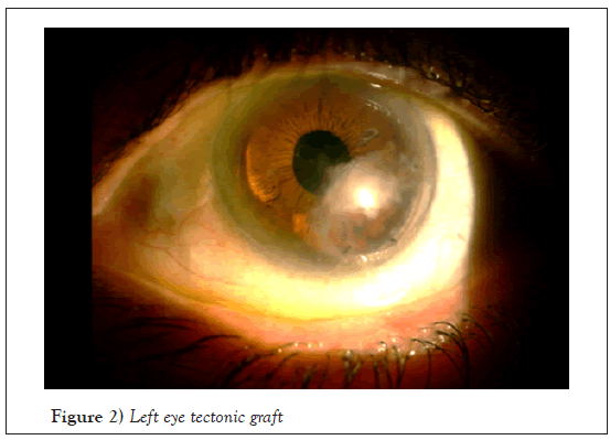
Slit lamp examination: Bilateral patch grafts inferiorly that were bulging forward (Figures 1 and 2) Pseudophakia, both eyes.
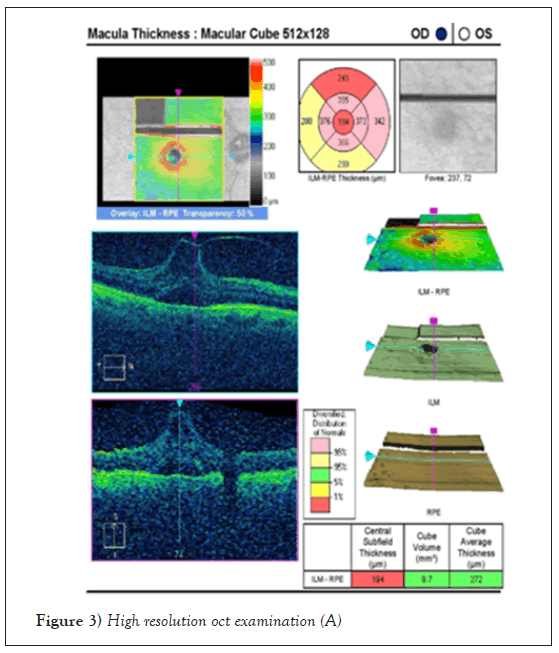
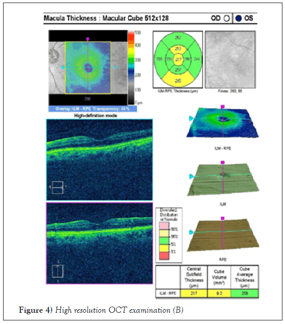
Dilated fundus examination with high resolution-retinal Optical Coherence Tomography (OCT) scan: Figure 3 showed Vitreo-macular traction (VMT) epiretinal membrane (ERM) right eye and Figure 4 showed Normal foveal contour in Left eye.
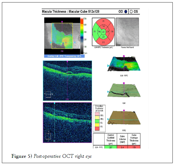
Patient has been advised and undergone right eye pars planavitrectomy with membrane peeling, endolaser and silicone oil (Figure 5). OCT shows sub- retinal fluid.
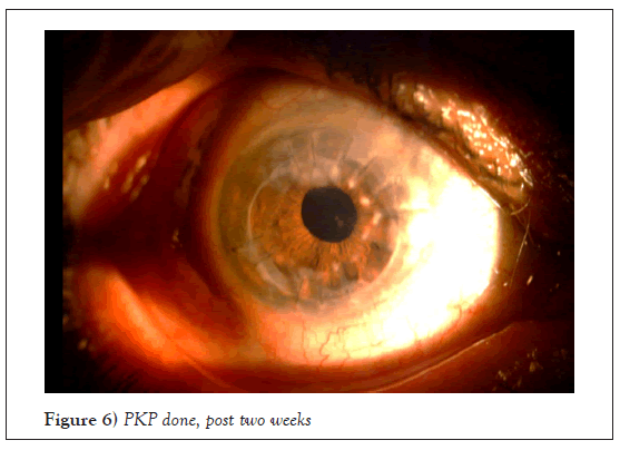
Two weeks later, he had undergone penetrating keratoplasty in the right eye (Figure 6).
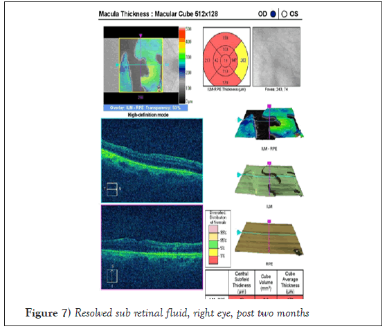
Post-operative 2 months follow up: Vision with multiple pin-holes in the right eye was fluctuating at (0.05) and OCT shows resolved sub-retinal fluid (Figure 7). The patient was referred to the contact lens clinic for the left eye. Atlantis Scleral Contact Lens fitting was done (Boston, XO materials).
Fitting from diagnostic set after few trials
OS: ATLANTIS X-Cel lens finally given to the patient with description below:
-4.00/D-steep/BC-7.34/Diameter-15.00/vault-std/scleral-zone-steep/center thickness 0.27. Over refraction +1.75.
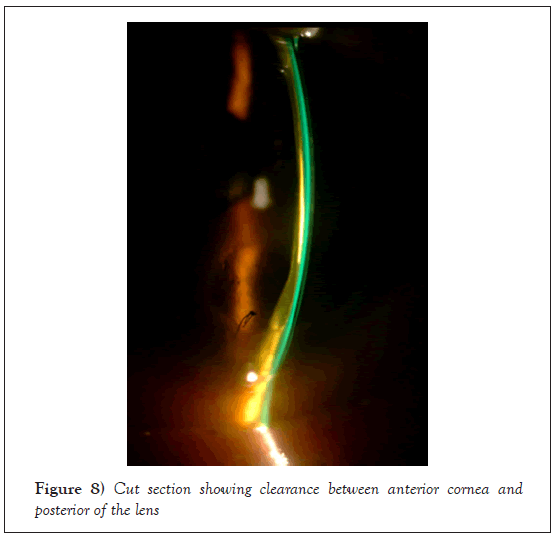
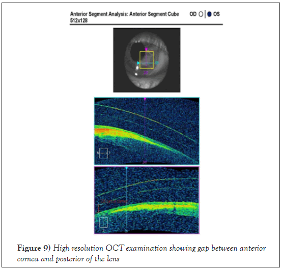
The patient’s vision with this contact lens reached up to (0.5). This is considered a reasonably good vision that is suitable for driving, according to international standards. And by addition +2.50 he was reading (20/40) with effort, and the patient has been advised to use reading glasses over the lenses for near task. NaFL (Sodium Fluorescein), examination was done after 30 minutes with the lens “on” (Figure 8 and Figure 9). After 30 minutes of lens on: inferior bulging area showing 230 micron clearance suggesting an optimal fit with no blanching and good tear exchange.
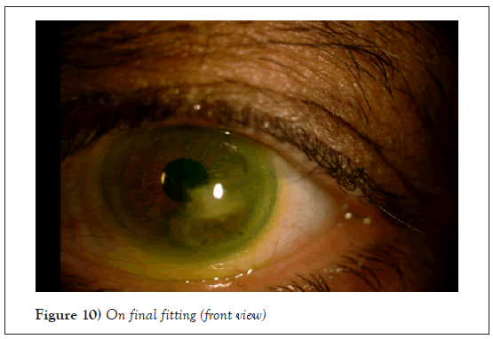
On final dispensing and teaching session, the patient was very happy and comfortable with the Atlantis Scleral Lens in left eye, with considerably good vision in Left eye: (0.5) and for reading with the reading glasses (20/40) (Figure 10). Patient was under follow-up after 1 week (1/12, 4/12) of using Atlantis Scleral lens and the reviews were happy and comfortable.
Conclusion
This patient had tectonic grafts and was initially considered legally blind. After his contact lens fitting in the left eye he was able to drive and to lead normal life. This case report shed the light on the importance of scleral contact lenses in management of severe corneal distortions. It also demonstrates that scleral contact lenses can provide reasonable vision even in patients with tectonic corneal grafts.
Disclosure
There is no proprietary or financial interest in any prosthetic device or contact lens.
REFERENCES
- Vanathi M, Sharma N, Titiyal JS, et al. Tectonic Jr Dіs for corneal thinning and perforations. Cornea. 2002; 21: 792-797.
- Jalie M. Modern spectacle lens design. Clin Exp Optom. 2020; 103: 3-10.
- AtlantisTM Scleral Lens Design. 2016.
- X-CelSpeciality Contacts. A Walman Company. 2020
- Nascimento H, Roque ADc. RPG Contact Lenses Adaptation: A Keratoconus Centring Problem. Optom Open Access. 2020; 5: 134.
- Ortiz-Toquero S, Rodriguez G, de Juan V, et al. Gas permeable contact lens fitting in keratoconus: Comparison of different guidelines to back optic zone radius calculations. Indian J Ophthalmol. 2019; 67(9): 1410.
- Vincent SJ, Fadel D. Optical considerations for scleral contact lenses: A review. Cont Lens Anterior Eye. 2019; 42: 598-613.


