Orbital teratoma
Received: 23-Jan-2019 Accepted Date: Feb 05, 2019; Published: 12-Feb-2019
This open-access article is distributed under the terms of the Creative Commons Attribution Non-Commercial License (CC BY-NC) (http://creativecommons.org/licenses/by-nc/4.0/), which permits reuse, distribution and reproduction of the article, provided that the original work is properly cited and the reuse is restricted to noncommercial purposes. For commercial reuse, contact reprints@pulsus.com
Abstract
Orbital teratoma is a rare tumour, usually benign congenital lesion that presents at birth with progressive growth and proptosis. The correct and early diagnosis is important for a better prognose. In this case report, we describe the clinical and radiological aspects of a case.
Keywords
Teratomas; Orbital tumours; Tooth
Introduction
Teratomas are tumours that originate from germ cells of the three embryological layers (ectoderm, mesoderm and endoderm) [1]. The extragonadal location is more frequent in childhood and the sacrococcygeal is the most common (59%-65%) [2]. Head and neck teratomas accounte for 5%-14% of all teratomas, in which only 0.8% of them is orbital. The majority of those tumours are benign, congenital, detect at birth and presents a rapid growth causing proptosis [3]. The radiological features are suggestive and appear as a heterogeneous multiloculated cystic mass with focus of others tissues, including ossification, fat and calcification [4]. Therefore, due to the rarity of this tumour, we present the clinical and radiological aspects of a case.
Case Report
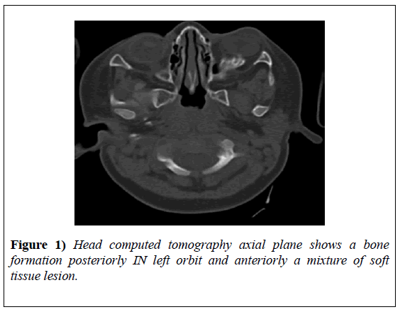
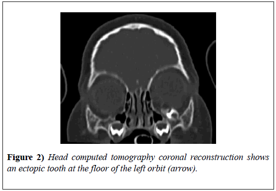
A 1-year-old boy, previously healthy, presented at the ophthalmology service with a nodulation in the left lower eyelid. The lesion was first seen at birth and since then grown progressively, impairing the visual axis. Physical examination showed a nodule in the left lower eyelid and lid conjunctiva hyperaemia. Computed tomography revealed a heterogeneous lesion composed of tissues of varied density, including adipose component and bone formation, as well as an ectopic tooth (Figures 1 and 2).
Figure 1) Head computed tomography axial plane shows a bone formation posteriorly IN left orbit and anteriorly a mixture of soft tissue lesion.
Figure 2): Head computed tomography coronal reconstruction shows an ectopic tooth at the floor of the left orbit (arrow).
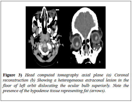
The lesion was extraconal in the floor of the orbit and dislocated the other structures superiorly (Figure 3). In view of these findings, the possibility of teratoma of the orbit was raised and the patient underwent excision of the lesion. The histopathological study confirmed the diagnostic hypothesis being classified as orbital teratoid tumour. After the surgical procedure the patient recovered well with ocular preservation.
Figure 3) Head computed tomography axial plane (a) Coronal reconstruction (b) Showing a heterogeneous extraconal lesion in the floor of left orbit dislocating the ocular bulb superiorly. Note the presence of the hypodense tissue representing fat (arrows).
Discussion
Orbital teratomas are rare tumours that clinically present with ocular proptosis, ranging from small lesions without involvement of the ocular bulb to massive lesions that occupy the entire orbit and enlarge the orbit to two or three times normal [5]. Females have predominance compared to males with a ratio of 2:1 and usually occur in the left orbit [6].
These tumours can be classified into four subtypes: complete fetus-infetus in orbit - "orbitopagus parasiticus"; parts of a second fetus within the orbit; true teratoma with the three germ layers; and a teratoid tumour that presents only two germ layers [2,5]. Our case consisted of a teratoid tumour presenting elements well differentiated from the mesoderm and endoderm.
The appearance of computed tomography imaging is similar in all teratomas, characterized by a heterogeneous multiloculated lesion with calcifications, fat tissue and bone formation [5]. Similar to our case, the presence of an ectopic tooth has already been described in literature [7]. The imaging exams are important because they aid in the therapeutic planning and allow the evaluation of the cranio-orbital invasion of the tumour [8]. Among the differential diagnoses should be considered: dermoid and epidermoid cyst, neuroblastoma, retinoblastoma, rhabdomyosarcoma, hemangioma and lymphangioma.
The treatment of choice is surgical excision [2]. Early surgery is recommended to avoid the risk of rapid growth resulting in necrosis, infection, hemorrhage and spontaneous rupture. Adjunctive chemotherapy or radiotherapy should be based on the pathologic findings for the individual case, but were believed to be ineffective especially in high grade immature teratomas [4,6]. The prognosis is usually good but depends on several factors incluind size and extension of the tumour, age, gender, histopathological components and degree of tissue differentiation. Patient follow-up is essential because of the possibility of recurrence with malignant degeneration.
Therefore, although rare, teratoma should be remembered in the presence of a congenital orbital lesion, since rapid diagnosis and appropriate treatment are essential for a better prognose and the preservation of vision.
Conflict of Interest
There are no conflicts of interest to disclose from all authors.
REFERENCES
- Smirniotopoulos JG, Chiechi MV. Teratomas, dermoids and dpidermoids of the head and neck. Radiographics. 1995;1437-55.
- Pellerano F, Guillermo E, Garrido G, et al. Congenital Orbital Teratoma. Ocul Oncol Pathol. 2016;11-6.
- Kivelä T, Tarkkanen, A. Orbital germ cell tumours revisited: A clinicopathological approach to classification. Surv Ophthalmol. 1994;541-54.
- Alkatan HM, AlObaidan OS, Kfoury H, et al. Orbital immature teratoma: A rare entity with diagnostic challenges. Saudi J Ophthalmol. 2018;75-8.
- Herman TE, Vachharajani A, Siegel MJ. Massive congenital orbital teratoma. J Perinatol. 2009;396-97.
- Alkatan HM, Chaudhry I, Alayoubi A. Mature teratoma presenting as orbital cellulitis in a 5-month-old baby. Ann Saudi Med. 2013;623-26.
- Hassan HMJ, Mc Andrew PT, Yagan A, et al. Mature orbital teratoma presenting as a recurrent orbital cellulitis with an ectopic tooth and sphenoid malformation- a case report. Orbit. 2008;309-12.
- Mahesh L, Krishnakumar S, Subramanian N, et al. Malignant teratoma of the orbit: A clinicopathological study of a case. Orbit. 2003;305-09.


