Persistent metopic suture with multiple sutural bones at unusual sites
Ambade HV*, Fulpatil MP and Kasote AP
Department of Anatomy, Government Medical College, Nagpur, Maharashtra State, India
- *Corresponding Author:
- Dr. H.V. Ambade
Department of Anatomy Government Medical College Nagpur
440 003 Maharashtra State, India
Tel: +91 (712) 2743588
E-mail: hemachimne@gmail.com
Citation: Ambade HV, Fulpatil MP, Kasote AP. Persistent metopic suture with multiple sutural bones at unusual sites. Int J Anat Var. 2017;10(3):69-70.
Copyright: This open-access article is distributed under the terms of the Creative Commons Attribution Non-Commercial License (CC BY-NC) (http://creativecommons.org/licenses/by-nc/4.0/), which permits reuse, distribution and reproduction of the article, provided that the original work is properly cited and the reuse is restricted to noncommercial purposes. For commercial reuse, contact reprints@pulsus.com
[ft_below_content] =>Keywords
Metopic suture; Sutural bones; Wormian bones; Skull; Unusual sites; Variations
Introduction
Metopic suture is present in between two frontal bones during fetal life and soon disappear after birth. The obliteration starts at the age of 2 years and completed at the age of 8 years from above downwards [1]. However, in 9% cases it remains persistent. The persistent metopic suture extends from nasion to bregma and is called metopism.
The sutural bones or ‘Wormian bones’ named after Ole Worm, professor of Anatomy, Copenhagen are small, irregular bone found at the sutures and fontanels of the skull, usually in the vicinity of lambdoid sutures [2]. They are usually present in 2 or 3 in number but can present in large numbers in hydrocephalic skulls. They are not named as different bones because they vary in number and shape from skull to skull [3]. The mechanism of formation of sutural bones is not clearly known. Pathological, mechanical and genetic factors have been proposed as the primary causal mechanism in the occurrence of sutural bones [4]. It is almost associated with abnormal development of the central nervous system and may serve as a useful marker for the early identification and treatment of the affected infant or child [5]. It may be the result of additional ossification centers in the fibrous tissue occurring during late fetal ages or post-natally; and which remain separated from the primary centers of ossification of cranial bones [5]. It is important to know about these sutural bones as they can mislead in the diagnosis of fracture of skull bones in medicolegal cases. Against this background present case is discussed.
Case Report
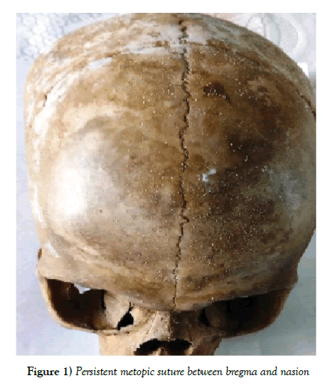
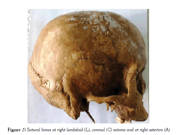
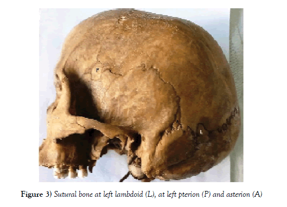
A partial skeletonised incomplete body was brought in the department of Anatomy for bone examination and opinion. The examined skeleton belonged to female of age about 30-40 years. The cranial sutures were started to fuse endocranially but not completely obliterated. The basi-occiput was fused with basi-sphenoid. However, the frontal bones were separated with persistent metopic suture present between bregma and nasion (Figure 1). Also, there were eight small and irregular shape sutural bones in the same skull. Of the eight sutural bones, four were present along the lambdoid suture, one along temporo-parietal suture at asterion on both side and one at left pterion and one along coronal suture on right side (Figures 2 and 3). There were no other abnormalities in the skull.
Discussion and Conclusion
Skull develops as chondrocranium and membranous neurocranium. Cartilaginous bars that develop in the basal region of the skull constitute the cartilaginous neurocranium and membrane bone develops in the region of the vault and side walls of the skull [1]. The growth of neurocranium is rapid during the first two years and by seventh year it reaches almost the adult size [1]. Most of the skull bones ossify during the first and second years at their apposed margins (sutures). The development of the skull continues until eighteenth to twenty-fifth year when occipito-sphenoid synchondrosis closes by synostosis [1]. The frontal bones of the fetal skull develop in two halves and are separated by the metopic sutures. The frontal bone ossifies at 8th week of intrauterine life from two primary centers [1]. The metopic suture usually disappears at the age of 2-3 years after birth. But it remains persistent in 5.1% of Asians and 8.7% in Europeans Caucasians skull [6]. The skull in present case belonged to female of age about 30-40 years having persistent metopic sutures. The incidence of sutural bone is variable, ranging from around 10% in Caucasian skull, through 40% in Indian skull to 80% in Chinese skull, more frequently in females [4]. Those fontanelles which close first are most likely to show sutural bones [2]. They are most commonly found in the vicinity of lambdoid suture [3,6,7]. Bergman et al [8] reported that almost 40% of skulls have sutural bones in the lambdoid suture. The next most common site of sutural bones is the epipteric bone at pterion [9,10]. Very rarely, they are seen in other sites especially, the coronal, sagittal and squamosal sutures. 4 However, Tewari et al who studied 1500 skull, could not find a single case of sutural bone in the coronal, sagittal and squamo-parietal sutures [11]. Hussain sahib and Nayak reported sutural bone at bregma [10,12]. In the present case, sutural bones were found in coronal suture, and at pterion and both asterion apart from the lambdoid suture.
Usually, 1-3 sutural bones at lambdoid suture or other site were found in the same skull [9,10]. However, a series of sutural bones ranging from 8-10 in a single skull was noted by Nayak, Verma et al, and Kaur et al; but all in the vicinity of lambdoid suture [3,6,8]. In the present case, sutural bones found were not only confined to lambdoid suture but also marked their presence at right coronal suture, left pterion and both asterion apart from the persistent metopic suture. Nayak, Verma also reported paired frontal bone in the Indian skull with sutural bone at bregma and lambda respectively [6,12]. But, persistent metopic suture with multiple sutural bones spreading beyond the lambdoid suture at asterion, left pterion, and right coronal suture in same skull is not reported previously.
References
- Sahana SN. Human anatomy. Vol I. 3rd edn., Howrah: KK Publishers (P) Ltd. 1993:462-9.
- Standring. Gray's anatomy. In: Williams PL, Warwick R, Dyson M, Bannister LH (eds.) London Churchill Livingstone. 2008: 417-8.
- Nayak SB. Multiple wormian bones at the lambdoid suture in an Indian skull. Neuroanatomy. 2008;7:52-3.
- Khan AA, Asari MA, Hassan A. Unusual presence of wormian (sutural ) bones in human skulls. Folia Morphol. 2011;70(4):291-4.
- Pryles CV, Khan AJ. Wormian bones. A marker of CNS abnormality? Am J Dis Child. 1979;133:380-2.
- Verma P, Seema, Mahajan A. Human skull with complete metopic suture and multiple sutural bones at lambdoid suture–a case report. International J Anat Var. 2014;7:7-9.
- Kaur J, Batra APS, Maahajan A. Skull with multiple sutural bones-A case report. Indian J Med Case Rep. 2013;2(3):47-9.
- Bergman RA, Afifi AK, Miyauchi R. Compendium of human anatomical variation. Baltimore, Urban and Schwarzenberg. 1988:197-205.
- Katikireddi RS, Sundara Setty SNR. Incidence of sutural bones at pterion in south Indian dried skulls. Int J Anat Res. 2016;4(1):2099-2101.
- Hussain Saheb S, Mavishetter GF, Thomas ST, et al. Unusual sutural bone at bregma-A case report. Biomed Res. 2010;21(4):369-70.
- Tewari PS, Malhotra VK, Agarwal SK, et al. Pre interparietal bone in man. Anatomischer Anzeiger. 1982;152:337-9.
- Nayak SB. Presence of wormian bones at bregma and paired frontal bone in an Indian skull. Neuroanatomy. 2006;5:42-3.
Ambade HV*, Fulpatil MP and Kasote AP
Department of Anatomy, Government Medical College, Nagpur, Maharashtra State, India
- *Corresponding Author:
- Dr. H.V. Ambade
Department of Anatomy Government Medical College Nagpur
440 003 Maharashtra State, India
Tel: +91 (712) 2743588
E-mail: hemachimne@gmail.com
Citation: Ambade HV, Fulpatil MP, Kasote AP. Persistent metopic suture with multiple sutural bones at unusual sites. Int J Anat Var. 2017;10(3):69-70.
Copyright: This open-access article is distributed under the terms of the Creative Commons Attribution Non-Commercial License (CC BY-NC) (http://creativecommons.org/licenses/by-nc/4.0/), which permits reuse, distribution and reproduction of the article, provided that the original work is properly cited and the reuse is restricted to noncommercial purposes. For commercial reuse, contact reprints@pulsus.com
Abstract
Sutural bones are small irregular bones found in the sutures and fontanels of the human skull. They are commonly found at lambda and lambdoid suture followed by pterion; and rarely at other sites. They vary from person to person in number and shape, hence not named. Usually, 1-3 sutural bones in one skull are present, but 8-10 sutural bones are also reported in the literature, all restricted in the vicinity of lambdoid sutures. In the present case, 8 sutural bones were present in a human skull at asterion, left pterion and right coronal suture apart from the lambdoid suture. Moreover, there was a persistent metopic suture between bregma to nasion in the same skull. The metopic suture with multiple sutural bones spreading beyond lambdoid suture at unusual sites is not reported previously. The knowledge of such variation and combination is rare and very important for forensic expert, radiologists, orthopedists, neurosurgeons and anthropologist point of view. It is very important to know about such variation because they can mislead the diagnosis of fracture of skull bones.
-Keywords
Metopic suture; Sutural bones; Wormian bones; Skull; Unusual sites; Variations
Introduction
Metopic suture is present in between two frontal bones during fetal life and soon disappear after birth. The obliteration starts at the age of 2 years and completed at the age of 8 years from above downwards [1]. However, in 9% cases it remains persistent. The persistent metopic suture extends from nasion to bregma and is called metopism.
The sutural bones or ‘Wormian bones’ named after Ole Worm, professor of Anatomy, Copenhagen are small, irregular bone found at the sutures and fontanels of the skull, usually in the vicinity of lambdoid sutures [2]. They are usually present in 2 or 3 in number but can present in large numbers in hydrocephalic skulls. They are not named as different bones because they vary in number and shape from skull to skull [3]. The mechanism of formation of sutural bones is not clearly known. Pathological, mechanical and genetic factors have been proposed as the primary causal mechanism in the occurrence of sutural bones [4]. It is almost associated with abnormal development of the central nervous system and may serve as a useful marker for the early identification and treatment of the affected infant or child [5]. It may be the result of additional ossification centers in the fibrous tissue occurring during late fetal ages or post-natally; and which remain separated from the primary centers of ossification of cranial bones [5]. It is important to know about these sutural bones as they can mislead in the diagnosis of fracture of skull bones in medicolegal cases. Against this background present case is discussed.
Case Report
A partial skeletonised incomplete body was brought in the department of Anatomy for bone examination and opinion. The examined skeleton belonged to female of age about 30-40 years. The cranial sutures were started to fuse endocranially but not completely obliterated. The basi-occiput was fused with basi-sphenoid. However, the frontal bones were separated with persistent metopic suture present between bregma and nasion (Figure 1). Also, there were eight small and irregular shape sutural bones in the same skull. Of the eight sutural bones, four were present along the lambdoid suture, one along temporo-parietal suture at asterion on both side and one at left pterion and one along coronal suture on right side (Figures 2 and 3). There were no other abnormalities in the skull.
Figure 1: Persistent metopic suture between bregma and nasion
Discussion and Conclusion
Skull develops as chondrocranium and membranous neurocranium. Cartilaginous bars that develop in the basal region of the skull constitute the cartilaginous neurocranium and membrane bone develops in the region of the vault and side walls of the skull [1]. The growth of neurocranium is rapid during the first two years and by seventh year it reaches almost the adult size [1]. Most of the skull bones ossify during the first and second years at their apposed margins (sutures). The development of the skull continues until eighteenth to twenty-fifth year when occipito-sphenoid synchondrosis closes by synostosis [1]. The frontal bones of the fetal skull develop in two halves and are separated by the metopic sutures. The frontal bone ossifies at 8th week of intrauterine life from two primary centers [1]. The metopic suture usually disappears at the age of 2-3 years after birth. But it remains persistent in 5.1% of Asians and 8.7% in Europeans Caucasians skull [6]. The skull in present case belonged to female of age about 30-40 years having persistent metopic sutures. The incidence of sutural bone is variable, ranging from around 10% in Caucasian skull, through 40% in Indian skull to 80% in Chinese skull, more frequently in females [4]. Those fontanelles which close first are most likely to show sutural bones [2]. They are most commonly found in the vicinity of lambdoid suture [3,6,7]. Bergman et al [8] reported that almost 40% of skulls have sutural bones in the lambdoid suture. The next most common site of sutural bones is the epipteric bone at pterion [9,10]. Very rarely, they are seen in other sites especially, the coronal, sagittal and squamosal sutures. 4 However, Tewari et al who studied 1500 skull, could not find a single case of sutural bone in the coronal, sagittal and squamo-parietal sutures [11]. Hussain sahib and Nayak reported sutural bone at bregma [10,12]. In the present case, sutural bones were found in coronal suture, and at pterion and both asterion apart from the lambdoid suture.
Usually, 1-3 sutural bones at lambdoid suture or other site were found in the same skull [9,10]. However, a series of sutural bones ranging from 8-10 in a single skull was noted by Nayak, Verma et al, and Kaur et al; but all in the vicinity of lambdoid suture [3,6,8]. In the present case, sutural bones found were not only confined to lambdoid suture but also marked their presence at right coronal suture, left pterion and both asterion apart from the persistent metopic suture. Nayak, Verma also reported paired frontal bone in the Indian skull with sutural bone at bregma and lambda respectively [6,12]. But, persistent metopic suture with multiple sutural bones spreading beyond the lambdoid suture at asterion, left pterion, and right coronal suture in same skull is not reported previously.
References
- Sahana SN. Human anatomy. Vol I. 3rd edn., Howrah: KK Publishers (P) Ltd. 1993:462-9.
- Standring. Gray's anatomy. In: Williams PL, Warwick R, Dyson M, Bannister LH (eds.) London Churchill Livingstone. 2008: 417-8.
- Nayak SB. Multiple wormian bones at the lambdoid suture in an Indian skull. Neuroanatomy. 2008;7:52-3.
- Khan AA, Asari MA, Hassan A. Unusual presence of wormian (sutural ) bones in human skulls. Folia Morphol. 2011;70(4):291-4.
- Pryles CV, Khan AJ. Wormian bones. A marker of CNS abnormality? Am J Dis Child. 1979;133:380-2.
- Verma P, Seema, Mahajan A. Human skull with complete metopic suture and multiple sutural bones at lambdoid suture–a case report. International J Anat Var. 2014;7:7-9.
- Kaur J, Batra APS, Maahajan A. Skull with multiple sutural bones-A case report. Indian J Med Case Rep. 2013;2(3):47-9.
- Bergman RA, Afifi AK, Miyauchi R. Compendium of human anatomical variation. Baltimore, Urban and Schwarzenberg. 1988:197-205.
- Katikireddi RS, Sundara Setty SNR. Incidence of sutural bones at pterion in south Indian dried skulls. Int J Anat Res. 2016;4(1):2099-2101.
- Hussain Saheb S, Mavishetter GF, Thomas ST, et al. Unusual sutural bone at bregma-A case report. Biomed Res. 2010;21(4):369-70.
- Tewari PS, Malhotra VK, Agarwal SK, et al. Pre interparietal bone in man. Anatomischer Anzeiger. 1982;152:337-9.
- Nayak SB. Presence of wormian bones at bregma and paired frontal bone in an Indian skull. Neuroanatomy. 2006;5:42-3.


