Prehilar renal artery division with supernumerary renal veins: a case series
Panchal P* and Singh S
Department of Anatomy, All India Institute of Medical Sciences, Patna, Phulwari Sharif, Patna, Bihar, India.
- *Corresponding Author:
- Dr. Padamjeet Panchal
All India Institute of Medical Sciences, Patna, Phulwari Sharif, Patna, Bihar-801507, India
Tel: +91 9905130001
E-mail: drpadamjeet@aiimspatna.org
Citation: Panchal P, Singh S. Prehilar renal artery division with supernumerary renal veins: a case series. Int J Anat Var. 2017;10(3):39-42.
Copyright: This open-access article is distributed under the terms of the Creative Commons Attribution Non-Commercial License (CC BY-NC) (http://creativecommons.org/licenses/by-nc/4.0/), which permits reuse, distribution and reproduction of the article, provided that the original work is properly cited and the reuse is restricted to noncommercial purposes. For commercial reuse, contact reprints@pulsus.com
[ft_below_content] =>Keywords
Renal artery; Renal vein; Hilum; Variations; Renal transplant
The renal arteries are large, paired arteries arising from the lateral aspect of aorta at the level of upper part of second lumbar vertebra and little below the origin of superior mesenteric artery. When the arteries arise at different cranio-caudal levels, the right ostium is more commonly higher than the left. The renal veins lie anterior to renal artery and open into the inferior vena cava at the level of L2 vertebra. The right renal vein is a shorter than the left renal vein. The left renal vein crosses anterior to aorta to open into left lateral aspect of inferior vena cava a little superior to the right. The relative positions of the hilar structures from anterior to posterior are renal vein, renal artery and the pelvis of the kidney [1]. Renal artery variations are divided into 2 groups: early division and extra renal artery. Branching of the main renal arteries into segmental branches more proximally than the renal hilus level is called early division [2]. Any extra vein other than renal vein emerging out of kidney and draining separately in the Inferior Vena Cava was named as “additional” renal vein [3]. Acquaintance to the renal vascular variations is of significance in diagnosis and treatment of renal trauma, renal transplantation, renal artery embolization, surgery for abdominal aortic aneurysm and conservative or radical renal surgery [4]. Since very few literatures are available on the early division of renal artery and additional renal veins in the past, the present study aims to report these incidental findings during routine dissection.
Case Presentation
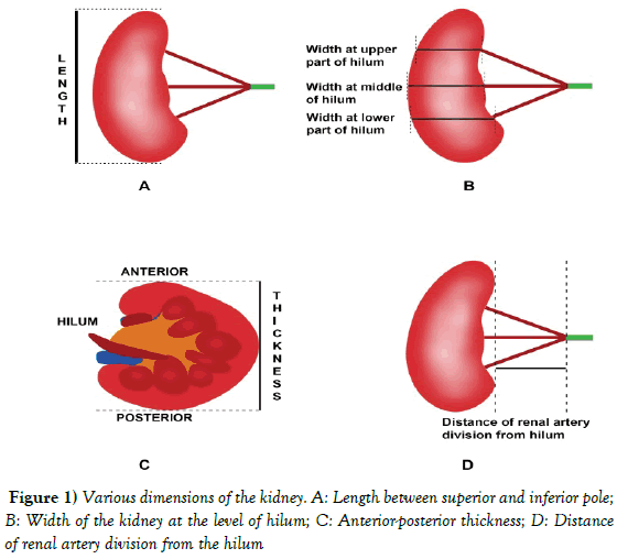
A routine abdominal dissection was conducted in two male and one female adult cadavers while teaching first year medical under graduate students in AIIMS, Patna. The retroperitoneal region was also dissected carefully as per the instructions in the Cunningham manual, the hilum of all the kidneys were cleaned for gross examination. The parameters such as, length, breadth and thickness of kidney and the distance of the division of the renal artery from middle of hilum of kidney along with number of renal veins was observed (Figure 1). Measurements were taken using a digital Vernier caliper (company: Aerospace, model: DVCA150, range: 0-150 mm, accuracy: +/- 0.03 mm/0.001). Five readings were taken and the average was calculated for each measurement and tabulated in Table 1.
| Cases | Side of kidney | Length (cm) | Width at the upper part of hilum (cm) | Width at the middle of hilum (cm) | Width at the lower part of hilum (cm) | Thickness (cm) |
|---|---|---|---|---|---|---|
| Case 1 | Right Left |
10.4 10.7 |
5.3 6.8 |
6.2 5.7 |
6.7 5.1 |
3.7 4.4 |
| Case 2 | Right Left |
9.7 8.5 |
5.3 5.1 |
5.2 5.1 |
5.4 3.98 |
4.8 4.2 |
| Case 3 | Right Left |
9.1 9.6 |
5.9 3.9 |
4.97 3.6 |
4.7 3.6 |
3.7 3.3 |
Table 1: Showing dimensions of the kidneys
Case 1
Right kidney
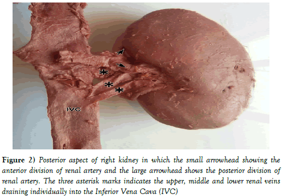
There is prehilar division of the renal artery into anterior and posterior divisions 2.5 cm from the hilum. The anterior division of renal artery again divided into six segmental arteries before entering the hilum. The posterior division of renal artery entered the hilum undivided. The three renal veins were observed as upper, middle and lower (Figure 2). The middle renal vein crossed behind the upper renal vein to reach the inferior vena cava from the hilum. The lower renal vein descended downward from the hilum to drain in inferior vena cava. The structures arranged at the hilum from before backward were segments of anterior division of renal artery, renal veins, posterior division of renal artery and the renal pelvis.
Figure 2: Posterior aspect of right kidney in which the small arrowhead showing the anterior division of renal artery and the large arrowhead shows the posterior division of renal artery. The three asterisk marks indicates the upper, middle and lower renal veins draining individually into the Inferior Vena Cava (IVC).
Left kidney
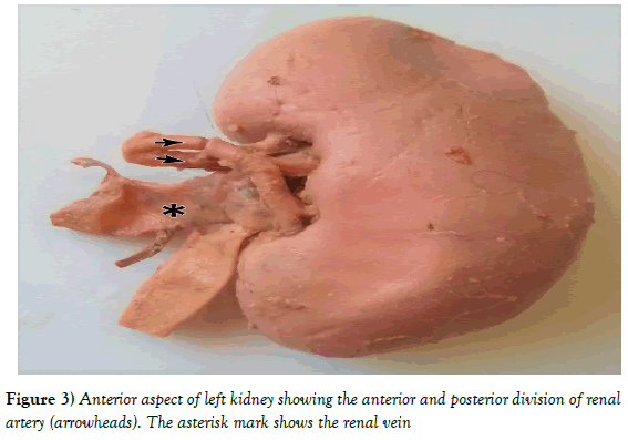
The early division of renal artery was observed as an anterior and a posterior division 35.77 mm from the hilum (Figure 3). The anterior division curved downwards and further divided into three segments in a ladder like pattern and the posterior division went horizontally into the hilum where it divided into two segments. The renal vein was present in between these two divisions of renal artery. The renal vein was formed by contribution of three segmental tributaries at 1.9 cm away from the hilum. The renal pelvis was dilated in this kidney. The structures arranged at the hilum from before backward were anterior division of renal artery, renal vein, posterior division of renal artery and the renal pelvis.
Case 2
Right kidney
The early division of renal artery was observed into an upper and a lower division 16.87 mm proximal to the hilum. The lower division of renal artery further divided into two segmental branches. On the anterior surface near the upper pole an artery was seen to enter the kidney. The structures at the hilum from before backward were renal vein, upper and lower division of renal arteries, the renal pelvis. The ureter was dilated and had a thickened wall with constricted lumen at 5.7 cm from the hilum.
Left kidney
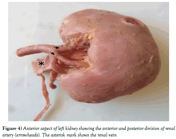
The anterior surface had a round cyst near its upper pole of one cm diameter; the posterior surface has another round cyst of 0.5 cm diameter (Figure 4). The renal artery divided into anterior and posterior divisions 29.51 mm proximal to the hilum. The anterior division of the artery gave three segmental branches outside hilum and the upper segment from it further gave three branches. The posterior division also gave three segmental branches outside the hilum. The left renal vein was formed by confluence of two tributaries which united at a distance of about 1 cm from the hilum. The renal vein was present between the two divisions of renal artery. The structures at hilum from before backward were anterior division of renal artery, renal vein and posterior division of renal artery along with renal pelvis. The posterior division took a course posterosuperiorly to renal pelvis before entering into the renal hilum.
Case 3
Right kidney
The renal artery divided into anterior and posterior divisions 27.31 mm from the hilum. The anterior division of renal artery further divided into upper and lower segment before reaching the hilum. The upper segment further divided into two branches and the lower segment also gave two branches before entering into the hilum. The posterior division of renal artery gave two segmental branches, the lower segment of which further gave two branches at the hilum. Two tributaries of renal vein arose from the hilum which joined at 2.5 mm away from the hilum to form the main right renal vein. Structures at the hilum from before backward were anterior division of renal artery, renal vein and posterior division of renal artery along with renal pelvis. The latter two were in same plane.
Left kidney
The early bifurcation of renal artery into anterior and posterior divisions was observed 33.19 mm before the hilum. The anterior division of renal artery followed a downward course to enter into the lower part of the hilum. The posterior division of renal artery gave three segmental branches, upper, middle and the lower segmental branches in fork pattern. The upper segment further gave three branches, and lower segment also gave two branches and the middle segmental branch entered hilum unbranched. The renal vein arose from the hilum as two tributaries and joined at a distance of 1.4 cm from the hilum. The upper segment of the posterior division of the renal artery came to the anterior part of the hilum through the gap between the two tributaries of the renal vein. The arrangement of structures at the hilum from before backward was: anterior division of renal artery, renal vein, the posterior division of renal artery and the renal pelvis.
Discussion
During embryonic life the kidney ascends from sacral position to reach the level of second lumber vertebrae at the 13 mm length of the embryo. During its ascent it receives its blood supply sequentially from neighboring arteries, i.e., middle sacral and common iliac arteries. In the beginning of third month the definitive renal artery is established, which arises from the most caudal of the three suprarenal arteries. There suprarenal artery represent the persistent mesonephric or lateral splanchnic artery [1].
The 15th day to 94th day of gestation is critical for renal vascular development in human embryo. The variations in renal vasculature can be due to multiple factors that interact with one another and may influence the timely occurrence of sequence of events during renal development [5]. Ferrara reported that Vascular Endothelial Growth Factor (VEGF) is one of the basic regulators of normal and abnormal angiogenesis. It also plays an important role in kidney development [6].
Early branching of renal artery is defined as the branches arising within 15 mm from the origin of the main renal artery ostium [7]. The stem of artery starting from its origin till its primary division is the main artery. The primary division of main artery not reaching the hilum is pre-segmental arteries and the secondary division reaching the hilum is segmental arteries [8]. Kang et al classified the individual cases with a single main renal artery into three types on the basis of number of division of the main renal artery into the pre-segmental branches (Table 2) [9].
| Type I | there is no pre-segmental branch |  |
| Type II | one pre-segmental branch is present |  |
| Type III | two pre-segmental branches are present |  |
Table 2: Showing the classification as given by Kang et al [9]
According to the classification given by Kang, in this study, out of the six kidneys, 4 were of type II and 2 kidneys were of type III variant. In the present study prehilar division of renal artery was present bilaterally in all the three cadavers dissected. Ozkan et al reported the incidence of early division of renal artery in 8% (32% of which occurred on the right side, 25% on the left, and 22% on the both sides) [2]. Budhiraja et al reported 61.8% (33.3% on right side and 28.5% on left side) [10]. In contrast to the study done by Budhiraja, the polar artery was not found in this study.
Similar findings were also reported by Arora et al, Dnyanesh et al, in the right side [11,12]. These branches may be interpreted falsely as being extra renal arteries in diagnostic imaging studies. The donor individuals with prehilar division of renal artery usually have a short pedicle for anastomosis with the recipient’s iliac artery and therefore are excluded from the criteria for renal transplant surgeries [13,14]. Similar to the present findings, Fernandes et al also reported a case with three renal veins draining the right kidney [15]. Satayapal reported incidence of additional right renal vein was 26% as compared to 2.6% on the left side. Second additional renal vein occurred infrequently on the right side (5%) [3]. Dnyanesh et al reported double renal vein on right side (1%) [12].
On the 8th week of intrauterine development, the bilaterally symmetrical cardinal venous system converts into unilateral right-sided inferior vena cava. Two renal veins are present on each side, one on ventral plane and another dorsal [16].
Confluence of the two tributaries occurs with further development producing a single vessel. In case of persistence of these two veins, an additional right renal vein results. The shifting of the venous arrangement to the right side may explain why right-sided anatomical variations are more common [17].
Variations of renal veins are usually asymptomatic and are discovered only during surgical procedures. Morphology of renal vein is significantly important for surgeons during transplantation, since variations restrict the availability of renal vein for mobilization procedures [18].
In the present case also three kidneys (two left kidney and one right kidney) showed two emerging veins at the hilum which joined to form the main renal vein outside the hilum, while one left kidney showed three emerging vein from the hilum which joined to form the main renal vein outside the hilum. Mukundan also reported a case where the right kidney drained into the inferior vena cava through two veins and the left kidney drained into the inferior vena cava through two veins which united to form a single large left renal vein and drained into the inferior vena cava [19].
Conclusion
Awareness of variations in the hilar region of kidney is necessary for surgical management during renal transplantation, urological procedures and for angiographic interventions. A thorough knowledge of anatomy of renal vasculature and its variations is important to avoid complications during these procedures.
References
- Standring S, Anatomy G. The Anatomical Basis of Clinical Practice. Elsevier. 2016;41:1243-4.
- Ugur O, Levent O, Fahri T, et al. Nihal Koca Renal artery origins and variations: angiographic evaluation of 855 consecutive patients. Diagn Interv Radiol. 2006;12:183-6.
- Satyapal KS. Classification of the drainage patterns of the renal veins. J Anat. 1995;186:329-33.
- Gupta V, Kotgirwar S, Trivedi S, et al. Bilateral variations in renal vasculature. International Journal of Anatomical Variations. 2010;3:53-5.
- Hodson CJ. The renal parenchyma and its blood supply. Curr Probl Diagn Radiol. 1978;7:1.
- Ferrara N. Role of vascular endothelial growth factor in the regulation of angiogenesis. Kidney Int. 1999;56:794-814.
- He B, Hamdorf JM. Clinical importance of anatomical variations of renal vasculature during laparoscopic donor nephrectomy. OA Anat. 2013;1:25.
- Weld KJ, Bhayani SB, Belani J, et al. Extrarenal vascular anatomy of kidney: assessment of variations and their relevance to partial nephrectomy. Urology. 2005;66:985-9.
- Kang WY, Sung DJ, Park BJ, et al. Perihilar branching patterns of renal artery and extrarenal length of arterial branches and tumour-feeding arteries on multidetector CT angiography. Br J Radiol. 2013;86:20120387
- Budhiraja V, Rastogi R, Jain V, et al. Anatomical variations of renal artery and its clinical correlations: a cadaveric study from central India. J Morphol Sci. 2013;30:228-33
- Arora AK, Verma P, Lalit, et al. Variant Segmental Renal Arteries in The Right Kidney- Clinical Correlations- A Case Report. Anat Physiol. 2012;2:103.
- Dnyanesh S, Dnyanesh DK, Dixit D, et al. A Study of Embryological Basis of Variations of Renal Vessels. Indian Journal of Anatomy. 2017;6:63-7.
- Weld KJ, Bhayani SB, Belani J, et al. Extrarenal vascular anatomy of kidney: assessment of variations and their relevance to partial nephrectomy. Urology. 2005;66:985-9.
- Madhyastha S, Suresh R, Rao R. Multiple variations of renal vessels and ureter. Indian J Urol. 2001;17:164-5.
- Fernandes RMP, Conte FHP, Favorito LA, et al. Triple right renal vein: An uncommon variation. Int J Morphol. 2005;23:231-3.
- Tatar I, Tore HG, H. Hamidi Celik, et al. Retroaortic and Circumaortic left renal veins with their finding and review of the literature. Anatomy. 2008;2:72-6
- Mankhause WS, Khalique A. The adrenal and renal mass and their connection with Azygos and lumber vein. J Anat. 1986;146:105-15.
- Satyapal KS, Kalideen JM, Haffejee AA, et al. Left renal vein variations. Surg Radiol Anat. 1999;21:77-81.
- Mukundan M, Vijaynath V, Ravi V. Bilateral Variation of Renal Vein: A Case Report. Int J Anat Res. 2016;4:3153-5.
Panchal P* and Singh S
Department of Anatomy, All India Institute of Medical Sciences, Patna, Phulwari Sharif, Patna, Bihar, India.
- *Corresponding Author:
- Dr. Padamjeet Panchal
All India Institute of Medical Sciences, Patna, Phulwari Sharif, Patna, Bihar-801507, India
Tel: +91 9905130001
E-mail: drpadamjeet@aiimspatna.org
Citation: Panchal P, Singh S. Prehilar renal artery division with supernumerary renal veins: a case series. Int J Anat Var. 2017;10(3):39-42.
Copyright: This open-access article is distributed under the terms of the Creative Commons Attribution Non-Commercial License (CC BY-NC) (http://creativecommons.org/licenses/by-nc/4.0/), which permits reuse, distribution and reproduction of the article, provided that the original work is properly cited and the reuse is restricted to noncommercial purposes. For commercial reuse, contact reprints@pulsus.com
Abstract
OBJECTIVES: To know the variations of renal vessels in human cadavers. METHODS: The study was performed on 3 cadaveric cases. The posterior abdominal wall was dissected to study the paired kidneys. RESULTS: The following observations were made: bilateral prehilar division of renal artery in all 3 cadavers, triple renal vein on right side in one kidney. CONCLUSION: Anatomical knowledge of the vascular variations is essential for the clinician to perform procedures such as renal transplantation, renal vascular operations more safely and efficiently.
-Keywords
Renal artery; Renal vein; Hilum; Variations; Renal transplant
The renal arteries are large, paired arteries arising from the lateral aspect of aorta at the level of upper part of second lumbar vertebra and little below the origin of superior mesenteric artery. When the arteries arise at different cranio-caudal levels, the right ostium is more commonly higher than the left. The renal veins lie anterior to renal artery and open into the inferior vena cava at the level of L2 vertebra. The right renal vein is a shorter than the left renal vein. The left renal vein crosses anterior to aorta to open into left lateral aspect of inferior vena cava a little superior to the right. The relative positions of the hilar structures from anterior to posterior are renal vein, renal artery and the pelvis of the kidney [1]. Renal artery variations are divided into 2 groups: early division and extra renal artery. Branching of the main renal arteries into segmental branches more proximally than the renal hilus level is called early division [2]. Any extra vein other than renal vein emerging out of kidney and draining separately in the Inferior Vena Cava was named as “additional” renal vein [3]. Acquaintance to the renal vascular variations is of significance in diagnosis and treatment of renal trauma, renal transplantation, renal artery embolization, surgery for abdominal aortic aneurysm and conservative or radical renal surgery [4]. Since very few literatures are available on the early division of renal artery and additional renal veins in the past, the present study aims to report these incidental findings during routine dissection.
Case Presentation
A routine abdominal dissection was conducted in two male and one female adult cadavers while teaching first year medical under graduate students in AIIMS, Patna. The retroperitoneal region was also dissected carefully as per the instructions in the Cunningham manual, the hilum of all the kidneys were cleaned for gross examination. The parameters such as, length, breadth and thickness of kidney and the distance of the division of the renal artery from middle of hilum of kidney along with number of renal veins was observed (Figure 1). Measurements were taken using a digital Vernier caliper (company: Aerospace, model: DVCA150, range: 0-150 mm, accuracy: +/- 0.03 mm/0.001). Five readings were taken and the average was calculated for each measurement and tabulated in Table 1.
| Cases | Side of kidney | Length (cm) | Width at the upper part of hilum (cm) | Width at the middle of hilum (cm) | Width at the lower part of hilum (cm) | Thickness (cm) |
|---|---|---|---|---|---|---|
| Case 1 | Right Left |
10.4 10.7 |
5.3 6.8 |
6.2 5.7 |
6.7 5.1 |
3.7 4.4 |
| Case 2 | Right Left |
9.7 8.5 |
5.3 5.1 |
5.2 5.1 |
5.4 3.98 |
4.8 4.2 |
| Case 3 | Right Left |
9.1 9.6 |
5.9 3.9 |
4.97 3.6 |
4.7 3.6 |
3.7 3.3 |
Table 1: Showing dimensions of the kidneys
Case 1
Right kidney
There is prehilar division of the renal artery into anterior and posterior divisions 2.5 cm from the hilum. The anterior division of renal artery again divided into six segmental arteries before entering the hilum. The posterior division of renal artery entered the hilum undivided. The three renal veins were observed as upper, middle and lower (Figure 2). The middle renal vein crossed behind the upper renal vein to reach the inferior vena cava from the hilum. The lower renal vein descended downward from the hilum to drain in inferior vena cava. The structures arranged at the hilum from before backward were segments of anterior division of renal artery, renal veins, posterior division of renal artery and the renal pelvis.
Figure 2: Posterior aspect of right kidney in which the small arrowhead showing the anterior division of renal artery and the large arrowhead shows the posterior division of renal artery. The three asterisk marks indicates the upper, middle and lower renal veins draining individually into the Inferior Vena Cava (IVC).
Left kidney
The early division of renal artery was observed as an anterior and a posterior division 35.77 mm from the hilum (Figure 3). The anterior division curved downwards and further divided into three segments in a ladder like pattern and the posterior division went horizontally into the hilum where it divided into two segments. The renal vein was present in between these two divisions of renal artery. The renal vein was formed by contribution of three segmental tributaries at 1.9 cm away from the hilum. The renal pelvis was dilated in this kidney. The structures arranged at the hilum from before backward were anterior division of renal artery, renal vein, posterior division of renal artery and the renal pelvis.
Case 2
Right kidney
The early division of renal artery was observed into an upper and a lower division 16.87 mm proximal to the hilum. The lower division of renal artery further divided into two segmental branches. On the anterior surface near the upper pole an artery was seen to enter the kidney. The structures at the hilum from before backward were renal vein, upper and lower division of renal arteries, the renal pelvis. The ureter was dilated and had a thickened wall with constricted lumen at 5.7 cm from the hilum.
Left kidney
The anterior surface had a round cyst near its upper pole of one cm diameter; the posterior surface has another round cyst of 0.5 cm diameter (Figure 4). The renal artery divided into anterior and posterior divisions 29.51 mm proximal to the hilum. The anterior division of the artery gave three segmental branches outside hilum and the upper segment from it further gave three branches. The posterior division also gave three segmental branches outside the hilum. The left renal vein was formed by confluence of two tributaries which united at a distance of about 1 cm from the hilum. The renal vein was present between the two divisions of renal artery. The structures at hilum from before backward were anterior division of renal artery, renal vein and posterior division of renal artery along with renal pelvis. The posterior division took a course posterosuperiorly to renal pelvis before entering into the renal hilum.
Case 3
Right kidney
The renal artery divided into anterior and posterior divisions 27.31 mm from the hilum. The anterior division of renal artery further divided into upper and lower segment before reaching the hilum. The upper segment further divided into two branches and the lower segment also gave two branches before entering into the hilum. The posterior division of renal artery gave two segmental branches, the lower segment of which further gave two branches at the hilum. Two tributaries of renal vein arose from the hilum which joined at 2.5 mm away from the hilum to form the main right renal vein. Structures at the hilum from before backward were anterior division of renal artery, renal vein and posterior division of renal artery along with renal pelvis. The latter two were in same plane.
Left kidney
The early bifurcation of renal artery into anterior and posterior divisions was observed 33.19 mm before the hilum. The anterior division of renal artery followed a downward course to enter into the lower part of the hilum. The posterior division of renal artery gave three segmental branches, upper, middle and the lower segmental branches in fork pattern. The upper segment further gave three branches, and lower segment also gave two branches and the middle segmental branch entered hilum unbranched. The renal vein arose from the hilum as two tributaries and joined at a distance of 1.4 cm from the hilum. The upper segment of the posterior division of the renal artery came to the anterior part of the hilum through the gap between the two tributaries of the renal vein. The arrangement of structures at the hilum from before backward was: anterior division of renal artery, renal vein, the posterior division of renal artery and the renal pelvis.
Discussion
During embryonic life the kidney ascends from sacral position to reach the level of second lumber vertebrae at the 13 mm length of the embryo. During its ascent it receives its blood supply sequentially from neighboring arteries, i.e., middle sacral and common iliac arteries. In the beginning of third month the definitive renal artery is established, which arises from the most caudal of the three suprarenal arteries. There suprarenal artery represent the persistent mesonephric or lateral splanchnic artery [1].
The 15th day to 94th day of gestation is critical for renal vascular development in human embryo. The variations in renal vasculature can be due to multiple factors that interact with one another and may influence the timely occurrence of sequence of events during renal development [5]. Ferrara reported that Vascular Endothelial Growth Factor (VEGF) is one of the basic regulators of normal and abnormal angiogenesis. It also plays an important role in kidney development [6].
Early branching of renal artery is defined as the branches arising within 15 mm from the origin of the main renal artery ostium [7]. The stem of artery starting from its origin till its primary division is the main artery. The primary division of main artery not reaching the hilum is pre-segmental arteries and the secondary division reaching the hilum is segmental arteries [8]. Kang et al classified the individual cases with a single main renal artery into three types on the basis of number of division of the main renal artery into the pre-segmental branches (Table 2) [9].
| Type I | there is no pre-segmental branch |  |
| Type II | one pre-segmental branch is present |  |
| Type III | two pre-segmental branches are present |  |
Table 2: Showing the classification as given by Kang et al [9]
According to the classification given by Kang, in this study, out of the six kidneys, 4 were of type II and 2 kidneys were of type III variant. In the present study prehilar division of renal artery was present bilaterally in all the three cadavers dissected. Ozkan et al reported the incidence of early division of renal artery in 8% (32% of which occurred on the right side, 25% on the left, and 22% on the both sides) [2]. Budhiraja et al reported 61.8% (33.3% on right side and 28.5% on left side) [10]. In contrast to the study done by Budhiraja, the polar artery was not found in this study.
Similar findings were also reported by Arora et al, Dnyanesh et al, in the right side [11,12]. These branches may be interpreted falsely as being extra renal arteries in diagnostic imaging studies. The donor individuals with prehilar division of renal artery usually have a short pedicle for anastomosis with the recipient’s iliac artery and therefore are excluded from the criteria for renal transplant surgeries [13,14]. Similar to the present findings, Fernandes et al also reported a case with three renal veins draining the right kidney [15]. Satayapal reported incidence of additional right renal vein was 26% as compared to 2.6% on the left side. Second additional renal vein occurred infrequently on the right side (5%) [3]. Dnyanesh et al reported double renal vein on right side (1%) [12].
On the 8th week of intrauterine development, the bilaterally symmetrical cardinal venous system converts into unilateral right-sided inferior vena cava. Two renal veins are present on each side, one on ventral plane and another dorsal [16].
Confluence of the two tributaries occurs with further development producing a single vessel. In case of persistence of these two veins, an additional right renal vein results. The shifting of the venous arrangement to the right side may explain why right-sided anatomical variations are more common [17].
Variations of renal veins are usually asymptomatic and are discovered only during surgical procedures. Morphology of renal vein is significantly important for surgeons during transplantation, since variations restrict the availability of renal vein for mobilization procedures [18].
In the present case also three kidneys (two left kidney and one right kidney) showed two emerging veins at the hilum which joined to form the main renal vein outside the hilum, while one left kidney showed three emerging vein from the hilum which joined to form the main renal vein outside the hilum. Mukundan also reported a case where the right kidney drained into the inferior vena cava through two veins and the left kidney drained into the inferior vena cava through two veins which united to form a single large left renal vein and drained into the inferior vena cava [19].
Conclusion
Awareness of variations in the hilar region of kidney is necessary for surgical management during renal transplantation, urological procedures and for angiographic interventions. A thorough knowledge of anatomy of renal vasculature and its variations is important to avoid complications during these procedures.
References
- Standring S, Anatomy G. The Anatomical Basis of Clinical Practice. Elsevier. 2016;41:1243-4.
- Ugur O, Levent O, Fahri T, et al. Nihal Koca Renal artery origins and variations: angiographic evaluation of 855 consecutive patients. Diagn Interv Radiol. 2006;12:183-6.
- Satyapal KS. Classification of the drainage patterns of the renal veins. J Anat. 1995;186:329-33.
- Gupta V, Kotgirwar S, Trivedi S, et al. Bilateral variations in renal vasculature. International Journal of Anatomical Variations. 2010;3:53-5.
- Hodson CJ. The renal parenchyma and its blood supply. Curr Probl Diagn Radiol. 1978;7:1.
- Ferrara N. Role of vascular endothelial growth factor in the regulation of angiogenesis. Kidney Int. 1999;56:794-814.
- He B, Hamdorf JM. Clinical importance of anatomical variations of renal vasculature during laparoscopic donor nephrectomy. OA Anat. 2013;1:25.
- Weld KJ, Bhayani SB, Belani J, et al. Extrarenal vascular anatomy of kidney: assessment of variations and their relevance to partial nephrectomy. Urology. 2005;66:985-9.
- Kang WY, Sung DJ, Park BJ, et al. Perihilar branching patterns of renal artery and extrarenal length of arterial branches and tumour-feeding arteries on multidetector CT angiography. Br J Radiol. 2013;86:20120387
- Budhiraja V, Rastogi R, Jain V, et al. Anatomical variations of renal artery and its clinical correlations: a cadaveric study from central India. J Morphol Sci. 2013;30:228-33
- Arora AK, Verma P, Lalit, et al. Variant Segmental Renal Arteries in The Right Kidney- Clinical Correlations- A Case Report. Anat Physiol. 2012;2:103.
- Dnyanesh S, Dnyanesh DK, Dixit D, et al. A Study of Embryological Basis of Variations of Renal Vessels. Indian Journal of Anatomy. 2017;6:63-7.
- Weld KJ, Bhayani SB, Belani J, et al. Extrarenal vascular anatomy of kidney: assessment of variations and their relevance to partial nephrectomy. Urology. 2005;66:985-9.
- Madhyastha S, Suresh R, Rao R. Multiple variations of renal vessels and ureter. Indian J Urol. 2001;17:164-5.
- Fernandes RMP, Conte FHP, Favorito LA, et al. Triple right renal vein: An uncommon variation. Int J Morphol. 2005;23:231-3.
- Tatar I, Tore HG, H. Hamidi Celik, et al. Retroaortic and Circumaortic left renal veins with their finding and review of the literature. Anatomy. 2008;2:72-6
- Mankhause WS, Khalique A. The adrenal and renal mass and their connection with Azygos and lumber vein. J Anat. 1986;146:105-15.
- Satyapal KS, Kalideen JM, Haffejee AA, et al. Left renal vein variations. Surg Radiol Anat. 1999;21:77-81.
- Mukundan M, Vijaynath V, Ravi V. Bilateral Variation of Renal Vein: A Case Report. Int J Anat Res. 2016;4:3153-5.


