Prepancreatic formation of portal vein associated with prepancreatic superior mesenteric artery and splenic vein
PS Chitra1*, K Maheshwari2 and V Anandhi1
1Department of Anatomy, K.A.P.V. Goverment Medical College, Tiruchirappalli, Tamilnadu, India
2Department of Anatomy, Vellore Medical College, Vellore, Tamilnadu, India
- *Corresponding Author:
- Dr. P. S. Chitra, MS
Associate Professor of Anatomy
K.A.P.V. Goverment Medical College
Tiruchirappalli – 620 001
Tamilnadu, INDIA.
Tel: +91 94438 64424
E-mail: pschitraezhilko@gmail.com
Date of Received: March 14th, 2013
Date of Accepted: November 24th, 2013
Published Online: June 1st, 2014
© Int J Anat Var (IJAV). 2014; 7: 35–36.
[ft_below_content] =>Keywords
prepancreatic postduodenal portal vein, prepancreatic superior mesenteric artery, prepancreatic splenic vein, embryological variant, surgical hazard
Introduction
Portal vein is formed by the convergence of superior mesenteric vein and splenic vein at the level of second lumbar vertebra. It lies anterior to the inferior venae cava and posterior to the neck of the pancreas and ascends behind the first part of the duodenum and enters right border of the lesser omentum anterior to the epiploic foramen to reach the right end of the porta hepatis. Usually the superior mesenteric artery is posterior to the body of pancreas and emerges out on the anterior aspect by passing between the uncinate process and body of the pancreas and crosses in front of the third part of the duodenum [1].
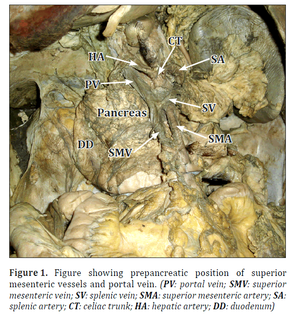
In the present study, the portal vein formation itself was found in front of the neck of the pancreas. For this the splenic vein came anterior to the pancreas by piercing the body of the pancreas. Crossing the superior mesenteric artery, it joined the superior mesenteric vein to form the portal vein. The superior mesenteric artery was also found in front of the neck and uncinate process of pancreas on the left of the superior mesenteric vein.
Case Report
During routine dissection for undergraduate students in one male cadaver aged about 50 years, at K.A.P.V. Govt. Medical College, Tiruchirappalli, Tamilnadu, India, the portal vein was found in front of the neck of the pancreas. Careful dissection was done to expose the superior mesenteric vein, splenic vein and superior mesenteric artery.
From the root of mesentery, the superior mesenteric vein passed anterior to the third part of duodenum and neck of the pancreas, where it joined the splenic vein to form the prepancreatic portal vein. It was seen that the splenic vein had pierced the body of the pancreas to reach the anterior surface where it crossed the superior mesenteric artery to join the superior mesenteric vein. The portal vein ascended up behind the 1st part of duodenum to enter the right free margin of lesser omentum (Figure 1).
Usually the superior mesenteric artery is posterior to the body of pancreas and emerges out on the anterior aspect by passing between the uncinate process and body of the pancreas and crosses in front of the third part of the duodenum before entering the root of mesentery. But in the present study the superior mesenteric artery, after its origin from the abdominal aorta at the level of L1, passed anterior to the neck of the pancreas and uncinate process and the third part of duodenum to enter the root of mesentery, which is highly unusual.
Discussion
The etiology of these variations can best be understood by reviewing the normal embryology. The development of the portal venous system occurs simultaneously with the development of pancreas and rotation of stomach.
The portal vein is formed from the right and left vilelline veins. These veins have three anastomoses, the cranial anastomosis intrahepatically, the middle anastamosis behind the duodenum and caudal anastomosis in front of duodenum. The superior mesenteric vein joins with the right vitelline vein, and the splenic vein joins with the left vitelline near its anastomosis. The proximal ventral anastomosis becomes the left branch of the portal vein, the dorsal anastomosis becomes the portal vein. The distal ventral anastomosis usually disappears [2].
A prepancreatic portal vein can form when the caudal ventral anastomosis persists instead of the middle one.
Around 100 cases of prepancreatic portal vein have been reported so far. The first case of prepancreatic portal vein was reported by Knight in 1921 during dissection of a cadaver [3]. Prepancreatic postduodenal portal vein is rare compared with prepancreatic preduodenal portal vein, such that only 12 cases including ours have been reported.
Inoue et al. reported a case of prepancreatic postduodenal portal vein in 2003 which was incidentally discovered during total gastrectomy [4].
In 2010 Tomizawa et al. also reported 2 cases of prepancreatic postduodenal portal vein found in an enhanced CT scan in cases of sigmoid colon cancer with liver metastasis and breast cancer [5]. In all the cases reported, the portal vein formation was caudal to the pancreatic head.
Our case is unique in that the portal vein formation itself was found in front of the neck of pancreas. It was also found that the superior mesenteric artery was passing anterior to the neck of the pancreas from its origin itself and then crossing the uncinate process and then entered the root of mesentery. This unusual course of the superior mesenteric artery has not been reported so far.
Conclusion
Though the prepancreatic postduodenal vein, prepancreatic superior mesenteric artery and prepancreatic splenic vein are seldom encountered, understanding of these variations is important to avoid surgical hazards including portal vein ligation, resection or intraoperative hemorrhage.
References
- Standring S, ed. Gray’s Anatomy. The Anatomical Basis of Clinical Practice. 40th Ed., Elsevier Churchill Livingstone Ltd. 2008; 1048, 1130.
- Patten BM. Human Embryology. New York and Toronto, The Blakiston Company Inc. 1953, 642–646.
- Knight HO. An anomalous portal vein with its surgical dangers. Ann Surg. 1921; 74: 697–699.
- Inoue M, Taenaka N, Nishimura S, Kawamura T, Aki T, Yamaki K, Enomoto H, Kosaka K, Yoshikawa K. Prepancreatic postduodenal portal vein: report of a case. Surg Today. 2003; 33: 956–959.
- Tomizawa N, Akai H, Akahane M, Ino K, Kiryu S, Ohtomo K. Prepancreatic postduodenal portal vein: a new hypothesis for the development of the portal venous system. Jpn J Radiol. 2010; 28: 157–161.
PS Chitra1*, K Maheshwari2 and V Anandhi1
1Department of Anatomy, K.A.P.V. Goverment Medical College, Tiruchirappalli, Tamilnadu, India
2Department of Anatomy, Vellore Medical College, Vellore, Tamilnadu, India
- *Corresponding Author:
- Dr. P. S. Chitra, MS
Associate Professor of Anatomy
K.A.P.V. Goverment Medical College
Tiruchirappalli – 620 001
Tamilnadu, INDIA.
Tel: +91 94438 64424
E-mail: pschitraezhilko@gmail.com
Date of Received: March 14th, 2013
Date of Accepted: November 24th, 2013
Published Online: June 1st, 2014
© Int J Anat Var (IJAV). 2014; 7: 35–36.
Abstract
Prepancreatic portal vein is an unusual condition. Around 100 cases of prepancreatic preduodenal portal vein have been reported. But only 11 cases of prepancreatic postduodenal portal vein have been reported till now. Here, we report a peculiar case of prepancreatic formation of portal vein associated with prepancreatic superior mesenteric artery. The formation of prepancreatic portal vein by the union of superior mesenteric vein with the splenic vein which came anteriorly by piercing the body of pancreas to form the portal vein in front of the neck of pancreas is an exceptional occurrence which has never been reported in world literature so far. Radiologists and surgeons need to be aware of these unfamiliar prepancreatic vessels to avoid major intraoperative injuries.
-Keywords
prepancreatic postduodenal portal vein, prepancreatic superior mesenteric artery, prepancreatic splenic vein, embryological variant, surgical hazard
Introduction
Portal vein is formed by the convergence of superior mesenteric vein and splenic vein at the level of second lumbar vertebra. It lies anterior to the inferior venae cava and posterior to the neck of the pancreas and ascends behind the first part of the duodenum and enters right border of the lesser omentum anterior to the epiploic foramen to reach the right end of the porta hepatis. Usually the superior mesenteric artery is posterior to the body of pancreas and emerges out on the anterior aspect by passing between the uncinate process and body of the pancreas and crosses in front of the third part of the duodenum [1].
In the present study, the portal vein formation itself was found in front of the neck of the pancreas. For this the splenic vein came anterior to the pancreas by piercing the body of the pancreas. Crossing the superior mesenteric artery, it joined the superior mesenteric vein to form the portal vein. The superior mesenteric artery was also found in front of the neck and uncinate process of pancreas on the left of the superior mesenteric vein.
Case Report
During routine dissection for undergraduate students in one male cadaver aged about 50 years, at K.A.P.V. Govt. Medical College, Tiruchirappalli, Tamilnadu, India, the portal vein was found in front of the neck of the pancreas. Careful dissection was done to expose the superior mesenteric vein, splenic vein and superior mesenteric artery.
From the root of mesentery, the superior mesenteric vein passed anterior to the third part of duodenum and neck of the pancreas, where it joined the splenic vein to form the prepancreatic portal vein. It was seen that the splenic vein had pierced the body of the pancreas to reach the anterior surface where it crossed the superior mesenteric artery to join the superior mesenteric vein. The portal vein ascended up behind the 1st part of duodenum to enter the right free margin of lesser omentum (Figure 1).
Usually the superior mesenteric artery is posterior to the body of pancreas and emerges out on the anterior aspect by passing between the uncinate process and body of the pancreas and crosses in front of the third part of the duodenum before entering the root of mesentery. But in the present study the superior mesenteric artery, after its origin from the abdominal aorta at the level of L1, passed anterior to the neck of the pancreas and uncinate process and the third part of duodenum to enter the root of mesentery, which is highly unusual.
Discussion
The etiology of these variations can best be understood by reviewing the normal embryology. The development of the portal venous system occurs simultaneously with the development of pancreas and rotation of stomach.
The portal vein is formed from the right and left vilelline veins. These veins have three anastomoses, the cranial anastomosis intrahepatically, the middle anastamosis behind the duodenum and caudal anastomosis in front of duodenum. The superior mesenteric vein joins with the right vitelline vein, and the splenic vein joins with the left vitelline near its anastomosis. The proximal ventral anastomosis becomes the left branch of the portal vein, the dorsal anastomosis becomes the portal vein. The distal ventral anastomosis usually disappears [2].
A prepancreatic portal vein can form when the caudal ventral anastomosis persists instead of the middle one.
Around 100 cases of prepancreatic portal vein have been reported so far. The first case of prepancreatic portal vein was reported by Knight in 1921 during dissection of a cadaver [3]. Prepancreatic postduodenal portal vein is rare compared with prepancreatic preduodenal portal vein, such that only 12 cases including ours have been reported.
Inoue et al. reported a case of prepancreatic postduodenal portal vein in 2003 which was incidentally discovered during total gastrectomy [4].
In 2010 Tomizawa et al. also reported 2 cases of prepancreatic postduodenal portal vein found in an enhanced CT scan in cases of sigmoid colon cancer with liver metastasis and breast cancer [5]. In all the cases reported, the portal vein formation was caudal to the pancreatic head.
Our case is unique in that the portal vein formation itself was found in front of the neck of pancreas. It was also found that the superior mesenteric artery was passing anterior to the neck of the pancreas from its origin itself and then crossing the uncinate process and then entered the root of mesentery. This unusual course of the superior mesenteric artery has not been reported so far.
Conclusion
Though the prepancreatic postduodenal vein, prepancreatic superior mesenteric artery and prepancreatic splenic vein are seldom encountered, understanding of these variations is important to avoid surgical hazards including portal vein ligation, resection or intraoperative hemorrhage.
References
- Standring S, ed. Gray’s Anatomy. The Anatomical Basis of Clinical Practice. 40th Ed., Elsevier Churchill Livingstone Ltd. 2008; 1048, 1130.
- Patten BM. Human Embryology. New York and Toronto, The Blakiston Company Inc. 1953, 642–646.
- Knight HO. An anomalous portal vein with its surgical dangers. Ann Surg. 1921; 74: 697–699.
- Inoue M, Taenaka N, Nishimura S, Kawamura T, Aki T, Yamaki K, Enomoto H, Kosaka K, Yoshikawa K. Prepancreatic postduodenal portal vein: report of a case. Surg Today. 2003; 33: 956–959.
- Tomizawa N, Akai H, Akahane M, Ino K, Kiryu S, Ohtomo K. Prepancreatic postduodenal portal vein: a new hypothesis for the development of the portal venous system. Jpn J Radiol. 2010; 28: 157–161.


