Presence of accessory coracobrachialis and its clinical importance – a case report
Naveen Kumar, Surekha D Shetty, Somayaji SN and Satheesha Nayak B*
Department of Anatomy, Melaka Manipal Medical College (Manipal Campus), International Centre for Health Sciences, Manipal University, Madhav Nagar, Manipal Karnataka State, India
- *Corresponding Author:
- Dr. Satheesha Nayak B
Professor and Head Department of Anatomy, MMMC Int. Centre for Health Sci. Manipal University, Madhav Nagar, Manipal, Udupi District Karnataka, 576 104, India
Tel: +91 820 2922519
E-mail: nayaksathish@yahoo.com
Date of Received: May 26th, 2011
Date of Accepted: April 16th, 2012
Published Online: July 11th, 2012
© Int J Anat Var (IJAV). 2012; 5: 27–28.
[ft_below_content] =>Keywords
coracobrachialis, accessory coracobrachialis, variation, arm, medial epicondyle
Introduction
Coracobrachialis is a weak flexor muscle of the arm. It originates from the tip of the coracoid process of scapula together with the short head of biceps brachii. Its tendon is inserted to the middle of the medial border of humerus where the nutrient foramen is located. The musculocutaneous nerve usually pierces the muscle and supplies it [1,2]. This muscle is functionally not so important in the arm and is said to be a weak flexor and adductor of the arm.
Coracobrachialis is known to show variations [3,4]. The variations reported include the presence of accessory slips that get attached to medial epicondyle, medial supracondylar ridge, lesser tubercle and the medial intermuscular septum. We saw an accessory coracobrachialis muscle which is quite unique in its attachments and has not been reported yet.
Case Report
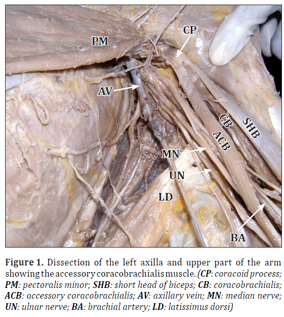
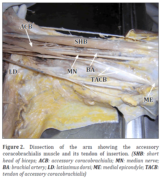
During routine dissections for medical undergraduate students, we observed the variation in an approximately 60-year-old male cadaver fixed in 10% formalin solution. The variation found was on the left limb and it was unilateral. There was an accessory coracobrachialis which took its origin from the coracoid process of scapula. At the origin, the muscle fused with the origin of pectoralis minor laterally and coracobrachialis and short head of biceps medially (Figure 1). The accessory muscle had a fleshy belly situated medial to the coracobrachialis and it crossed superficial to the median nerve and brachial artery from lateral to medial side (Figures 1, 2). In the middle of the arm, the fleshy part gradually became a tendon and the tendon partially fused with the medial intermuscular septum and finally got inserted to the medial epicondyle of humerus (Figure 2). The muscle was supplied by a branch of the musculocutaneous nerve. The musculocutaneous nerve in this case, did not pierce the coracobrachialis. It entered the arm between the coracobrachialis and the accessory coracobrachialis. Its further course and distribution in the arm were as usual.
Figure 1: Dissection of the left axilla and upper part of the arm showing the accessory coracobrachialis muscle. (CP: coracoid process; PM: pectoralis minor; SHB: short head of biceps; CB: coracobrachialis; ACB: accessory coracobrachialis; AV: axillary vein; MN: median nerve; UN: ulnar nerve; BA: brachial artery; LD: latissimus dorsi)
Discussion
The coracobrachialis muscle morphologically represents the adductor compartment of the arm. But its role as an adductor of the arm is insignificant in humans. In some mammals it is tricipital in origin. Upper two heads are fused to take origin from the coracoid process and enclose musculocutaneous nerve between them. The lower head is usually suppressed in man. In some cases it is represented by ligament of Struther which extends from an occasional bony projection called supratrochlear spur; from the anteromedial surface of the lower part of the humerus to the medial epicondyle. In such cases the median nerve and brachial artery pass deep to the ligament. The compression of these structures by the ligament may lead to vascular spasm and median nerve palsy [5]. Variations of the coracobrachialis are common [6,7]. The most common variation is the downward extension of its superficial part. It sometimes extends as far as medial epicondyle [7]. Coracobrachialis may have a third head called coracobrachialis brevis. It arises from the coracoid process and gets inserted to the capsule of the shoulder joint or crest of the lesser tubercle [6]. An accessory coracobrachialis has been reported by Kopuz et al. [8]. The muscle they observed originated from the coracoid process and had a fleshy belly that passed in front of the biceps muscle. It was inserted to the antebrachial fascia and the medial epicondyle. Entrapment of median nerve and brachial artery by a tendinous arch of coracobrachialis has been reported recently [9]. The muscle we observed had a sufficiently large fleshy belly that crossed the median nerve and brachial artery. This may result in the compression of these structures. This muscle can be used in muscle graft surgeries as it is an accessory muscle and its removal may not cause any functional problems. In high level median nerve palsies, the presence of accessory coracobrachialis cannot be ruled out. When the accessory coracobrachialis muscle is large, it may restrict the abduction of the arm also. It may also lead to confusions in MRI and CT scan evaluations.
References
- McMinn RMH, ed. Last’s Anatomy: Regional and Applied. 8th Ed., Edinburgh, Churchill Livingstone. 1990; 79.
- Williams PL, Warwick R, Dyson M, Bannister LH, eds. Gray’s Anatomy. 37th Ed., Edinburgh-London, Churchill Livingstone. 1989; 614–615.
- Wood J. On human muscular variations and their relation to comparative anatomy. J Anat Physiol. 1867; 1: 44–59.
- Howell AB, Straus WL. The brachial flexor muscles in primates. Proceedings of the United States National Museum. 1931; 80: 1–31.
- AK Datta. Essentials of Human Anatomy. 3rd Ed., Kolkata, Current Books International. 2004; 56–59.
- Beattie PH. Description of bilateral coracobrachialis brevis muscle, with a note on its significance. Anat Rec. 1947; 97: 123–126.
- Warner JJ, Paletta GA, Warren RF. Accessory head of the biceps brachii. Case report demonstrating clinical relevance. Clin Orthop Relat Res. 1992; 280: 179–181.
- Kopuz C, Icten N, Yildirim M. A rare accessory coracobrachialis muscle: a review of the literature. Surg Radiol Anat. 2003; 24: 406–410.
- Rodrigues V, Nayak S, Nagabhooshana S, Vollala VR. Median nerve and brachial artery entrapment in the tendinous arch of coracobrachialis muscle. Int J Anat Var (IJAV). 2008, 1: 28–29.
Naveen Kumar, Surekha D Shetty, Somayaji SN and Satheesha Nayak B*
Department of Anatomy, Melaka Manipal Medical College (Manipal Campus), International Centre for Health Sciences, Manipal University, Madhav Nagar, Manipal Karnataka State, India
- *Corresponding Author:
- Dr. Satheesha Nayak B
Professor and Head Department of Anatomy, MMMC Int. Centre for Health Sci. Manipal University, Madhav Nagar, Manipal, Udupi District Karnataka, 576 104, India
Tel: +91 820 2922519
E-mail: nayaksathish@yahoo.com
Date of Received: May 26th, 2011
Date of Accepted: April 16th, 2012
Published Online: July 11th, 2012
© Int J Anat Var (IJAV). 2012; 5: 27–28.
Abstract
Coracobrachialis muscle is a muscle of the arm and is known to show several variations in its attachments. During routine dissections, we found an accessory coracobrachialis muscle. It took origin from coracoid process of scapula and got inserted to the medial intermuscular septum and medial epicondyle of the humerus. It crossed the median nerve and brachial artery in the middle of the arm. This accessory muscle can be used in muscle transplants. Since it crosses the median nerve and brachial artery in the arm, compression of these structures by the muscle cannot be ruled out.
-Keywords
coracobrachialis, accessory coracobrachialis, variation, arm, medial epicondyle
Introduction
Coracobrachialis is a weak flexor muscle of the arm. It originates from the tip of the coracoid process of scapula together with the short head of biceps brachii. Its tendon is inserted to the middle of the medial border of humerus where the nutrient foramen is located. The musculocutaneous nerve usually pierces the muscle and supplies it [1,2]. This muscle is functionally not so important in the arm and is said to be a weak flexor and adductor of the arm.
Coracobrachialis is known to show variations [3,4]. The variations reported include the presence of accessory slips that get attached to medial epicondyle, medial supracondylar ridge, lesser tubercle and the medial intermuscular septum. We saw an accessory coracobrachialis muscle which is quite unique in its attachments and has not been reported yet.
Case Report
During routine dissections for medical undergraduate students, we observed the variation in an approximately 60-year-old male cadaver fixed in 10% formalin solution. The variation found was on the left limb and it was unilateral. There was an accessory coracobrachialis which took its origin from the coracoid process of scapula. At the origin, the muscle fused with the origin of pectoralis minor laterally and coracobrachialis and short head of biceps medially (Figure 1). The accessory muscle had a fleshy belly situated medial to the coracobrachialis and it crossed superficial to the median nerve and brachial artery from lateral to medial side (Figures 1, 2). In the middle of the arm, the fleshy part gradually became a tendon and the tendon partially fused with the medial intermuscular septum and finally got inserted to the medial epicondyle of humerus (Figure 2). The muscle was supplied by a branch of the musculocutaneous nerve. The musculocutaneous nerve in this case, did not pierce the coracobrachialis. It entered the arm between the coracobrachialis and the accessory coracobrachialis. Its further course and distribution in the arm were as usual.
Figure 1: Dissection of the left axilla and upper part of the arm showing the accessory coracobrachialis muscle. (CP: coracoid process; PM: pectoralis minor; SHB: short head of biceps; CB: coracobrachialis; ACB: accessory coracobrachialis; AV: axillary vein; MN: median nerve; UN: ulnar nerve; BA: brachial artery; LD: latissimus dorsi)
Discussion
The coracobrachialis muscle morphologically represents the adductor compartment of the arm. But its role as an adductor of the arm is insignificant in humans. In some mammals it is tricipital in origin. Upper two heads are fused to take origin from the coracoid process and enclose musculocutaneous nerve between them. The lower head is usually suppressed in man. In some cases it is represented by ligament of Struther which extends from an occasional bony projection called supratrochlear spur; from the anteromedial surface of the lower part of the humerus to the medial epicondyle. In such cases the median nerve and brachial artery pass deep to the ligament. The compression of these structures by the ligament may lead to vascular spasm and median nerve palsy [5]. Variations of the coracobrachialis are common [6,7]. The most common variation is the downward extension of its superficial part. It sometimes extends as far as medial epicondyle [7]. Coracobrachialis may have a third head called coracobrachialis brevis. It arises from the coracoid process and gets inserted to the capsule of the shoulder joint or crest of the lesser tubercle [6]. An accessory coracobrachialis has been reported by Kopuz et al. [8]. The muscle they observed originated from the coracoid process and had a fleshy belly that passed in front of the biceps muscle. It was inserted to the antebrachial fascia and the medial epicondyle. Entrapment of median nerve and brachial artery by a tendinous arch of coracobrachialis has been reported recently [9]. The muscle we observed had a sufficiently large fleshy belly that crossed the median nerve and brachial artery. This may result in the compression of these structures. This muscle can be used in muscle graft surgeries as it is an accessory muscle and its removal may not cause any functional problems. In high level median nerve palsies, the presence of accessory coracobrachialis cannot be ruled out. When the accessory coracobrachialis muscle is large, it may restrict the abduction of the arm also. It may also lead to confusions in MRI and CT scan evaluations.
References
- McMinn RMH, ed. Last’s Anatomy: Regional and Applied. 8th Ed., Edinburgh, Churchill Livingstone. 1990; 79.
- Williams PL, Warwick R, Dyson M, Bannister LH, eds. Gray’s Anatomy. 37th Ed., Edinburgh-London, Churchill Livingstone. 1989; 614–615.
- Wood J. On human muscular variations and their relation to comparative anatomy. J Anat Physiol. 1867; 1: 44–59.
- Howell AB, Straus WL. The brachial flexor muscles in primates. Proceedings of the United States National Museum. 1931; 80: 1–31.
- AK Datta. Essentials of Human Anatomy. 3rd Ed., Kolkata, Current Books International. 2004; 56–59.
- Beattie PH. Description of bilateral coracobrachialis brevis muscle, with a note on its significance. Anat Rec. 1947; 97: 123–126.
- Warner JJ, Paletta GA, Warren RF. Accessory head of the biceps brachii. Case report demonstrating clinical relevance. Clin Orthop Relat Res. 1992; 280: 179–181.
- Kopuz C, Icten N, Yildirim M. A rare accessory coracobrachialis muscle: a review of the literature. Surg Radiol Anat. 2003; 24: 406–410.
- Rodrigues V, Nayak S, Nagabhooshana S, Vollala VR. Median nerve and brachial artery entrapment in the tendinous arch of coracobrachialis muscle. Int J Anat Var (IJAV). 2008, 1: 28–29.


