Prevalence Summary for Five Aberrant Muscles of the Hand: A Literature Review and Clinical and Pedagogical Resource
Received: 07-Dec-2021, Manuscript No. ijav-21-4035; Editor assigned: 08-Jan-2022, Pre QC No. ijav-21-4035(PQ); Accepted Date: Dec 21, 2021; Reviewed: 25-Jan-2022 QC No. ijav-21-4035; Revised: 27-Mar-2022, Manuscript No. ijav-21-4035(R); Published: 04-Feb-2022, DOI: 10.37532/1308-4038.14(12).179
Citation: Smith LM. Prevalence Summary for Five Aberrant Muscles of the Hand: A Literature Review and Clinical and Pedagogical Resource. Int J Anat Var. 2021;14(12):146-149.
This open-access article is distributed under the terms of the Creative Commons Attribution Non-Commercial License (CC BY-NC) (http://creativecommons.org/licenses/by-nc/4.0/), which permits reuse, distribution and reproduction of the article, provided that the original work is properly cited and the reuse is restricted to noncommercial purposes. For commercial reuse, contact reprints@pulsus.com
Abstract
Being aware of muscle anomalies and the prevalence of these anomalies is critically important for surgeons but equally useful for anatomy instructors. The purpose of this study was to perform a literature review and then create a clinical and pedagogical resource that provides descriptions and prevalence values for aberrant muscles and tendons on the hand. Bilateral dissections of a cadaver hand revealed an anomalous extensor medii proprius muscles. Twenty journal articles were chosen from an interdisciplinary database. Molded clay muscles and ribbon tendons were added to hand bone models to create visual representations. Variations of the extensor indicis proprius have a collective prevalence rate of 96.5%. The variations of the extensor digitorum brevis manus have a collective prevalence rate of 4%, the extensor medii proprius 3.7%, extensor indicis et medii communis 1.6%, and the three variations of the extensor pollicis et indicis have the lowest collective prevalence rate of 0.75%.
Keywords
Anomalous hand muscles; Extensor medii proprius; Extensor digitorum brevis manus; Extensor indicis et medii communis; Extensor pollicis et indicis; Anatomical variations
Introduction
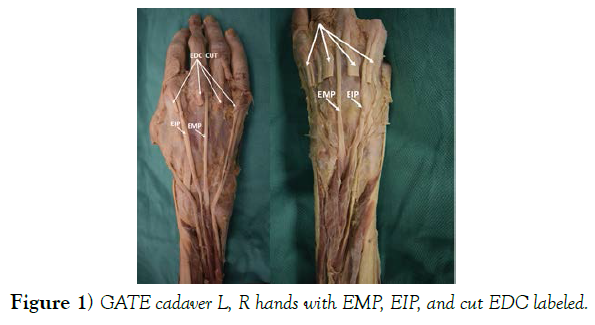
The muscle and tendon variations of the hand are not uncommon and certainly not a new phenomenon. The first description of the Extensor Digitorum Brevis Manus (EDBM). Current published research seems to emphasize having accurate knowledge of aberrant muscles and tendon variations for clinical reasons, for understanding donor tendon availability, for surgical planning, and for “Radiologists to better assess the structures under the Magnetic Resonance Imaging (MRI)”. This includes knowing those characteristics most important to surgeons for treating traumatized and diseased hands and for tendon grafting and transfer. Yet, these anomalous variations are not typically mentioned in textbooks or reference. The Gross Anatomy for Teacher Education (GATE) program at the University of Alabama at Birmingham (UAB) Department of Cell, Developmental, and Integrative Biology provides teachers/instructors with cadaver dissection experience that they may not otherwise have had in the academic career or job experience. In the July 2019 GATE experience one student group discovered an anomalous muscle in the hand. It was determined to be and extensor medii proprius (EMP) muscle (Figure 1). A question was raised as to the prevalence of this anomaly. A literature review did not reveal any one resource with cumulative prevalence and description information for all anomalous muscles and tendon variations of the hand. Having such a resource would be an asset for clinical and pedagogical purposes. The objective of this study was to perform a literature review and create a pedagogical resource that provides a list and description of aberrant muscles of the hand and their relative prevalence.
Materials and Methods
Bilateral dissections were performed on the cadaver hand. This was important because “the pattern observed in one hand is not necessarily that noted for the other hand”. The skin and superficial fascia were removed and the extensor digitorum communis (EDC) tendons were cut exposing the deep tendons. Twenty digital photographs were taken of both hands. Periodical investigation revealed many publications about the EMP and other anomalous muscles of the hand. However, no one article provided prevalence data for all five of the anomalous muscles.
Between September 2019 and January 2020 articles were collected from literature searches using a major university discovery system which included 890 interdisciplinary databases and typing in “Anomalous muscles of the hand” and by typing in various cited references titles. Twenty full text articles with copyright dates from 1991 to 2020 were selected. Articles were selected based on the following criteria [1]. It provides a description of an anomalous muscle and its tendons on the hand [2] it provides a description of an anomalous muscle and its tendons on the hand and discusses usefulness to clinical and/or pedagogical situations [3] it provides a description of an anomalous muscle and its tendons on the hand and provides prevalence values.
Photographs of the cadaver hands and of hand replicates, and prevalence tables were chosen as visual aids to enhance interpretations of the written anatomical location descriptions [4]. Two of the twenty photographs provided by Dr. Brooks of the cadaver hand were chosen and labeled using a photo paint software. Replicates of hands were created by molding red clay as muscles and white clay as the extensor retinaculum, using white ¼ inch ribbon cut in half as tendons, and then positioning these onto a handforearm bone model. Photographs were taken and then labeled using a photo paint software. Nearly all articles chosen provided some degree of prevalence information. However, stood out from the rest as having the most extensive and conclusive prevalence data for all five muscles. Furthermore, Yammine’s work incorporated values from many of the other articles chosen for this review. Yammine’s sample sizes were impressive. For the EI his sample size was 3,858 hands. For the EDBM his samples size was 5,989 hands. For the EIMC his samples size was 3,984 hands. For the EPI his sample size was 4,911 hands. Samples of this size can be considered representative for the prevalence estimates for this anomalous muscle in the greater human population. For this reason, prevalence values, presented as pooled prevalence estimates (PPE) provided by Yammine were used in the results of this literature review [5].
Some, but not all, of the literature reviewed addressed prevalence as it related to ancestry, gender, and right or left sides. Although worthy of representation, because this information was not inclusive of all five anomalous muscles it was not included in this review.
There is potential limitation and bias with this review because this author cannot concede that all possible relevant articles were discovered [6]. To reduce ambiguity of terms associated with naming of the fingers, the terms first, second, third, fourth, and fifth were used rather than thumb, index, middle, ring, and little. Although the term “thumb” is seldom called into question, in an effort to remain consistent the term “first” was used [7].
Discussion
The five most aberrant muscles and their tendons revealed by the literature review are: variations of the extensor indicis proprius (aEIP), the extensor digitorum brevis manus (EDBM), the extensor medii proprius (EMP), extensor indicis et medii communis (EIMC), and the extensor pollicis et indicis (EPI) [8].
The objective of this article was to provide a summary of the prevalence rates for five aberrant muscles of the hand. If you are a clinician preparing for surgery or patient consultation, or you are an educator teaching with cadavers, you may want to know the likelihood of seeing one or more of these muscles (Table 1) [9].
| Anomalous Muscle | PPE |
|---|---|
| aEIP | 96.5% |
| EDBM | 4.0% |
| EMP | 3.7% |
| EIMC | 1.6% |
| EPI | 0.75% |
TABLE 1 Summary PPEs for five anomalous muscles of the hand (values respectfully borrowed from Yammine).
EIP
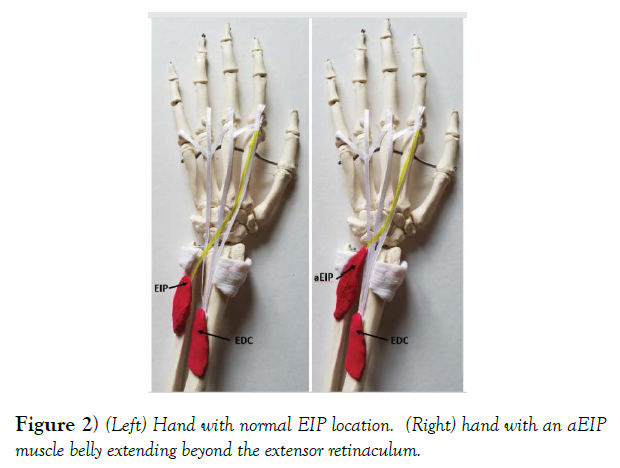
The EIP has an origin arising from the posterior surface of the distal ulna and below the extensor pollicis longus. This origin is similar to the EMP and suggests a common embryological origin. The insertion for the EIP is on the dorsal aspect of the second metacarpo-phalangeal joint. The EIP muscle has anomalies (aEIP) of muscle belly location (Figure 2) and with the number of slips (Table 2). Slip “as a separation of a tendon that forms two discrete insertions.” The prevalence for having an EIP is 96.5%. When it is present, 4.0% of the time the muscle belly will extend beyond the extensor retinaculum. If an EIP is present, 92.6% of the time it will have a single slip [10]. There are also three variations to the single slip EIP based on insertion location on the ulnar side, radial side, or palmar side of the second finger. Less likely to be seen are aEIP muscles having either double or triple slips. These have PPEs of 7.2% and 0.3% respectivel. According to some hand surgeons the EIP is “Considered best substitute for surgical reconstruction in hand surgery particularly of the abductor pollicis longus” other surgeons prefer to use the extensor pollicis longus. The EIP is also the most common tendon used in tendon transfer. Anomalous muscles are mostly asymptomatic except for the aEIP which has been associated with dorsal wrist pain, dorsal wrist ganglion, and a condition called extensor indicis proprius syndrome. Wrote about two variants to the EIP, the extensor indicis radialis and the extensor indicis ulnaris, but because he found the origins of the muscle bellies to be fused with that of the EIP, suggests that these tendons are a “Result of splitting rather than variants [11].”
| PPE | Single slip | Double Slip | Triple slip | |
|---|---|---|---|---|
| (normal location) | (aEIP) | (aEIP) | ||
| EIP | 96.5% | 92.6% | 7.2% | 0.3% |
TABLE 2 PPEs for the presence of an EIP and for anomalous EIP slips.
EDBM
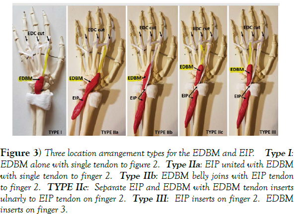
The origin of the EDBM is near the lunate bone and frequently as far proximal as the distal margin of the radius, but without direct attachment to the carpal bones”. Insertion is either on the second or third finger. The EDBM has several variations classified into three types based on their insertions and proximity to the EIP (Figure 3). Type I EDBM inserts as the EIP typically would on the ulnar side of the dorsal expansion of the second finger. No EIP is present with Type I. Type II EDBM and an EIP insert on the second finger. There are three subtypes of Type II. For Type IIa the EIP arises from the ulna but joins with the EDBM belly which inserts on the index finger. In type IIb, the distal end of the EDBM belly joins the EIP tendon. In type IIc, the EIP inserts normally, but the EDBM tendon inserts more ulnarly than the EIP tendon, often with a membranous accessory slip inserting on the third finger. “In type III, the EIP inserts on the index finger [second finger], but the EDBM inserts on the middle [third finger] finger with or without an accessory EIP to the middle finger”. For type III, insertions to the fourth and fifth fingers have been reported but none to the first finger. The EDBM has a crude overall cadaveric prevalence of 4.0% (Table 3). It was found to insert more often on the index finger (Type I and II 77%) than on the middle finger (Type III 23%). If the subtypes of Type II are considered individually then the highest PPE is with Type I (31%) [12].
Figure 3) Three location arrangement types for the EDBM and EIP. Type I:
EDBM alone with single tendon to figure 2. Type IIa: EIP united with EDBM
with single tendon to finger 2. Type IIb: EDBM belly joins with EIP tendon
to finger 2. TYPE IIc: Separate EIP and EDBM with EDBM tendon inserts
ulnarly to EIP tendon on finger 2. Type III: EIP inserts on finger 2. EDBM
inserts on finger 3.
| PPE | Type I | Type II a | Type II b | Typed II c | Type III | |
|---|---|---|---|---|---|---|
| EDBM | 4.00% | 31.00% | 4.60% | 15.00% | 26.40% | 23% |
TABLE 3) PPEs for three location arrangements (Types) of the EDBM
EMP
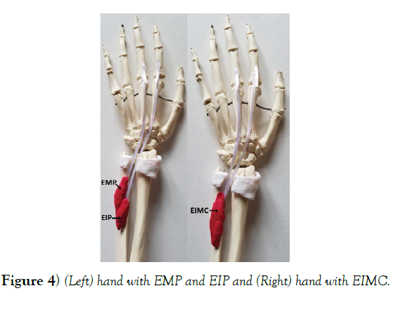
The EMP, as seen in the GATE cadaver, arises from the posterior surface of the distal ulna and interosseous membrane, and passes distally deep to the extensor digitorum communis (EDC) tendon and inserting on the dorsal expansion of the proximal phalanx of the third finger. The EMP muscle has a pooled prevalence estimate (PPE) of 3.7% and is most often accompanied by the EIP (Figure 4). In some cases, if the EIP tendon is doubled having one slip to the second finger and another slip passing to the third finger then the EMP is considered a “differentiated portion of the EIP”. Klena suspects that the EMP has been underreported due to its location under the extensor digitorum communis tendon and a more proximal insertion [13].
EIMC
The EIMC has an origin from the posterior surface of the distal ulna similar as the EIP. An EIMC is essentially an EMP and EIP sharing one belly. Tan describe it as being like an EIP muscle with its tendon that splits to insert into both the second and third fingers. von Schroeder and Botte (1991) suggest that the EIMC is an anomaly of the EIP. The EIMC has a PPE of 1.6%. There are no cases in their study in which the EMP or the EIMC split and inserted on the fourth and fifth fingers [14].
EPI
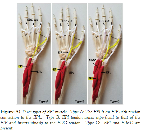
Fingers The EPI, similar as the EIP, has an origin from the dorsal ulna, medial and below the extensor pollicis longus (EPL), or it can have an origin located between the EIP and EPL. As its name implies, insertion in on the first (medial side) and second fingers (radial dorsum side). Some authors say that the EIP, EIMC, EMP, and EPI are variants of the EI. Yoshida found “that both the EPL and the EIP muscles were always present in all upper limbs in which the EPI was identified; therefore, this muscle might not be considered as a variant of the EI.” Recorded three types of EPI (Figure 5) Type A is a tendon connection from the EIP to the EPL. Type B is a tendon arising from the EIP belly and for Type C the EPI is present along with the EIMC. The EPI is rare in humans. It has a PPE of 0.75%. If it is present, it may cause difficulty for thumb adduction. It is more common in dogs, fox, wolves, jackals, panthers, and dingoes [15].
The EIP and EMP having common origins and the occurrence of the EIMC suggests a single embryological origin. The EMP and EIMC are frequently found in Old World Monkeys but are variable in humans. The EIP is constant in chimpanzees and gorillas [16].
Knowledge of anomalous muscles may not be as critical to anatomy and physiology instructors as it is for the professionals in the clinical and surgical arenas. Nevertheless, an instructor may find themselves addressing a student asking about a muscle that is certainly in the cadaver in front of them but not in the available muscle atlas. The literature review represented in this article fulfills a need for one resource containing descriptions and prevalence values for the five commonly encountered anomalous muscles and tendons of the hand.
Acknowledgement
Respect and thanks are extended to Doctors John P. Dupaix and MaryBeth Cermak, hand surgeons at Hamot Medical Center in Erie Pennsylvania, for taking time out of their busy schedules to review and make comments about my manuscript. Dr. Cermak also asked permission to provide the article to her residences.
Great appreciation is extended to the donor families of the cadavers at the University of Alabama at Birmingham.
Much gratitude is extended to William Brooke PhD at the University of Alabama at Birmingham Department of Cell, Developmental, and Integrative Biology for taking the digital photographs, answering many questions via email, and for his encouragement to proceed with this project.
Many thanks to the instructors and teaching assistants at the GATE program at the University of Alabama at Birmingham Department of Cell, Developmental, and Integrative Biology.
FUNDING: The research received no external funding.
CONFLICTS OF INTEREST: The author declares no conflict of interest.
ETHICS APPROVAL: Not applicable.
CONSENT TO PARTICIPATE: Not applicable.
CONSENT FOR PUBLICATION: Not applicable.
AVAILAIBILITY OF DATA AND MATERIAL: Not applicable.
CODE AVAILAIBILITY: Not applicable.
AUTHOR’S CONTRIBUTION: Only one author.
REFERENCES
- Dupaix J. Orthopedic Surgeon of the Hand and Upper Extremity. Hamot Medical Center, Erie Pennsylvania. J Hand Surg. 2017;52(4):76-77.
- Gonzalez S, Mark H. MD. Anatomy of the extensor tendons to the index finger. J Hand Surg. 1996;21: 988 – 991.
- Gray R, Henry. Gray’s Anatomy. Fifteenth Edition. Barnes & Noble Books, Inc. 1995.
- Klena S, Joel C, Riehl E. Anomalous extensor tendons to the long finger: A cadaveric study of incidence. J Hand Surg 2012;37: 938-941.
- Komiyama M, NWE TM, Toyota. Variations of the extensor indicis muscle and tendon. J Hand Surg. 1999;24:575-578.
- Li Jing R, Zhen Feng. Bilateral exntensor medii proprius with split tendon of extensor indicis proprius, a rare anatomical variant. Rom J Morphol Embryol. 2013;54(3): 639-641.
- Marieb H, Elaine N. Human Anatomy & Physiology. Tenth Edition. Pearson Education, Inc. USA. 2016.
- Netter D, Frank H. Atlas of Human Anatomy. Third Edition. ICON Learning Systems, LLL a subsidiary of MediMedia USA, Inc. 2003.
- Suwannakhan A, Tawonsawatruk T. Extensor tendons and variations of the medial four digits of hand: a cadaveric study. Surg Radiol Anat. 2016;38:1083-1093.
- Tan T, Swee M. Anomalous extensor muscles of the hand: A review. J Hand Surg. 1999;24A(3): 449-455.
- Von S, Herbert P. The extensor medii proprius and anomalous extensor tendons to the long finger. J Hand Surg. 1991;16A (6): 1141-1145.
- Yammine K. Evidence- based anatomy. Clin Anat. 2014;27:847–852.
- Yammine K. The prevalence of the extensor digitorum brevis manus and its variants in humans: a systematic review and meta-analysis. Surg Radiol Anat. 2015;37:3-9.
- Yammine K. The Prevalence of the extensor indicis tendon and its variants: a systematic review and meta-analysis. Surg Radiol Anat 2015 37:247-254.
- Yoshida Y. Anatomical studies on the extensor pollicis et indicis accessories muscle and the extensor indicis radialis muscle in Japanese. Okajimas Folia Anat J. 1990;71:355-363.
- Zilber V. Sebastien MD. Anatomical variations of the extensor tendons to the fingers over the dorsum of the hand: A study of 50 hands and a review of the Literature. Plast Reconst Surg. 2004; 3:245-246.
Google Scholar, Crossref, Indexed at
Google Scholar, Crossref, Indexed at
Google Scholar, Crossref, Indexed at
Google Scholar, Crossref, Indexed at
Google Scholar, Crossref, Indexed at
Google Scholar, Crossref, Indexed at
Google Scholar, Crossref, Indexed at
Google Scholar, Crossref, Indexed at
Google Scholar, Crossref, Indexed at
Google Scholar, Crossref, Indexed at
Google Scholar, Crossref, Indexed at


