Rarely encountered aneurysm of ophthalmic artery in cadaver – a case report
Kishore Kumar Agarwal*, Alok Saxena, Amal Rani Das and Sudhakar
Department of Anatomy, Veer Chandra Singh Garhwali Government Medical Sciences and Research Institute, Srinagar, Uttarakhand, India
- *Corresponding Author:
- Dr. Kishore Kumar Agarwal, MBBS, MS (Anatomy)
Ganga Vihar, Gauja Jali, Bichli Bareilley Road, Haldwani Distt-Nainital, Uttarakhand, India
Tel: +91 999 7973377
E-mail: drkkanatomy@gmail.com
Date of Received: May 20th, 2011
Date of Accepted: September 16th, 2011
Published Online: November 21st 2011
© Int J Anat Var (IJAV). 2011; 4: 180–181.
[ft_below_content] =>Keywords
optic canal, optic nerve, left anterior clinoid process
Introduction
Ophthalmic artery is the branch of internal carotid artery as it emerges from the cavernous sinus medial to anterior clinoid process. Thereafter it traverses in optic canal to enter in the orbit, lies inferolaterally to the optic nerve. It continues forward over the optic nerve (80%) and below the nerve (20%). Main trunk of the artery ends by dividing in to supratrochlear and dorsal nasal branch [1].
Intracranial and intracanalicular part of ophthalmic artery is more prone for the diseases and injuries. Therefore it becomes necessary to have knowledge about its anatomy, relations with the surrounding structures, variations and pathology [2]. One of the pathological conditions is carotid-ophthalmic aneurysm. Finding of aneurysm at the commencement of ophthalmic artery is rare. Location of this type of aneurysm may hinder the diagnostic as well as surgical procedures because of its close proximity with cavernous sinus, carotid siphon and optic nerve. Angiography is the diagnostic tool for carotid-ophthalmic aneurysm [3]. An insight into such particular type of aneurysm might prove fruitful towards neurosurgeries with minimal incidence of injury to surrounding structures.
Case Report
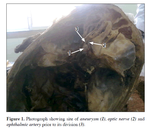
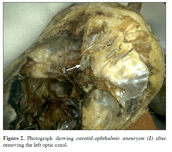
During routine dissection a dilatation was incidentally observed unilaterally in one male cadaver at the beginning of left ophthalmic artery after carefully removing the anterior clinoid process and frontal plate of orbit. Course of the ophthalmic artery was traced and noticed that this artery having lateral relation with optic nerve in the optic canal. Thereafter this artery gave one lateral branch running to the lacrimal gland and one medial branch crossing the optic nerve. We enclosed photographs to define the size and site of aneurysm and course of artery (Figures 1, 2). Morphometric analysis of aneurysm was taken in to consideration, which included the size, shape and location of aforesaid aneurysm. An oval dilatation of 21 mm diameter was measured using caliper.
Discussion
There are various schools of thoughts on the sites of this aneurysm. As per the previous literatures, Drake et al. suggested the site of aneurysm at the commencement of the ophthalmic artery on the basis of angiography [4]. Study conducted by the Kothandaram et al. propounds the presence of dilatation between the ophthalmic artery and internal carotid artery at its division [5]. Observations recorded by Guidetti and La Torre revealed the locus of this dilatation superior to the cavernous sinus and inferior to the origin of posterior communicating artery [3].
Findings of present report were consistent with Drake et al., in terms of position of aneurysm but a remarkable difference was noticed with the findings of Kothandaram et al. and Guidetti et al.
Females are preponderant to the carotid-ophthalmic aneurysm, more frequent on left side [3]. We found an aneurysm on the left side as mentioned by aforesaid author but in opposite gender (in male cadaver).
There is a high risk of rupturing of the aneurysm. We did not encounter this situation in present report probably due to its presence within the bony canal, which prevented its expansion. We presumed this aneurysm might cause the compression of optic nerve leading to the visual impairment.
Knowledge of types of aneurysms and their morphometry may be a crucial milestone for neurosurgeons to perform safe surgery with limited injury to related structures.
Acknowledgement
We acknowledge all the staff members of anatomy department as well Mr. Rakesh Singh Negi, technical staff of this department for his cooperation in completion of this study.
References
- Standering S, ed. Gray’s Anatomy. 40th Ed., London, Churchill Livingstone. 2005; 665.
- Erdogmus S, Govsa F. Anatomic features of the intracranial and intracanalicular portions of ophthalmic artery: for the surgical procedures. Neurosurg Rev. 2006; 29: 213–218.
- Sengupta RP, Gryspeerdt GL, Hankinson J. Carotid-ophthalmic aneurysms. J Neurol Neurosurg Psychiatry. 1976; 39: 837–853.
- Drake CG, Vanderlinden RG, Amacher AL. Carotid-opthalmic aneurysms. J Neurosurg. 1968; 29: 24–31
- Kothandaram P, Dawson BH, Kruyt RC. Carotid-ophthalmic aneurysms. A study of 19 patients. J Neurosurg. 1971; 34: 544–548.
Kishore Kumar Agarwal*, Alok Saxena, Amal Rani Das and Sudhakar
Department of Anatomy, Veer Chandra Singh Garhwali Government Medical Sciences and Research Institute, Srinagar, Uttarakhand, India
- *Corresponding Author:
- Dr. Kishore Kumar Agarwal, MBBS, MS (Anatomy)
Ganga Vihar, Gauja Jali, Bichli Bareilley Road, Haldwani Distt-Nainital, Uttarakhand, India
Tel: +91 999 7973377
E-mail: drkkanatomy@gmail.com
Date of Received: May 20th, 2011
Date of Accepted: September 16th, 2011
Published Online: November 21st 2011
© Int J Anat Var (IJAV). 2011; 4: 180–181.
Abstract
An incidental finding of ophthalmic artery aneurysm was noticed during routine dissection of cadavers in Anatomy department of Veer Chandra Singh Garhwali Government Medical Sciences and Research Institute, Srinagar, Uttarakhand. A sacculated oval dilatation was observed on the left side of floor of optic canal while breaking the roof of aforementioned canal. Manifestations were made on the basis of size of swelling, source, location and structures related. We also assumed the possible symptoms caused by aneurysm/swelling on the basis of anatomical relation.
-Keywords
optic canal, optic nerve, left anterior clinoid process
Introduction
Ophthalmic artery is the branch of internal carotid artery as it emerges from the cavernous sinus medial to anterior clinoid process. Thereafter it traverses in optic canal to enter in the orbit, lies inferolaterally to the optic nerve. It continues forward over the optic nerve (80%) and below the nerve (20%). Main trunk of the artery ends by dividing in to supratrochlear and dorsal nasal branch [1].
Intracranial and intracanalicular part of ophthalmic artery is more prone for the diseases and injuries. Therefore it becomes necessary to have knowledge about its anatomy, relations with the surrounding structures, variations and pathology [2]. One of the pathological conditions is carotid-ophthalmic aneurysm. Finding of aneurysm at the commencement of ophthalmic artery is rare. Location of this type of aneurysm may hinder the diagnostic as well as surgical procedures because of its close proximity with cavernous sinus, carotid siphon and optic nerve. Angiography is the diagnostic tool for carotid-ophthalmic aneurysm [3]. An insight into such particular type of aneurysm might prove fruitful towards neurosurgeries with minimal incidence of injury to surrounding structures.
Case Report
During routine dissection a dilatation was incidentally observed unilaterally in one male cadaver at the beginning of left ophthalmic artery after carefully removing the anterior clinoid process and frontal plate of orbit. Course of the ophthalmic artery was traced and noticed that this artery having lateral relation with optic nerve in the optic canal. Thereafter this artery gave one lateral branch running to the lacrimal gland and one medial branch crossing the optic nerve. We enclosed photographs to define the size and site of aneurysm and course of artery (Figures 1, 2). Morphometric analysis of aneurysm was taken in to consideration, which included the size, shape and location of aforesaid aneurysm. An oval dilatation of 21 mm diameter was measured using caliper.
Figure 2: Photograph showing carotid-ophthalmic aneurysm (1) after removing the left optic canal.
Discussion
There are various schools of thoughts on the sites of this aneurysm. As per the previous literatures, Drake et al. suggested the site of aneurysm at the commencement of the ophthalmic artery on the basis of angiography [4]. Study conducted by the Kothandaram et al. propounds the presence of dilatation between the ophthalmic artery and internal carotid artery at its division [5]. Observations recorded by Guidetti and La Torre revealed the locus of this dilatation superior to the cavernous sinus and inferior to the origin of posterior communicating artery [3].
Findings of present report were consistent with Drake et al., in terms of position of aneurysm but a remarkable difference was noticed with the findings of Kothandaram et al. and Guidetti et al.
Females are preponderant to the carotid-ophthalmic aneurysm, more frequent on left side [3]. We found an aneurysm on the left side as mentioned by aforesaid author but in opposite gender (in male cadaver).
There is a high risk of rupturing of the aneurysm. We did not encounter this situation in present report probably due to its presence within the bony canal, which prevented its expansion. We presumed this aneurysm might cause the compression of optic nerve leading to the visual impairment.
Knowledge of types of aneurysms and their morphometry may be a crucial milestone for neurosurgeons to perform safe surgery with limited injury to related structures.
Acknowledgement
We acknowledge all the staff members of anatomy department as well Mr. Rakesh Singh Negi, technical staff of this department for his cooperation in completion of this study.
References
- Standering S, ed. Gray’s Anatomy. 40th Ed., London, Churchill Livingstone. 2005; 665.
- Erdogmus S, Govsa F. Anatomic features of the intracranial and intracanalicular portions of ophthalmic artery: for the surgical procedures. Neurosurg Rev. 2006; 29: 213–218.
- Sengupta RP, Gryspeerdt GL, Hankinson J. Carotid-ophthalmic aneurysms. J Neurol Neurosurg Psychiatry. 1976; 39: 837–853.
- Drake CG, Vanderlinden RG, Amacher AL. Carotid-opthalmic aneurysms. J Neurosurg. 1968; 29: 24–31
- Kothandaram P, Dawson BH, Kruyt RC. Carotid-ophthalmic aneurysms. A study of 19 patients. J Neurosurg. 1971; 34: 544–548.


