Renal artery origins, destinations and variations: Cadaveric study in Ethiopian population
2 Department of Human Anatomy, College of Medicine and Health Sciences, University of Gondar, Ethiopia, Email: ayanaw.worku@gmail.com
3 Department of Neuroscience (PhD), College of Medicine and Health Sciences, University of Gondar, Ethiopia
Received: 06-Dec-2017 Accepted Date: Dec 22, 2017; Published: 02-Jan-2018, DOI: 10.37532/1308-4038.18.11.001
Citation: Animaw Z, Worku A, Muche A. Renal artery origins, destinations and variations: Cadaveric study in ethiopian population. Int J Anat Var. 2018;11(1):001-003.
This open-access article is distributed under the terms of the Creative Commons Attribution Non-Commercial License (CC BY-NC) (http://creativecommons.org/licenses/by-nc/4.0/), which permits reuse, distribution and reproduction of the article, provided that the original work is properly cited and the reuse is restricted to noncommercial purposes. For commercial reuse, contact reprints@pulsus.com
Abstract
Introduction: Renal arteries are paired end arteries branched from abdominal aorta at the level of first lumbar vertebra (L1) and second lumbar vertebra (L2), which consumes around 20% of the cardiac output. Due to embryological and racial reasons variation in distribution of the renal artery is common. The aim of the present study is to describe the origin, distribution and possible variations of renal arteries.
Methods and materials: Observational based study was conducted by using 30 cadavers (60 sides) obtained from the Department of Human Anatomy of six Ethiopian Medical Schools. Data analysis was conducted using thematic approaches.
Results: Forty-seven percent of the main renal artery originates at the level of L1 from the abdominal aorta. Of the 10 (33.3%) accessory renal arteries, 7(63.63%) and 3 (27.27%) were identified on the left and right sides respectively.
Conclusions: The variations of the main renal arteries mainly revolved around the level of origin and manifested prehilar branching pattern while entering into the kidney parenchyma. Another commonly observed variation was the existence of an accessory renal artery in different patterns. Hence, knowledge of renal vascular pattern and possible variations is very important in clinical and surgical procedures mainly during renal transplantation and other vascular interventions.
Keywords
Main renal artery; Abdominal aorta; Variatio; Accessory renal artery
Introduction
Renal arteries are paired end arteries branched from abdominal aorta. Between the 6th and 9th week of embryonic period, the kidneys ascend from the pelvis to the retroperitoneum. During their path they obtain blood supply from temporary arteries which arise from the abdominal aorta, but these arteries will be degenerated and replaced by new arteries till the final paired definitive renal arteries are established. When these transient arteries failed to degenerate and remain persistent, they will appear as accessory renal arteries [1]. These accessory renal arteries are also called aberrant renal arteries. They may cross the corresponding ureter causing obstruction of urine flow [2].
Renal arteries consume around 20% of the total cardiac output. Each of these arteries divided into anterior and posterior divisions at the hilum of the kidney and further into segmental arteries supplying the renal vascular segments [3].
There are variations regarding renal artery number, origin and distribution. Among these accessory renal arteries are the commonest, presumed to be found 30% of the time [4]. These accessory renal arteries can also be classified as superior polar, hilar and inferior polar based on their point of entry into the kidney parenchyma [4].
A study done in Greek revealed that renal vasculature is highly dependent on individual’s racial origin [5]. Therefore, due to the current advanced renal vascular surgery, including renal transplantation, it is important to state the vascular distribution and variation of renal arteries among different population.
Several studies have been reported racial differences in the anatomy of the main renal and accessory renal arteries among different ethnic groups in different populations throughout the globe. However, to the best of our knowledge, no study was conducted in Ethiopia. Hence, the aim of this study is to describe the origin, distribution and possible variations of renal arteries among Ethiopian cadavers.
Materials and Methods
Study design and study area: The observational study design was conducted to assess the anatomical variation in origin and distribution of the renal artery on 30 (60 sides) Ethiopian cadavers in the Department of Human Anatomy dissection rooms at the University of Gondar, Bahir Dar University, Debre Tabor University, Debre Markos University, St. Paul’s Medical School and Jimma University. The study was conducted from September 2016 to June 2017.
Material and methods: A total of 30 cadavers were obtained from the Department of Human Anatomy of University of Gondar, Bahir Dar University, Debre Tabor University, Debre Markos University, St. Paul’s Medical School and Jimma University. The cadavers were embalmed according to conventional method using formalin, alcohol, glycerin and phenol for routine dissection sessions.
During routine abdominal dissection course conducted for human anatomy postgraduate and undergraduate medical students, the abdominal viscera except kidneys were removed and preserved for teaching purpose. Then, the kidneys and the branches of abdominal aorta were exposed. All the branches of the abdominal aorta were identified. The branch of abdominal aorta with larger caliber that directed to the kidneys named as renal arteries. However, the arteries that enter into the kidney parenchyma with smaller caliber (3 mm–5 mm) were accessory renal arteries. Then, the anatomy of main and accessory renal arteries including their origin and distribution were photographed with a Canon (Melville, NY, USA) digital camera with a resolution of 16 mega pixels and well noted accordingly. The origin of main renal arteries with respect to the lumbar vertebrae and number of accessory renal arteries were presented as frequency and percentage.
Ethical considerations: Ethical clearance was obtained from the School of Medicine Ethical Review Committee of University of Gondar. Dead human bodies “cadavers” were prepared for teaching purposes in the Departments of Human Anatomy. An official letter was submitted to the University of Gondar, Bahir Dar University, Debre Tabor University, Debre Markos University, St. Paul’s Medical School, and Jimma University to obtain permission of access to examine the cadavers. Confidentiality was strictly maintained at all levels of the study.
Results
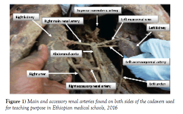
In the present study a total of 30 cadavers, 25 (83.3%) male and 5 (16.7%) female cadavers were observed. The total number of renal arteries examined, including accessory renal arteries were 73 of which 33 (45.21%) were on the right side and 40 (54.79%) on the left side, originating from abdominal aorta. Among the total 30 cadavers, 10 (33.33%) of the cases had accessory renal arteries in 7 (63.63%) on the left side and 3 (27.27%) on the right side (Table 1 and Figure 1).
| Number of accessory renal arteries | Right side | Left side | Total |
|---|---|---|---|
| One | 3 (27.27%) | 4 (36.36%) | 7 (63.63%) |
| Two | _____ | 3 (27.27%) | 3 (27.27%) |
| Total | 3 (27.27%) | 7 (63.63%) | 10 (100%) |
Table 1: Number and percentage of accessory renal arteries found on both sides among cadavers used for teaching purpose in Ethiopian medical schools, 2016
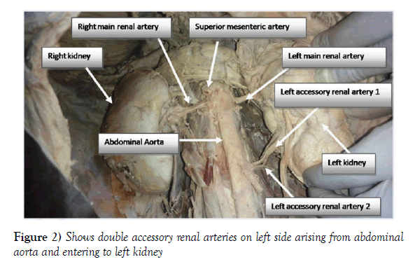
The accessory renal arteries were found in 10 cadavers. Of these, bilateral accessory renal artery was found in a single case (Figure 1). The remaining cases possess unilateral accessory renal arteries (Table 1). A single accessory renal artery was observed in 7 (63.63%) of the cases, while double accessory renal arteries were identified in 3 (27.27%) of the case (Table 1 and Figure 2).
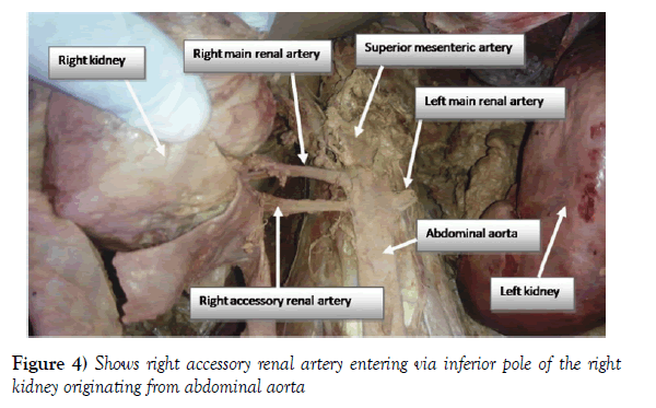
Among the total 10 accessory renal arteries, 8 (80%) of them enter into the kidney parenchyma in the inferior pole of the kidney while 2 (20%) of accessory renal arteries enter through the hilum of the kidney (Figures 3 and 4).
All accessory renal arteries were originated from the abdominal aorta. The average distance of accessory renal arteries from the superior mesenteric artery is 3.5 cm.
The main renal artery originated at the level of L1, L1-L2 and L2 in 46.7%, 43.3% and 10.0% of the cases, respectively (Table 2). The average distance of the right and left renal artery from the superior mesenteric artery was 0.6 cm and 0.7 cm, respectively. In 23 cases the main renal artery arose at the same level of superior mesenteric artery. However, in 16.7% and 6.7% of the study subjects the right main renal arteries originated higher than the left main renal arteries and vice versa, respectively.
| Vertebral level | Right side | Left side | Total |
|---|---|---|---|
| L1 | 13 (46.4%) | 15 (53.6%) | 28 (46.7% a) |
| L1-L2 | 15 (57.7%) | 11 (42.3%) | 26 (43.3% a) |
| L2 | 2 (33.3%) | 4 (66.7%) | 6 (10.0% a) |
Table 2: Origin of main renal artery with respect to the lumbar (L) vertebral level among cadavers used for teaching purpose in Ethiopian medical schools, 2016
Discussion
Even though the majority of renal artery variations are due to the presence of accessory renal arteries, the main renal arteries also have variation vis-à-vis their level of origin and pattern to enter into the renal parenchyma [5-7].
Our study showed that the main renal arteries were mainly arisen from the abdominal aorta. Similarly, studies conducted elsewhere support the notion that main renal arteries have had an abdominal aortic origin [6-8]. However, computed tomography angiogram and CT angiography studies reported common iliac artery as the origin of the main renal arteries [7,9].
In addition, the origin of main renal arteries had variation at the level of lumbar vertebrae. Consequently, the right main renal arteries originated at the level of L1, L1-L2 and L2 in 46.4%, 57.7% and 33.3% of the cases, respectively. The left main renal arteries on the other hand originated at the level of L1 in 53.6% of subjects, L1-L2 in 42.3% of the subjects and L2 in about 66.7% the subjects. However, a clinical oriented textbook clearly stated common level of origin of the main renal arteries at the level of L1-L2 [10]. A recent cadaver based study reported maximum incidence of origin of the main renal artery at the level of L1 (right side - 86%; left side - 80%) [8]. Conversely, Bhandari et al., reported that the right renal artery had L1, L1-L2 and L2 origin in 77.5%, 19.67% and 2.28% of the cases respectively. But, on the left side the main renal arteries have only L1 and L1-L2 level of origin in 36.76% and 44.2% of the cases, respectively [11].
The distance of the main renal artery from the superior mesenteric artery can be an option to demonstrate its variation. Various findings support the notion of right and left main renal arteries variability at site of their origin [8,11-13]. According to our findings, in 76.67% of the study subjects the origin of the right and left main renal arteries was at the same level. However, the right main renal artery is higher than the left and the left is higher than the right in about 16.7% and 6.7% of the subjects, respectively. Emerging evidence reported that right main renal arteries originated from abdominal aorta superior to the left main renal artery [8,11-13]. A study conducted in India also revealed that the right main renal artery originated at the same level of origin to left renal artery (26.7%), superior to the left (63.33%) and left higher than the right one (10%) [12].
In the present study, all main renal arteries are manifested by prehilar branching pattern while entering into the kidney parenchyma. On the other hand, a study done in India revealed that hilar and pre-hilar branch patterns of main renal arteries in 88.33% and 11.67% of the cases [12].
Our results indicated that 33.33% of the cadavers had accessory renal arteries. Computed tomographic and cadaveric based studies reported that the prevalence of accessory renal artery might be ranged from 13.3% up to 36.1% [3,12,14]. Besides study conducted from Greece population reported that 27.4% of the cases had accessory renal artery [5]. This variation might be due to contextual factors including race, study type and sample size.
About sixty four percent of accessory renal arteries in this study were found on the left side while the rest 27.27% were on the right side. Similarly, computed tomography angiogram studies claimed that left side was the common site for the occurrence of accessory renal arteries [3,4,7]. However, reports from Brazil and India revealed that in most of the cases accessory renal arteries appeared on right side [15-17].
Study conducted in Colombian mestizo population reported that all accessory renal arteries originated from the abdominal aorta [4]. Similarly, our finding identified abdominal aorta as sole origin to the accessory renal arteries. Although the commonest origin to accessory renal arteries is the abdominal aorta, there are other rare extra aortic origins identified in different literatures, such as common iliac, inferior mesenteric, main renal artery and celiac trunk [3,12,18].
As far as this study is concerned, 3 (27.27%) cases had double accessory renal arteries on the left side. The rest 7 (63.63%) were single arteries found either on the right side 3 (27.27%) or left side 4 (36.36%). Angiographic evaluation of renal artery variation amongst Greeks identified one, two and three accessory renal arteries in 21.4%, 5.6% and 0.4% of the examined cases, respectively [5]. These results, therefore, indicate the proportion of accessory renal arteries decreases as the number of accessory renal arteries increases.
Our findings revealed the occurrence of bilateral accessory renal artery in a single case and the rest have unilateral (Figure 1 and Table 1). Population and cadaveric based studies reported the incidence of unilateral 11.6% (4) and 24.2% (12) and bilateral 6.67% (4) and 7.7% (12) accessory renal arteries. Of note, the mentioned findings declared left unilateral dominance over the bilateral accessory renal arteries.
Like the main renal arteries, accessory renal arteries observed in the present study also possess different path to enter into the kidney. Among accessory renal arteries 8 (80%) of them are inferior polar arteries. There are only two accessory renal arteries which entered into the kidney through the hilum. Our findings are in line with reports from Colombia and Trinidad and Tobago where the majority of accessory renal arteries enter through the inferior pole of the kidney [3,4]. This is due to the fact that kidneys ascend from the pelvic region during embryonic life; the most likely accessory renal arteries which fail to degenerate and persist will remain in the lower pole of the kidney [12,18]. Nonetheless, there is a uniquely stated report from India that concludes the superior polar accessory renal arteries are the most frequently found type [15].
In conclusion, the variations of the main renal arteries mainly revolved around the level of origin and manifested prehilar branching pattern while entering into the kidney parenchyma. Another commonly observed variation was the existence of an accessory renal artery in different patterns. Hence, prior knowledge of renal vascular pattern and possible variations is very important in clinical and surgical procedures mainly during renal transplantation and other vascular interventions. Our study will open up avenues for further research in the field in Ethiopia.
Acknowledgement
The authors would like to extend their deepest appreciation to the staff members of Department of Human Anatomy in University of Gondar, Bahir Dar University, Debre Tabor University, Debre Markos University, St. Paul’s Medical School and Jimma University for their unreserved cooperation. Zelalem Animaw acknowledges University of Gondar and Debre Tabor University for providing financial support.
REFERENCES
- Moore K, Persaud T. The developing human. Saunders. 2003;P: 250.
- Hegazy A. Clinical embryology for medical students and postgraduate doctors. Lambert Academic Publishing, Berlin. 2014.
- Johnson PB, Cawich SO, Shah SD, et al. Accessory renal arteries in a Caribbean population: a computed tomography based study. Springer plus. 2013;2:443.
- Saldarriaga B, Perez AF, Ballesteros LE. A direct anatomical study of additional renal arteries in a Colombian mestizo population. Folia Morphol (Warsz). 2008;67:129-34.
- Papaloucas C, Fiska A, Pistevou-Gombaki K, et al. Angiographic evaluation of renal artery variation amongst Greeks. Aristotle Univ Med J. 2007;34:43-7.
- Saritha S, Jyothi N, Kumar MP, et al. Cadaveric study of accessory renal arteries and its surgical correlation. Int J Med Sci. 2013;1:19-22.
- Sasikala P, Sulochana S, Rajan T, et al. Comparative Study of Anatomy of Renal Artery in Correlation with the Computed Tomography Angiogram. WJMS. 2013;8:300-5.
- Gupta R, Chawla K, Aggarwal A, et al. Clinical aspects of renal artery variation. IJSRM. 2014;2:1657-60.
- Hazirolan T, Oz M, Turkbey B, et al. CT angiography of the renal arteries and veins: normal anatomy and variants. Diagn Interv Radiol. 2009;17:67-73.
- Moore K, Dalley F, Agur AR. Clinical oriented anatomy. Lippincott Williams & Wilkins. 2010;295-6.
- Bhandari K, Acharya S, Mane P, et al. Study on Variation in the Origin of Renal Artery. IOSR-JDMS. 2014;13:55-7.
- Ankolekar V, Sengupta R. Renal artery variations: A cadaveric study with clinical relevance. IJCRR. 2013;5:154-61.
- Ugur O, Levent O, Fahri T, et al. Renal artery origins and variations: angiographic evaluation of 855 consecutive patients. Diagn Interv Radiol. 2006;12:183-6.
- Aristotle S. Anatomical Study of Variations in the Blood Supply of Kidneys. J Clin Diagn Res 2013;7:1555-7.
- Budhiraja V, Rastogi R, Asthana A. Renal artery variations: embryological basis and surgical correlation. Rom J Morphol Embryol. 2016;51:533-6.
- Soares T, Ferraz J, Dartibale C, et al. Variations in human renal arteries. Acta Sci Biol Sci. 2013;35:277-2.
- Nagato AC, Rocha CLJV, Bandeira ACB, et al. Morphometric and quantitative analysis of the afferent renal artery variation. J Morphol Sci 2013;30:82-5.
- He B, Hamdorf J. Clinical importance of anatomical variations of renal vasculature during laparoscopic donor nephrectomy. OA Anatomy. 2013;1:25.


