Reversed palmaris longus forming a mass in the distal forearm
Fikret Eren, Cenk melikoglu*, Deniz Kok and Salim Iskender
Etimesgut Military Hospital, Department of Plastic Surgery, Ankara, TURKEY.
- *Corresponding Author:
- Cenk Melikoglu, MD
Fevzipasa Bul. No: 172/2 Basmane Izmir, TURKEY.
Tel: +90 (505) 276 70 69
E-mail: cenkmelikoglu@gmail.com
Date of Received: January 1st,2014
Date of Accepted: February 18th, 2015
Published Online: December 30th, 2015
© Int J Anat Var (IJAV). 2015; 8: 37–39.
[ft_below_content] =>Keywords
Guyon’s Canal,nerve compression,palmaris longus
Introduction
The palmaris longus muscle originates from the medial epicondyle of the humerus with other flexor muscles, and is involved in the structure of the palmar fascia. The palmaris longus muscle is the most anatomically variable muscle of the upper extremity. Muscle agenesis was determined in 12% of cases, which were included in the cadaver studies. In addition to cases in which the muscle body was one-third centrally or one-third distally (oppositely) located, there was a case in which double palmaris longus muscles were located both distally and proximally, and the tendon was also located between these two muscle bodies. Again, a normally located muscle can be present in advanced hypertrophic conditions. The palmaris longus muscle can have variations in the form of duplication, accessory slips, and substitute structures, completely missing in the muscle as well. Additionally, its origin or insertion can have frequent variations. These anatomical variations have clinical implications including possible median and ulnar neuropathies, in addition to mimicking soft tissue tumors sometimes [1].
Case Report
A 21-year-old male patient applied to our outpatient clinic with complaints of swelling and mild discoloration in the distal portion of his right forearm and on the volar side of his wrist that was present since childhood.
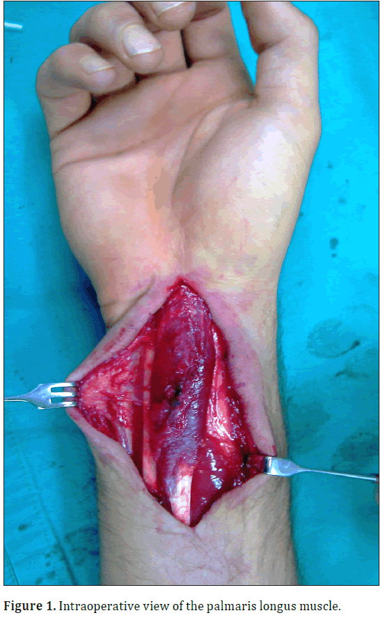
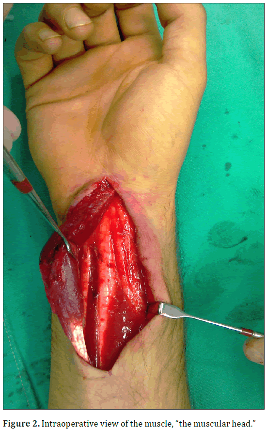
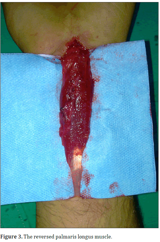
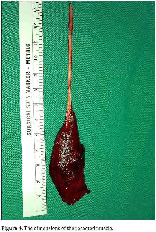
In the physical examination, a mass was observed with pale purple reflections on the skin, which was located on the volar side of the distal one-third of the right forearm, homogenous in structure, without definite borders, soft when compared to the opposite side, without any change in its location and size during wrist movements, and did not become discolored when pressure was applied. Motor and sensory examinations of the extremity were within normal limits. Tinel’s sign and Phalen’s maneuver were negative in the trace of the median nerve. No radiological or neurophysiological evaluation was performed. The patient underwent to explorative surgery under regional anesthesia. Applying a zigzag incision on the mass traversed skin and subcutaneous tissue. The mass appeared as a muscle body that was covered by the forearm fascia. When the muscle mass was completely dissected, it was observed that it was single-headed, reversed palmaris longus. The variant muscle had no marked pressure on the median nerve. It was observed that the muscle did not extend to the Guyon’s canal. The muscle body and tendon were excised (Figures 1-4).
Discussion
Functionally, the palmaris longus muscle is primarily a wrist flexor. This muscle flexes the wrist and tenses palmar aponeurosis joint; however, it provides no substantial flexion force that would inhibit range of the motion in the wrist if its tendon was removed.
It has been reported in the literature that the variants of palmaris longus were most commonly accompanied by neuropathies related to the presence of a mass and nerve compression caused during exertion. In some of those cases, it was shown that the muscle extended to the Guyon’s canal [2].
The absence of the muscle has been described as ranging from a high of about 25% to 16% in white Caucasians [3–4]. An otherwise unimportant muscle, it provides a very useful graft in hand surgery due to the length and diameter of the palmaris longus tendon, and the fact that it can be used without producing any functional deformities in the hand or wrist.
In the current case, no electrophysiological study was performed due to the absence of complaints or signs from the physical examination indicating nerve compression; however, there were many signs of compression detected during effort in many of the reported cases in the literature.
The variations of the palmaris longus muscle are of higher prevalence than any other muscle in the forearm. Double and reverse palmaris longus muscles have been reported, which are associated with median nerve compression [5].
Simple and effective treatment via surgical excision is suggested in cases where the mass has been present for a significant amount of time, and the clinical manifestation of nerve compression is absent [6]. Hand surgeons should be well informed about the anatomic variations of the upper extremity to be comfortable during their surgical practice.
References
- Saadeh FA, Bergman RA. An unusual accessory flexor (opponens) digiti minimi muscle.Anat Anz. 1998; 165: 327–329.
- Ogun TC, Karalezli N, Ogun CO. The concomitant presence of two anomalous muscles in the forearm. Hand (N Y). 2007; 2: 120–122.
- Troha F, Baibak GJ, Kelleher JC. Frequency of the palmaris longus tendon in North American Caucasians. Ann Plast Surg. 1990; 25: 477–478.
- Wehbé MA. Tendon graft donor sites. J Hand Surg Am. 1992; 17: 1130–1132.
- Schuurman AH, van Gils AP. Reversed palmaris longus muscle on MRI: report of four cases. Eur Radiol. 2000; 10: 1242–1244.
- Acikel C, Ulkur E, Karagoz H, Celikoz B. Effort-related compression of median and ulnar nerves as a result of reversed three-headed and hypertrophied palmaris longus muscle with extension of Guyon’s canal. Scand J Plast Reconstr Surg Hand Surg. 2007; 41: 45–47.
Fikret Eren, Cenk melikoglu*, Deniz Kok and Salim Iskender
Etimesgut Military Hospital, Department of Plastic Surgery, Ankara, TURKEY.
- *Corresponding Author:
- Cenk Melikoglu, MD
Fevzipasa Bul. No: 172/2 Basmane Izmir, TURKEY.
Tel: +90 (505) 276 70 69
E-mail: cenkmelikoglu@gmail.com
Date of Received: January 1st,2014
Date of Accepted: February 18th, 2015
Published Online: December 30th, 2015
© Int J Anat Var (IJAV). 2015; 8: 37–39.
Abstract
The palmaris longus muscle has significant anatomical variance compared to other muscles of the upper extremity. The most frequent variation is the complete absence of the muscle, but a number of other variations exist. These variations include reversed, duplicated, bifid, or hypertrophied palmaris longus muscles. Anomalous muscles usually do not cause pathologic symptoms, but are a diagnostic dilemma. They become a surgical problem when they cause symptoms or are difficult to differentiate from soft-tissue masses. This article describes the presence of a reversed palmaris longus muscle in a 21-year-old man, diagnosed during exploration of the soft tissue mass on his right forearm.
-Keywords
Guyon’s Canal,nerve compression,palmaris longus
Introduction
The palmaris longus muscle originates from the medial epicondyle of the humerus with other flexor muscles, and is involved in the structure of the palmar fascia. The palmaris longus muscle is the most anatomically variable muscle of the upper extremity. Muscle agenesis was determined in 12% of cases, which were included in the cadaver studies. In addition to cases in which the muscle body was one-third centrally or one-third distally (oppositely) located, there was a case in which double palmaris longus muscles were located both distally and proximally, and the tendon was also located between these two muscle bodies. Again, a normally located muscle can be present in advanced hypertrophic conditions. The palmaris longus muscle can have variations in the form of duplication, accessory slips, and substitute structures, completely missing in the muscle as well. Additionally, its origin or insertion can have frequent variations. These anatomical variations have clinical implications including possible median and ulnar neuropathies, in addition to mimicking soft tissue tumors sometimes [1].
Case Report
A 21-year-old male patient applied to our outpatient clinic with complaints of swelling and mild discoloration in the distal portion of his right forearm and on the volar side of his wrist that was present since childhood.
In the physical examination, a mass was observed with pale purple reflections on the skin, which was located on the volar side of the distal one-third of the right forearm, homogenous in structure, without definite borders, soft when compared to the opposite side, without any change in its location and size during wrist movements, and did not become discolored when pressure was applied. Motor and sensory examinations of the extremity were within normal limits. Tinel’s sign and Phalen’s maneuver were negative in the trace of the median nerve. No radiological or neurophysiological evaluation was performed. The patient underwent to explorative surgery under regional anesthesia. Applying a zigzag incision on the mass traversed skin and subcutaneous tissue. The mass appeared as a muscle body that was covered by the forearm fascia. When the muscle mass was completely dissected, it was observed that it was single-headed, reversed palmaris longus. The variant muscle had no marked pressure on the median nerve. It was observed that the muscle did not extend to the Guyon’s canal. The muscle body and tendon were excised (Figures 1-4).
Discussion
Functionally, the palmaris longus muscle is primarily a wrist flexor. This muscle flexes the wrist and tenses palmar aponeurosis joint; however, it provides no substantial flexion force that would inhibit range of the motion in the wrist if its tendon was removed.
It has been reported in the literature that the variants of palmaris longus were most commonly accompanied by neuropathies related to the presence of a mass and nerve compression caused during exertion. In some of those cases, it was shown that the muscle extended to the Guyon’s canal [2].
The absence of the muscle has been described as ranging from a high of about 25% to 16% in white Caucasians [3–4]. An otherwise unimportant muscle, it provides a very useful graft in hand surgery due to the length and diameter of the palmaris longus tendon, and the fact that it can be used without producing any functional deformities in the hand or wrist.
In the current case, no electrophysiological study was performed due to the absence of complaints or signs from the physical examination indicating nerve compression; however, there were many signs of compression detected during effort in many of the reported cases in the literature.
The variations of the palmaris longus muscle are of higher prevalence than any other muscle in the forearm. Double and reverse palmaris longus muscles have been reported, which are associated with median nerve compression [5].
Simple and effective treatment via surgical excision is suggested in cases where the mass has been present for a significant amount of time, and the clinical manifestation of nerve compression is absent [6]. Hand surgeons should be well informed about the anatomic variations of the upper extremity to be comfortable during their surgical practice.
References
- Saadeh FA, Bergman RA. An unusual accessory flexor (opponens) digiti minimi muscle.Anat Anz. 1998; 165: 327–329.
- Ogun TC, Karalezli N, Ogun CO. The concomitant presence of two anomalous muscles in the forearm. Hand (N Y). 2007; 2: 120–122.
- Troha F, Baibak GJ, Kelleher JC. Frequency of the palmaris longus tendon in North American Caucasians. Ann Plast Surg. 1990; 25: 477–478.
- Wehbé MA. Tendon graft donor sites. J Hand Surg Am. 1992; 17: 1130–1132.
- Schuurman AH, van Gils AP. Reversed palmaris longus muscle on MRI: report of four cases. Eur Radiol. 2000; 10: 1242–1244.
- Acikel C, Ulkur E, Karagoz H, Celikoz B. Effort-related compression of median and ulnar nerves as a result of reversed three-headed and hypertrophied palmaris longus muscle with extension of Guyon’s canal. Scand J Plast Reconstr Surg Hand Surg. 2007; 41: 45–47.


