Right ectopic kidney with rare vascular variations
Amudha Govindarajan* and Jamuna Meenakshisundaram
Department of Anatomy, PSG Institute of Medical Sciences and Research, Peelamedu, Coimbatore, Tamilnadu, India
- *Corresponding Author:
- Dr. Amudha Govindarajan, MS
Associate Professor, Department of Anatomy, PSG Institute of Medical Sciences and Research, Peelamedu, Coimbatore, Tamilnadu, 641402, India
Tel: +91 422 2570170
E-mail: ammuramesh@gmail.com
Date of Received: July 5th, 2010
Date of Accepted: December 23rd, 2010
Published Online: January 18th, 2011
© IJAV. 2011; 4: 12–14.
[ft_below_content] =>Keywords
renal artery, renal vein, abdominal aorta, inferior vena cava
Introduction
Variations in the position and vasculature of the kidneys are well known and often reported by both anatomists and radiologists. Three to four percent of newborns show some type of abnormality in the urinary tract [1]. Since the kidneys are pelvic organs developmentally, failure or malformations in the ascent of kidneys are common [2]. The ectopic kidneys have been found in a frequency of 1:500 to 1:110, and one normal kidney and one ectopic kidney in 1:3000 [3]. The ectopic kidneys are usually found in the pelvis. As the kidneys ascend up, they derive their blood supply from the segmental mesonephric arteries. The development of the renal veins is closely related to the development of the inferior vena cava [4]. So, variations in the renal vessels are frequently found or they may go unnoticed. Ectopic kidneys with associated vascular anomalies are very common [1]. The mesonephric arteries may fail to regress and persist as supernumerary vessels [5]. Up to 25 % of adult kidneys have accessory renal arteries [1]. An aberrant renal artery present in the poles is called as polar artery. Superior polar arteries were found in 7% of patients [6].
The variation found in the present case is right ectopic kidney with rare vascular variations. This variant has significant clinical importance in the aspect of the recent advances in the urological and interventional radiological procedures.
Case Report
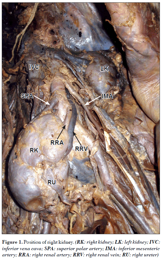
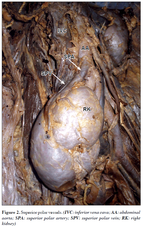
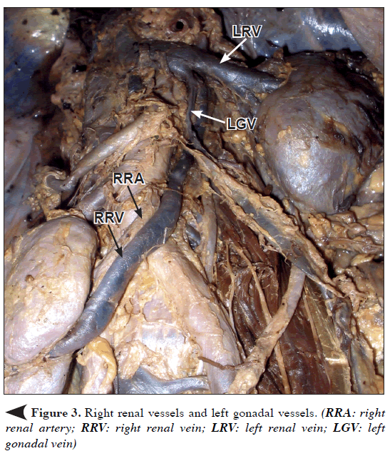
During routine dissection of 73-year-old adult male cadaver, multiple variations were found in the right kidney. The right kidney was located at the pelvic brim between the fourth lumbar vertebra and second sacral vertebra (Figure 1). The right renal hilar artery was observed to emerge from the left side of the abdominal aorta above its bifurcation (Figure 1). There was an aberrant superior polar artery emerging from the left side of the abdominal aorta (Figures 1, 2). The right renal vein emerging from the hilum of the right kidney was observed to take a long course (Figure 3). It was observed to cross the abdominal aorta at its bifurcation and ascend up along the left side of the abdominal aorta behind the inferior mesenteric artery, left gonadal vein and left renal vessels (Figures 2, 3). Above the left renal vein, it joined with the left suprarenal vein and opened into the left renal vein. There was an additional renal vein in the upper pole of the right kidney, which was present posterior to the superior polar artery. The superior polar vein was found to drain into the left side of the inferior vena cava (Figure 2). The right ureter was found anterior to the hilar renal vessels (Figure 1). The left kidney was as usual. The left renal vein was observed to cross the abdominal aorta anterior to it and open in to the inferior vena cava. It was also as usual.
Discussion
The failure in the ascent of the kidneys is often reported in the literature. They are commonly found in the pelvic region [1]. Since the kidneys ascend up from the pelvis during fetal life to reach posterior abdominal wall, they are supplied by segmental mesonephric arteries [2]. The presence of superior polar vessels may be because of the persistent mesonephric arteries that failed to regress. But the anomaly/variation related to the origin of the right hilar renal artery –like in this case– is very rare. Since the right ureter was anterior to the right hilar renal vessels, it may be due to duplication and rotation of left kidney to right and right renal agenesis. The drainage of the right hilar renal vein substantiates this theory. Since the right kidney, a duplicated part of the left kidney, the right renal hilar vein drains in to the left renal vein which is the natural course of that vein.
Supernumerary veins, double renal arteries and veins and accessory veins have been reported from time to time. Additional renal veins are seen more frequently in the right side (27.8%) than the left side (1%) [3]. The way of drainage of the right superior polar vein in the present case also substantiates the origin of the right kidney from the left side, since it was seen to open into the left side of the inferior vena cava.
During clinical procedures like removal of donor kidneys, veins need close attention because variations in the pattern of veins may lead to severe vascular complications [7]. During urological procedures and in removal of donor kidneys, awareness regarding these kind of variations will help the surgeons to prevent vascular complications. It is also important for radiologists to interpret angiograms. In selective angiography, the radiologists must be aware of these kind of unusual origins of renal vessels to insert catheter into the correct vessel, which is very important for the accuracy of diagnosis [6].
References
- Moore KL, Persaud TVN. The Developing Human, Clinically Oriented Embryology. 8th Ed., Philadelphia, Saunders. 2008; 244–260.
- Konya A. An unusual congenital abnormality of the renal artery: left renal artery arising from the right renal artery. Br J Radiol. 1985; 58: 891–893.
- Belsare SM, Chimmalgi M, Vaidya SA, Sant SM. Ectopic kidney and associated anomalies: A case report. J Anat Soc India. 2002; 51: 236–238.
- Baum S, Bousen E, Arams HL, Faitelson BB, eds. Abrams’ Angiography, Anomalies and Malformations. Volume II. 4th Ed., United States of America, Little, Brown and Company. 1997; 1217–1229.
- Redman JF. Anatomy of the Genitourinary System. In: Gillenwater JY, Grayhack JT, Howards SS, Mitchell ME, eds. Adult and Pediatric Urology, 3rd Ed., Missouri, Mosby. 1996; 25–26.
- Dhar P. An additional renal vein. Clin Anat. 2002; 15: 64–66.
- El Fettouh HA, Herts BR, Nimeh T, Wirth SL, Caplin A, Sands M, Ramani AP, Kaouk J, Goldfarb DA, Gill IS. Prospective comparison of 3-dimensional volume rendered computerized tomography and conventional renal arteriography for surgical planning in patients undergoing laparoscopic donor nephrectomy. J Urol. 2003; 170: 57–60.
Amudha Govindarajan* and Jamuna Meenakshisundaram
Department of Anatomy, PSG Institute of Medical Sciences and Research, Peelamedu, Coimbatore, Tamilnadu, India
- *Corresponding Author:
- Dr. Amudha Govindarajan, MS
Associate Professor, Department of Anatomy, PSG Institute of Medical Sciences and Research, Peelamedu, Coimbatore, Tamilnadu, 641402, India
Tel: +91 422 2570170
E-mail: ammuramesh@gmail.com
Date of Received: July 5th, 2010
Date of Accepted: December 23rd, 2010
Published Online: January 18th, 2011
© IJAV. 2011; 4: 12–14.
Abstract
During routine dissection of 73-year-old adult male cadaver, multiple variations were found in the right kidney. The right kidney was located at the level between fourth lumbar vertebra and second sacral vertebra. There was a superior polar artery, and the right renal artery emerged from the left side of the abdominal aorta at the level of fourth lumbar vertebra. There were two renal veins. The vein from superior pole of the kidney drained into the inferior vena cava. The renal vein from the hilum of the right kidney had a long course along the left side of the abdominal aorta and joined with the left suprarenal vein behind the left renal vein and drained into the left renal vein. Knowledge regarding the variations in renal vessels is very important for urologists and interventional radiologists.
-Keywords
renal artery, renal vein, abdominal aorta, inferior vena cava
Introduction
Variations in the position and vasculature of the kidneys are well known and often reported by both anatomists and radiologists. Three to four percent of newborns show some type of abnormality in the urinary tract [1]. Since the kidneys are pelvic organs developmentally, failure or malformations in the ascent of kidneys are common [2]. The ectopic kidneys have been found in a frequency of 1:500 to 1:110, and one normal kidney and one ectopic kidney in 1:3000 [3]. The ectopic kidneys are usually found in the pelvis. As the kidneys ascend up, they derive their blood supply from the segmental mesonephric arteries. The development of the renal veins is closely related to the development of the inferior vena cava [4]. So, variations in the renal vessels are frequently found or they may go unnoticed. Ectopic kidneys with associated vascular anomalies are very common [1]. The mesonephric arteries may fail to regress and persist as supernumerary vessels [5]. Up to 25 % of adult kidneys have accessory renal arteries [1]. An aberrant renal artery present in the poles is called as polar artery. Superior polar arteries were found in 7% of patients [6].
The variation found in the present case is right ectopic kidney with rare vascular variations. This variant has significant clinical importance in the aspect of the recent advances in the urological and interventional radiological procedures.
Case Report
During routine dissection of 73-year-old adult male cadaver, multiple variations were found in the right kidney. The right kidney was located at the pelvic brim between the fourth lumbar vertebra and second sacral vertebra (Figure 1). The right renal hilar artery was observed to emerge from the left side of the abdominal aorta above its bifurcation (Figure 1). There was an aberrant superior polar artery emerging from the left side of the abdominal aorta (Figures 1, 2). The right renal vein emerging from the hilum of the right kidney was observed to take a long course (Figure 3). It was observed to cross the abdominal aorta at its bifurcation and ascend up along the left side of the abdominal aorta behind the inferior mesenteric artery, left gonadal vein and left renal vessels (Figures 2, 3). Above the left renal vein, it joined with the left suprarenal vein and opened into the left renal vein. There was an additional renal vein in the upper pole of the right kidney, which was present posterior to the superior polar artery. The superior polar vein was found to drain into the left side of the inferior vena cava (Figure 2). The right ureter was found anterior to the hilar renal vessels (Figure 1). The left kidney was as usual. The left renal vein was observed to cross the abdominal aorta anterior to it and open in to the inferior vena cava. It was also as usual.
Discussion
The failure in the ascent of the kidneys is often reported in the literature. They are commonly found in the pelvic region [1]. Since the kidneys ascend up from the pelvis during fetal life to reach posterior abdominal wall, they are supplied by segmental mesonephric arteries [2]. The presence of superior polar vessels may be because of the persistent mesonephric arteries that failed to regress. But the anomaly/variation related to the origin of the right hilar renal artery –like in this case– is very rare. Since the right ureter was anterior to the right hilar renal vessels, it may be due to duplication and rotation of left kidney to right and right renal agenesis. The drainage of the right hilar renal vein substantiates this theory. Since the right kidney, a duplicated part of the left kidney, the right renal hilar vein drains in to the left renal vein which is the natural course of that vein.
Supernumerary veins, double renal arteries and veins and accessory veins have been reported from time to time. Additional renal veins are seen more frequently in the right side (27.8%) than the left side (1%) [3]. The way of drainage of the right superior polar vein in the present case also substantiates the origin of the right kidney from the left side, since it was seen to open into the left side of the inferior vena cava.
During clinical procedures like removal of donor kidneys, veins need close attention because variations in the pattern of veins may lead to severe vascular complications [7]. During urological procedures and in removal of donor kidneys, awareness regarding these kind of variations will help the surgeons to prevent vascular complications. It is also important for radiologists to interpret angiograms. In selective angiography, the radiologists must be aware of these kind of unusual origins of renal vessels to insert catheter into the correct vessel, which is very important for the accuracy of diagnosis [6].
References
- Moore KL, Persaud TVN. The Developing Human, Clinically Oriented Embryology. 8th Ed., Philadelphia, Saunders. 2008; 244–260.
- Konya A. An unusual congenital abnormality of the renal artery: left renal artery arising from the right renal artery. Br J Radiol. 1985; 58: 891–893.
- Belsare SM, Chimmalgi M, Vaidya SA, Sant SM. Ectopic kidney and associated anomalies: A case report. J Anat Soc India. 2002; 51: 236–238.
- Baum S, Bousen E, Arams HL, Faitelson BB, eds. Abrams’ Angiography, Anomalies and Malformations. Volume II. 4th Ed., United States of America, Little, Brown and Company. 1997; 1217–1229.
- Redman JF. Anatomy of the Genitourinary System. In: Gillenwater JY, Grayhack JT, Howards SS, Mitchell ME, eds. Adult and Pediatric Urology, 3rd Ed., Missouri, Mosby. 1996; 25–26.
- Dhar P. An additional renal vein. Clin Anat. 2002; 15: 64–66.
- El Fettouh HA, Herts BR, Nimeh T, Wirth SL, Caplin A, Sands M, Ramani AP, Kaouk J, Goldfarb DA, Gill IS. Prospective comparison of 3-dimensional volume rendered computerized tomography and conventional renal arteriography for surgical planning in patients undergoing laparoscopic donor nephrectomy. J Urol. 2003; 170: 57–60.


