Right sternalis muscle
Raghu Jetti1,Narendra Pamidi1*,Venkata Ramana Vollala1,Rakesh Vasavi2 and Venu M.Nerella1
1Department of Anatomy, Melaka Manipal Medical College (Manipal Campus), International Centre for Health Sciences, Manipal,India.
2Department of Anatomy, KMC International Center, Manipal,India.
- *Corresponding Author:
- Narendra Pamidi
Lecturer, Department of Anatomy,Melaka Manipal Medical College (Manipal Campus), International Centre for Health Sciences, Manipal, 576104, Karnataka, India.
Tel: +91 820 2922642
Fax: +91 820 2571905
E-mail: pommidi_narendra@yahoo.co.in
Date of Received: December 10th, 2008
Date of Accepted: March 22nd, 2009
Published Online: Marcah 24th, 2009
© IJAV. 2009; 2: 41–42.
[ft_below_content] =>Keywords
sternalis muscle,location,nerve supply,tumor,breast surgery
Introduction
Myotome portion of the somites give rise to most of the skeletal muscles of the body. Muscles of the extremities develop from somatic mesoderm. Splanchnic mesoderm gives rise to cardiac muscle and most of the smooth muscles; whereas mesoderm of the pharyngeal arches gives rise to some of the head and neck muscles. The sternalis muscle is a derivative of superficial part of the rectus abdominis muscle [1]. Sternalis is a flat, ribbon-shaped muscle that begins from the lower part of the ribs, rectus sheath and then courses upward, finally inserting into the upper part of the sternum and ribs or the sternocleidomastoid muscle [2]. Various authors have classified sternalis muscle under 4 main categories regarding the structure it has been derived: (a) from pectoralis major, (b) from rectus abdominis, (c) from sternomastoid and (d) from the panniculus carnosus [3].
It is necessary to record and discuss unusual anatomical variants with the use of advanced diagnostic and therapeutic tools as these variants could present a challenge to the radiologist or surgeon in establishing a diagnosis [4].
Case Report
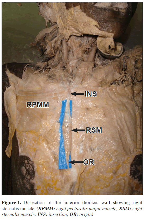
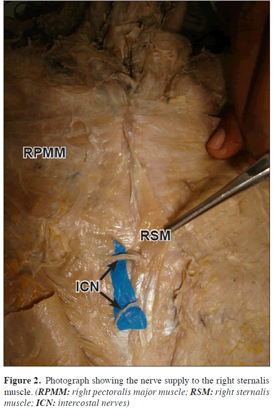
During the dissection classes for MBBS students in the Department of Anatomy at Melaka Manipal Medical College, the right sternalis muscle has been noticed in a 65-year-old male cadaver (Figure 1). It was located at the anterior thoracic wall, along the right lateral side of the sternum. It originated as a tendon from the fascia of the right rectus abdominis and external oblique aponeurosis, ran upwards on the right lateral side of the sternum, finally inserted into the tendon of the right sternocleidomastoid muscle. It was measured 10 cm in length and 1.5 cm in width in the middle of the muscle. It was innervated by the 3rd and 4th anterior intercostal nerves (Figure 2).
Discussion
According to Bergman et al., this muscle is present in humans only. It is reportedly found in 3-5% of the population and is classified with the pectoral group of muscles [5]. Generally it is unilateral [6]. It is less common among men (6.4%) than women (8.7%) [7]. The incidence is 4-8% in Indians [8], 13.1% in Japanese, 3.3% in Filipinos and 1% in Chinese [9].
In a literature survey by O’Neill and J Folan-Curran, 55% of the sternalis muscles were innervated by branches of the internal or external thoracic nerves, 43% by branches of the intercostal nerves and 2% by both from the intercostal and the thoracic nerves [3].
The existence and nerve supply of the sternalis muscle is still confusing anatomists. In a 15 years experience of Kida et al. on 40 cases, they found that the sternalis muscle is supplied by the pectoral nerves. Branches of the intercostal nerves may pierce the muscle to become cutaneous but do not directly supply the sternalis [10]. In the present case, however, the sternalis muscle is not supplied by the pectoral nerves but it was pierced by the intercostals nerves.
Sternalis muscle appears as an unusual bulge in the median breast in mammography, which could be mistaken either for a tumor on initial investigation or as a recurrence of cancer during post-treatment checkups.
Sternalis muscle can be easily identified by the use of CT or MRI [11]. Many surgeons and radiologists are not familiar with the sternalis muscle [6]. Early identification of sternalis muscle, its location is necessary to proceed in an appropriate plane during surgical dissection in breast surgeries [4].
References
- Sadler TW. Langman’s medical embryology. 9th Ed., Baltimore, Lippincott Williams and Wilkins. 2004; 199–209.
- Williams PL, Bannister LH, Berry MM, Collins P, Dyson M, Dussek JE, Ferguson MW, eds. Gray’s Anatomy. 38th Ed., Edingburg, Churchill Livingstone. 1995; 838.
- O’Neill MN, Folan-Curran J. Case report: bilateral sternalis muscles with a bilateral pectoralis major anomaly. J Anat. 1998; 193: 289–292.
- Kabay B, Akdogan I, Ozdemir B, Adiguzel E. The left sternalis muscle variation detected during mastectomy. Folia Morphol (Warsz). 2005; 64: 338–340.
- Bergman RA, Thompson SA, Afifi AK, Saadeh FA. Compendium of Human Anatomic Variation. Munich, Urban and Schwarzenberg. 1988; 7–8.
- Bailey PM, Tzarnas CD. The sternalis muscle: a normal finding encountered during breast surgery. Plast Reconstr Surg. 1999; 103: 1189–1190.
- Scott-Conner CE, Al-Jurf AS. The sternalis muscle. Clin Anat. 2002; 15: 67–69.
- Harish K, Gopinath KS. Sternalis muscle: importance in surgery of the breast. Surg Radiol Anat. 2003; 25: 311–314.
- Jeng H, Su SJ. The sternalis muscle: an uncommon anatomical variant among Taiwanese. J Anat. 1998; 193: 287–288.
- Kida MY, Izumi A, Tanaka S. Sternalis muscle: topic for debate. Clin Anat. 2000; 13: 138–140.
- Bradley FM, Hoover HC Jr, Hulka CA, Whitman GJ, McCarthy KA, Hall DA, Moore R, Kopans DB. The sternalis muscle: an unusual normal finding seen on mammography. AJR Am J Roentgenol. 1996; 166: 33–36.
Raghu Jetti1,Narendra Pamidi1*,Venkata Ramana Vollala1,Rakesh Vasavi2 and Venu M.Nerella1
1Department of Anatomy, Melaka Manipal Medical College (Manipal Campus), International Centre for Health Sciences, Manipal,India.
2Department of Anatomy, KMC International Center, Manipal,India.
- *Corresponding Author:
- Narendra Pamidi
Lecturer, Department of Anatomy,Melaka Manipal Medical College (Manipal Campus), International Centre for Health Sciences, Manipal, 576104, Karnataka, India.
Tel: +91 820 2922642
Fax: +91 820 2571905
E-mail: pommidi_narendra@yahoo.co.in
Date of Received: December 10th, 2008
Date of Accepted: March 22nd, 2009
Published Online: Marcah 24th, 2009
© IJAV. 2009; 2: 41–42.
Abstract
Knowledge regarding the muscular variations of the chest and their identification for the proper dissection planes through radiological examination is important. Sternalis is an occasional muscle, which lies along the side of the sternum. It may be confused as a tumor. The existence of sternalis muscle, its location, orientation and early identification are necessary in breast surgeries. Presence of sternalis muscle adjacent to the breast is of clinical importance.
-Keywords
sternalis muscle,location,nerve supply,tumor,breast surgery
Introduction
Myotome portion of the somites give rise to most of the skeletal muscles of the body. Muscles of the extremities develop from somatic mesoderm. Splanchnic mesoderm gives rise to cardiac muscle and most of the smooth muscles; whereas mesoderm of the pharyngeal arches gives rise to some of the head and neck muscles. The sternalis muscle is a derivative of superficial part of the rectus abdominis muscle [1]. Sternalis is a flat, ribbon-shaped muscle that begins from the lower part of the ribs, rectus sheath and then courses upward, finally inserting into the upper part of the sternum and ribs or the sternocleidomastoid muscle [2]. Various authors have classified sternalis muscle under 4 main categories regarding the structure it has been derived: (a) from pectoralis major, (b) from rectus abdominis, (c) from sternomastoid and (d) from the panniculus carnosus [3].
It is necessary to record and discuss unusual anatomical variants with the use of advanced diagnostic and therapeutic tools as these variants could present a challenge to the radiologist or surgeon in establishing a diagnosis [4].
Case Report
During the dissection classes for MBBS students in the Department of Anatomy at Melaka Manipal Medical College, the right sternalis muscle has been noticed in a 65-year-old male cadaver (Figure 1). It was located at the anterior thoracic wall, along the right lateral side of the sternum. It originated as a tendon from the fascia of the right rectus abdominis and external oblique aponeurosis, ran upwards on the right lateral side of the sternum, finally inserted into the tendon of the right sternocleidomastoid muscle. It was measured 10 cm in length and 1.5 cm in width in the middle of the muscle. It was innervated by the 3rd and 4th anterior intercostal nerves (Figure 2).
Discussion
According to Bergman et al., this muscle is present in humans only. It is reportedly found in 3-5% of the population and is classified with the pectoral group of muscles [5]. Generally it is unilateral [6]. It is less common among men (6.4%) than women (8.7%) [7]. The incidence is 4-8% in Indians [8], 13.1% in Japanese, 3.3% in Filipinos and 1% in Chinese [9].
In a literature survey by O’Neill and J Folan-Curran, 55% of the sternalis muscles were innervated by branches of the internal or external thoracic nerves, 43% by branches of the intercostal nerves and 2% by both from the intercostal and the thoracic nerves [3].
The existence and nerve supply of the sternalis muscle is still confusing anatomists. In a 15 years experience of Kida et al. on 40 cases, they found that the sternalis muscle is supplied by the pectoral nerves. Branches of the intercostal nerves may pierce the muscle to become cutaneous but do not directly supply the sternalis [10]. In the present case, however, the sternalis muscle is not supplied by the pectoral nerves but it was pierced by the intercostals nerves.
Sternalis muscle appears as an unusual bulge in the median breast in mammography, which could be mistaken either for a tumor on initial investigation or as a recurrence of cancer during post-treatment checkups.
Sternalis muscle can be easily identified by the use of CT or MRI [11]. Many surgeons and radiologists are not familiar with the sternalis muscle [6]. Early identification of sternalis muscle, its location is necessary to proceed in an appropriate plane during surgical dissection in breast surgeries [4].
References
- Sadler TW. Langman’s medical embryology. 9th Ed., Baltimore, Lippincott Williams and Wilkins. 2004; 199–209.
- Williams PL, Bannister LH, Berry MM, Collins P, Dyson M, Dussek JE, Ferguson MW, eds. Gray’s Anatomy. 38th Ed., Edingburg, Churchill Livingstone. 1995; 838.
- O’Neill MN, Folan-Curran J. Case report: bilateral sternalis muscles with a bilateral pectoralis major anomaly. J Anat. 1998; 193: 289–292.
- Kabay B, Akdogan I, Ozdemir B, Adiguzel E. The left sternalis muscle variation detected during mastectomy. Folia Morphol (Warsz). 2005; 64: 338–340.
- Bergman RA, Thompson SA, Afifi AK, Saadeh FA. Compendium of Human Anatomic Variation. Munich, Urban and Schwarzenberg. 1988; 7–8.
- Bailey PM, Tzarnas CD. The sternalis muscle: a normal finding encountered during breast surgery. Plast Reconstr Surg. 1999; 103: 1189–1190.
- Scott-Conner CE, Al-Jurf AS. The sternalis muscle. Clin Anat. 2002; 15: 67–69.
- Harish K, Gopinath KS. Sternalis muscle: importance in surgery of the breast. Surg Radiol Anat. 2003; 25: 311–314.
- Jeng H, Su SJ. The sternalis muscle: an uncommon anatomical variant among Taiwanese. J Anat. 1998; 193: 287–288.
- Kida MY, Izumi A, Tanaka S. Sternalis muscle: topic for debate. Clin Anat. 2000; 13: 138–140.
- Bradley FM, Hoover HC Jr, Hulka CA, Whitman GJ, McCarthy KA, Hall DA, Moore R, Kopans DB. The sternalis muscle: an unusual normal finding seen on mammography. AJR Am J Roentgenol. 1996; 166: 33–36.


