Sea urchin disease: Case study on Venezuelan shores
Received: 13-Jan-2018 Accepted Date: Jan 18, 2018; Published: 24-Jan-2018
Citation: Loffler SG. Sea urchin disease: case study on Venezuelan shores. J Mar Microbiol. 2018;2(1):1-2.
This open-access article is distributed under the terms of the Creative Commons Attribution Non-Commercial License (CC BY-NC) (http://creativecommons.org/licenses/by-nc/4.0/), which permits reuse, distribution and reproduction of the article, provided that the original work is properly cited and the reuse is restricted to noncommercial purposes. For commercial reuse, contact reprints@pulsus.com
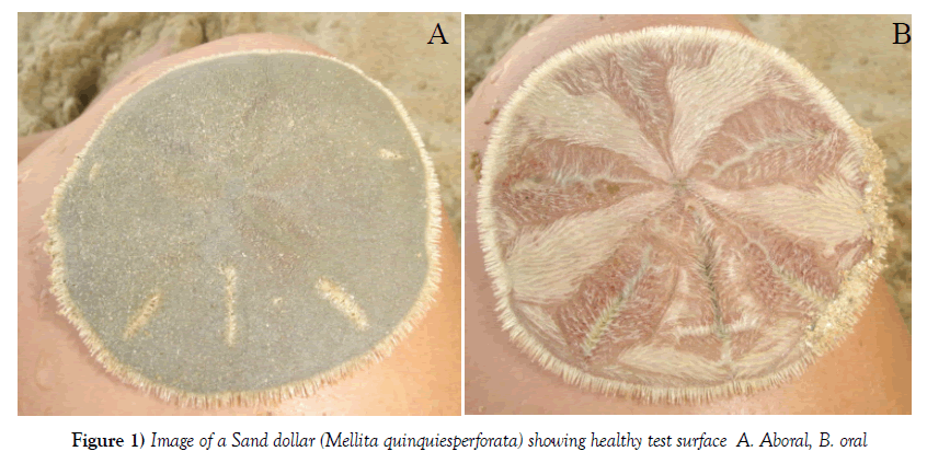
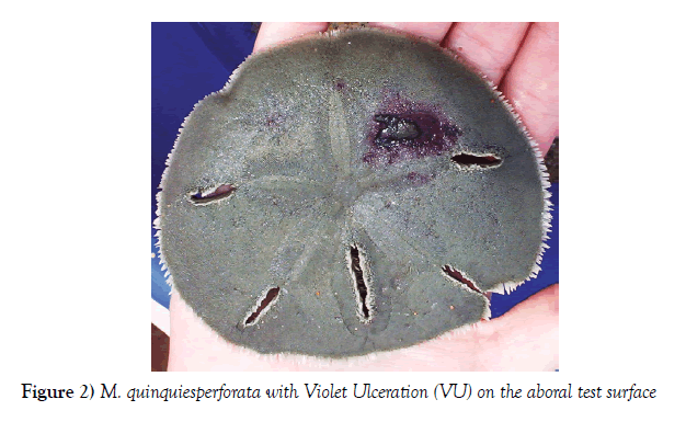
The prevalence of clinical signs in echinoids and other marine invertebrates has been reported since the 70`s in the Caribbean sea. However, the presence of clinical signs in echinoids has not been reported in Venezuela until now. We carried out a visual census of the sand dollar population (Mellita quinquiesperforata) on Margarita Island and described several clinical signs observed. The clinical signs observed with higher prevalence are listed in decreasing order: Black Dots (BD), Violet Ulceration (VU), Brown Patches (BP), Green Erosion (GE), Red Dots (RD), Decolored Patches (DP), Pink Band (PB) and Black Ulceration (BU). The clinical signs with higher grade affecting the aboral surface found were VU and BU. Gram coloration of the affected test surface revealed elevated presence Gram+ cocci and bacilli. This work pretends to expand the baseline for future studies of pathologies of echinoids in the Caribbean sea, specially Venezuela. At the time no molecular studies were carried out and microbiology isolation was unsuccessful. Sand dollars (Mellita quinquiesperforata) can be found on sandy beaches with oxygenated water, with high concentration of calcium, potassium, sodium, phosphorus, chlorides and with low presence of organic matter. These equinoids belong to the Order Clypeasteroidea and form dense patches (sometimes barried for protection) were the adults live in the intertidial zone and juveniles live near to the shallow intertidial zone. The principal food source is infauna comprising benthic phyto and zooplankton. Equinoids have been reported to be affected by the “Black Sea Urchin disease” [1-4] “ Bald Sea urchin disease” also called “Red Spot disease” [5-9]. This disease is caused by bacteria (Vibrio anguillarum and Aeromonas salmonicida) [6]. Bower reported that Gram-negative bacteria belonging to Acinetobackter spp. and Alcaligenes spp. were more abundant on lesion surfaces than on healthy test surfaces. Another disease reported in equinoids is the “Green Sea Urchin Amoebasis” [10-13], affecting mainly Strongylocentrotus droebachiensis, this disease is caused by Paramoeba invadens and caused mass mortalities of this sea urchin in Nova Scotia. In the Caribbean sea, mass mortalities of Diadema antillarum were reported to the caused by Clostridium perfringens and C. sordelli [4]. During 1983-1991 mass mortality almost extinguished these populations affecting 98% from Panama to the Bermudas. Lessios [2] studied the recovery rate of the populations of D. antillarum and found only a slight recovery after the mass mortality event. Sand dollars (M. quinquiesperforata) express signs of stress caused by environmental changes releasing coelomic cells with violet coloration Figures 1 and 2. These cells have substances with antibiotic and antimycotic properties and are released by stress-induced effects [14,15]. Further studies of the observed clinical signs of sand dollar populations have a great scientific interest since these animals are indicators of environmental changes of biotic and abiotic factors of the coast, caused by contamination and global warming with seem to promote development of these pathologies [16,17]. In the Venezuelan shore have been reported disease affecting corals of the Los Roques archipelago and the National Park of Morrocoy. The signs described by several authors were yellow band and black spot syndromes, black bands, white plague and bleaching [18-21] describes six clinical signs observed in M. quinquiesperforata populations on Margarita Island and classifies them into levels of affection (low, regular and severe). In a more recent study the same author studied seven localities on Margarita Island and censed M. quinquiesperforata individual during 45 minutes in the shallow subtidial zone, all individuals were measured in length (mm) and high (mm). The objective of this census was to analyze where the higher prevalence could be observed and if there are risk factors for these populations that could be established using statistics, such as: size, dry season or wet season, beaches with touristic traffic and beaches without. The results revealed that bigger sand dollars have 4,4 times more probability to have at least one clinical sign than smaller ones (juveniles) and that in the rain season the animals have 1,8 times more probability to have clinical signs. The comparison between beaches with touristic activity and the one without, revealed that the sand dollars censed on touristic beaches have 3.25 times more probability to have at least one clinical sign that the ones censed on beaches without. These studies are first to report pathologies in sand dollars in Venezuela and the Caribbean sea. Further studies have to be made to isolate and characterize the etiological agents of these clinical signs.
REFERENCES
- Bak R, Carpay M, Steveninck E. Densities of the sea urchin Diadema antillarum before and after mass mortalities on the coral reefs of Curacao. Marine Ecology-Progress Series. 1984;17:105-8.
- Lessios HA. Diadema antillarum populations in Panama twenty years following mass mortality. Coral Reefs. 2005;24:125-27.
- Lessios HA, Robertson DR, Cubit JD. Spread of Diadema antillarum mass mortality through the Caribbean. Sciences New Series. 1984;226:335-37.
- Peters E. Disease of Other Invertebrate Phyla: Porifera, Cnidaria, Ctenophora, Annelida, Echinodermata. Pathobiology of Marine and Estaurine Organisms. CRC Press Bca Ratón. 1999;393-449.
- Höbaus E, Fenaux L, Hignette M. Premitères observations on lesions caused by a disease affecting the test of sea urchins in the western Mediterranean. Rapp PV Meeting Commiss Int Explor Sci Mediterranean Sea. 1981;27:221-22.
- Gilles KW, Pearse JS. Disease in sea urchins Strongylocentrotus purpuratus: experimental infection and bacterial virulence. Diseases of Aquatic Organisms. 1986;1:105-14.
- Maes P, Jangroux M. The bald-sea urchin disease: A biopathological approach. Helgoländer Meeresuntersuchungen. 1984;37:217-24.
- Maes P, Jangroux M. The bald-sea urchin disease: a bacterial infection. Proceedings of the Fifth International Echinoderm Conference Galway, Escocia. 1994;2429:313-314.
- Maes P, M Jangoux. The bald-sea-urchin disease: a biopathological approach. Helgoländer Meeresuntersuchungen. 1984;37:217-24.
- Scheibling R. Echinoids, epizootics and ecological stability in the rocky subtidal off Nova Scotia, Canada. Helgoländer Wissenschaftliche Meeresuntersuchungen. 1984;37:233-42.
- Brady SM, Scheibling RE. Repopulation of the shallow subtidal zone by green sea urchins (Strongylocentrotus droebachiensis) following mass mortality in Chapter 3. Nova Scotia Canada J Mar Biol Assoc UK. 2005;85:1511-17.
- Brady SM, Scheibling RE. Changes in growth and reproduction of green sea urchins, Strongylocentrotus droebachiensis (Müller), during repopulation of the shallow subtidal zone after mass mortality. J Experimental Marine Biology and Ecology. 2006;335:277–91.
- Jangoux M. Diseases of Echinodermata. In: Kinne, O. (eds), Diseases of Marine Animals. Volume III: Introduction, Cephalopoda, Annelida, Crustacea, Chaetognatha, Echinodermata, Urochordata. Biologische Anstalt Helgoland Germany. 1990;440-44.
- Wardlaw A, Unkles S. Bactericidal Activity of Coelomic Fluid from the Sea Urchin Echinus esculentus. J Invertebrate Pathology. 1978;32: 25-34.
- Smith A, Smith L. The sand dollar as a possible indicator of environmental stress. J Aquariculture & Aquatic Sciences. 1987;5:1-4.
- Kinne O. Disease of marine animals. Volume 1 General Aspects, protozoa to gastropoda. Edit. John Wiley and Sons. Bath, Gran Bretaña. 1980;466.
- Harvell C, Kim K, Burkholder J, et al. Emerging Marine Diseases-Climate Links and Anthropogenic Factors. Science. 1999;285:1505-10.
- Croquer A, Bone D. Diseases in scleractinid corals: A new problem in the reef of Cayo Sombrero, Morrocoy National Park, Venezuela? J Tropical Biology 51 Supl. 2003;4:167-72.
- Croquer A, Pauls S, Zubillaga A. White plague disease outbreak in a coral reef at Los Roques National Park, Venezuela. Revista de Biología Tropical. 2003;51:39-45.
- García A, Croquer A, Sheila M. Relationship between disease incidence and size and species structure in corals of Los Roques Archipelago National Park Venezuela. 2006;9:1-12.
- Grune S, González D, Lopez P, et al. Description of some pathological symptoms in irregular Urchin Mellita quinquiesperforata (Leske, 1778) obtained on beaches of the island of Margarita, Venezuela Summary. XI Latin American Congress of Marine Sciences Viña del Mar Chile 2005.


