Septate gallbladder: Gross and histological perspectives in an uncommon occurrenceglass
Niladri Kumar Mahato*
Department of Anatomy, Sri Aurobindo Institute of Medical Sciences (SAIMS), Indore, Madhya Pradesh, India
- *Corresponding Author:
- Dr. Niladri Kumar Mahato
Associate Professor, Department of Anatomy, Sri Aurobindo Institute Of Medical Sciences (SAIMS), Indore-Ujjain Highway, Bhawrasala Indore, Madhya Pradesh, 452 111, India
Tel: +91 731 4231000 ext: 433
E-mail: mahatonk@gmail.com
Date of Received: September 29th, 2009
Date of Accepted: March 30th, 2010
Published Online: May 17th, 2010
© Int J Anat Var (IJAV). 2010; 3: 70–72.
[ft_below_content] =>Keywords
cystic bud, cholelithiasis, Rokitansky-Aschoff sinuses
Introduction
Anatomical variations of the gallbladder are not uncommon and usually present as a wide variety of malformations pertaining to its size, shape, number and position [1]. They have been reported to occur in 0.1% of the population [2]. Septations occurring inside the gallbladder, though, occur fairly rarely and are also reported quite infrequently. Septations in the gallbladder have been reported to be single [3] or multiple [4,5]. Septations in gallbladder form due to incomplete and improper vacuolization or resolution of the solid cystic bud during development around the third month of intra-uterine life as reported previously [4], due to excessive ‘wrinkling’ of the wall of the organ giving rise to more than one septa [6]. Septate gallbladders are by and large asymptomatic and usually detected during a post mortem examination [7]. These anomalies may also be significantly associated with recurrent abdominal colic [8], with jaundice [7] or with stone formation due to stagnation of bile and acute cholecystitis [3] requiring early removal of the bladder [8] or recommended cholecystectomy even in absence of stones in patients with inflammation of gallbladder having such septum [3].
The present case report is based on the morphological and histological study in a gallbladder with a single septum. Single septum inside gallbladders may be oriented longitudinally (referred to as bi-lobed gallbladder) or may be transverse to oblique in orientation (called an ‘hour-occurrenceglass’ gallbladder) [9]. Detailed classification of ‘lobed’ gallbladder (Boyden) has been documented [10]. The hour-glass gallbladders usually feature a transversely oblique septum that separates the gallbladder fundus from the rest of the body. The septum bears a minute to moderate opening that connects the two isolated chambers. Reports reveal that compartmentalization of the cavity of the gallbladder with narrow connection leads to biliary stasis and cholelithiasis [3]. Investigating modalities such as ultrasound [11] and other techniques [12] are applied pre-operatively to detect these abnormalities, with varying degree of success.
Case Report
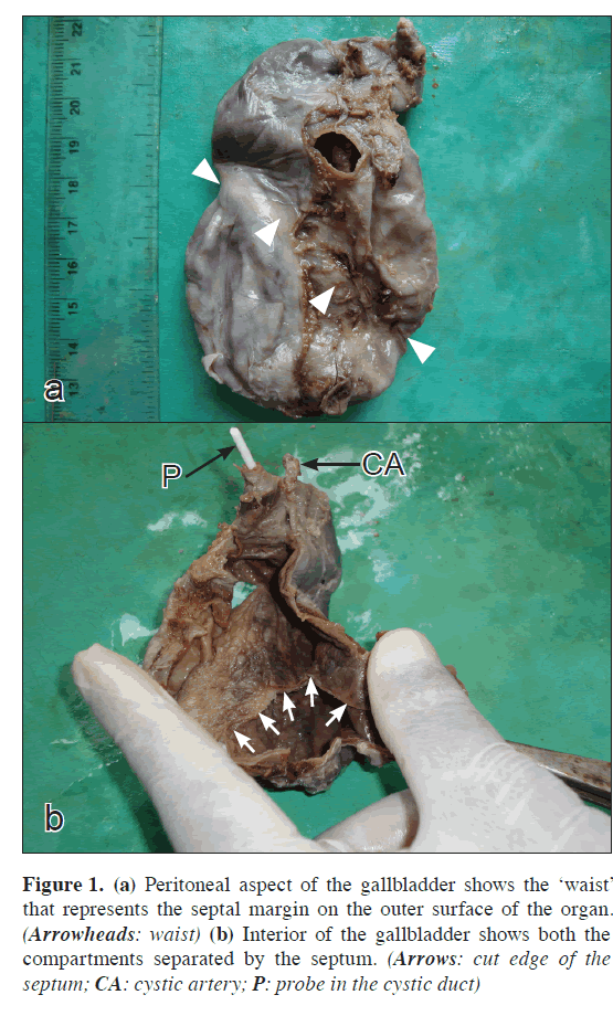
A 48-year-old lady was diagnosed with cholelithiasis. A septate gallbladder was discovered after the surgery following complaint of abdominal colic. Pre-operative ultrasonography revealed multiple calculi, distention and mild thickening of the gallbladder wall. The organ, after its removal, was opened from its posterior (peritoneal) aspect and inspected. Examination of the gallbladder showed a well-formed septa within. Observation of the gallbladder from outside revealed an external fibrous band on the serosa of the bladder. This band like structure coincided with the margins of the septa inside. It represented a constriction between the fundus (below) and the remaining part of the gallbladder (above) and appeared as something like a ‘waist’ of the organ (Figure 1a). Parts of the gallbladder, both above and below the ‘waist’ were enlarged. Positions of the cystic duct and artery were normally oriented at the upper end of the organ (Figure 1a). Morphology of the interior demonstrated a transversely positioned septa (inclined a shade downwards towards the left) that divided the cavity almost equally into two enlarged cavities as upper and lower (Figure 1b). The septa presented a minute opening (about 3mm in diameter) at its left extreme, connecting the two cavities. Both the cavities contained multiple stones. The compartments contained fairly large rounded stones that were unlikely to pass through the opening. The texture and the appearance of the epithelium with its folds seemed to look normal. The organ had a single cystic duct and artery (Figure 1b).
Figure 1: (a) Peritoneal aspect of the gallbladder shows the ‘waist’ that represents the septal margin on the outer surface of the organ. (Arrowheads: waist) (b) Interior of the gallbladder shows both the compartments separated by the septum. (Arrows: cut edge of the septum; CA: cystic artery; P: probe in the cystic duct)
Further Consideration
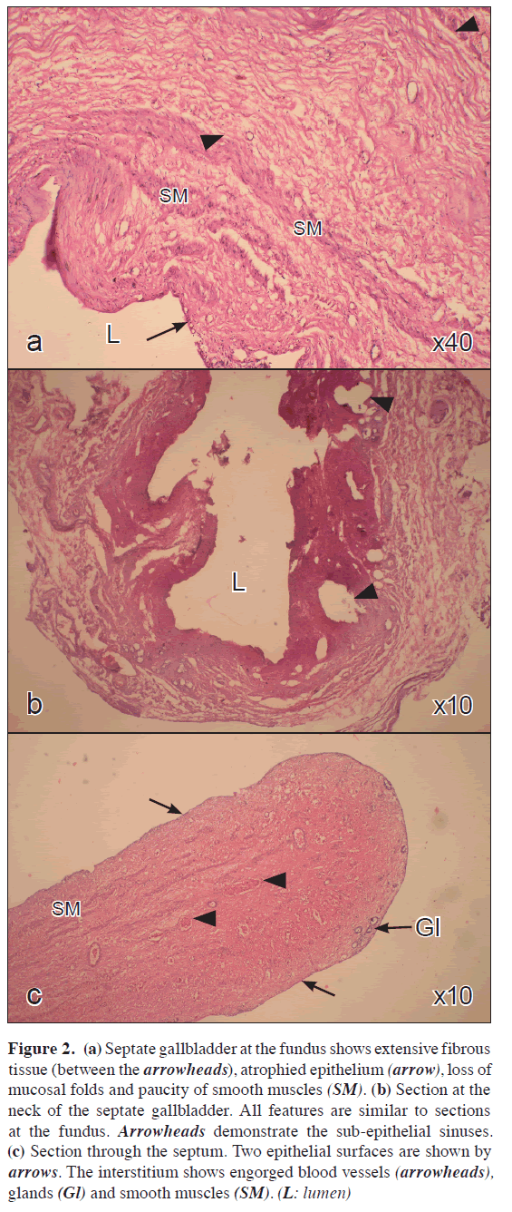
Five microns thick sections were taken from tissues at the fundus, neck, septum and the cystic duct of the organ. Sections were studied after staining with routine hematoxylin & eosin stains, for histology. Histological examination of the tissues featured a generalized thinning of the epithelium throughout the gallbladder and its septum, and glandular atrophy (Figures 2a,b,c). Excessive fibrous tissue was observed in the interstitium with reduction in the mass of smooth muscle in the walls and the septa. Signs of formation of Rokitansky-Aschoff SMSMsinuses were observed below the mucosa and were confined to the wall of the gallbladder (Figure 2b).
Figure 2: (a) Septate gallbladder at the fundus shows extensive fibrous tissue (between the arrowheads), atrophied epithelium (arrow), loss of mucosal folds and paucity of smooth muscles (SM). (b) Section at the neck of the septate gallbladder. All features are similar to sections at the fundus. Arrowheads demonstrate the sub-epithelial sinuses. (c) Section through the septum. Two epithelial surfaces are shown by arrows. The interstitium shows engorged blood vessels (arrowheads), glands (Gl) and smooth muscles (SM). (L: lumen)
Discussion
Gallbladders bearing transverse septa with minimal communication between the two cavities are liable to produce bile stasis, stones and increased pressure symptoms in the organ. Septation predisposes cholelithiasis in these isolated compartments with supervening chronic or acute inflammation. All these features were evident in the gallbladder discussed in the study. Pre-operative investigations, though confirmed the presence of multiple stones in the organ, they were unable to comment on the presence of the transverse septa. Studies have documented that radiological detection of the condition is often difficult [11]. It is important to note that the gallbladder possessed a single cystic artery that accompanied a normal, single cystic duct. Such information is lacking in available literature. Since septate gallbladders develop from incomplete or segmental compartmentalization of a solid cystic bud, it may be conceived that structures proximal to the neck of the organ, such as the cystic artery and the duct would remain normal, as seen in the present case. These organs demonstrate septa that are oriented transverse to the axes of the organ. Longitudinally oriented septa give rise to ‘bi-lobed’ gallbladders with more than one independently draining gallbladder compartments provided with more than one trunk of main cystic vessels. As found in the present specimen, isolation of the inferior compartment predisposes formation of stones in them due to sequestration of bile in the lower part of the gallbladder. Once stones are formed in the isolated inferior section of the organ, it is trapped in the compartment due to the narrow communication between the two cavities.
Infection and distention, therefore, are common presentations associated with these variations. Distention of the gallbladder, as observed in this case, resulted in excessive stretching of the organ walls and presented as grossly thinned smooth muscle and the epithelium. There smooth muscle component of the organ wall was considerably lost out. There was, however, no evidence of desquamation of the epithelial lining in the present case, as sometimes found with acute cholecystites; but sub-epithelial, intramural sinuses could be localized easily. The septum resembled the rest of the organ both in gross appearance and in histology. This fact further substantiates the common embryological origins of the gallbladder wall and the septa (found as an embryological variant). The septum contains fibroareolar tissue, smooth muscle and glandular epithelium. It is important to note that the septum bears histological characteristics similar to any other area of the rest of the organ and such reports are rare to come across in literature reviews [5].
Acknowledgement
I would like to convey my sincere thanks and gratitude to Dr. S. S. Nandedkar (Professor and Head, Department of Pathology, SAIMS, Indore) for his invaluable guidance in appreciating the microscopic perspectives of the study and for his overall encouragement.
References
- Gross RE. Congenital anomalies of the gallbladder: A review of 148 cases, with report of double gallbladder. Arch Surg. 1936; 32: 131–162.
- Johnson SR. Development of the liver and biliary apparatus. In: Romanes GJ, ed. Cunninghams TextBook of Anatomy. 12th Ed., Oxford, Oxford University Press. 1987; 485.
- Deutsch AA, Englestein D, Cohen M, Kunichevsky M, Reiss R. Septum of the gallbladder, clinical implications and treatment. Postgrad Med J. 1986; 62: 453–456.
- Haslam RH, Gayler BW, Ebert PA. Multiseptate gallbladder. A cause of recurrent abdominal pain in childhood. Am J Dis Child. 1966; 112: 600–603.
- Simon M, Tandon BN. Multiseptate gallbladder. A case report. Radiology.1963; 80: 84–86.
- Bhagavan BS, Amin PB, Land AS, Weinberg T. Multiseptate gallbladder. Embryogenetic hypotheses. Arch Pathol. 1970; 89: 382–385.
- Martinoli C, Derchi LE, Pastorino C, Cittadini G Jr. Martino C, Derchi LE, Pastronno C, Cittadim G. Case report: imaging of a bilobed gallbladder. Br J Radiol. 1993; 66: 734–736.
- Alawneh I, Baranzanchi I, Jacobi K. Multi-septate gallbladder as a cause of colic. Med Klin. 1981; 76: 190–191. (German)
- Flannery MG, Caster MP. Congenital hourglass gall bladder. South Med J. 1957; 50: 1255–1258.
- Gigot J, Van Beers B, Goncette L, Etienne J, Collard A, Jadoul P, Therasse A, Otte JB, Kestens P. Laparoscopic treatment of gallbladder duplication. A plea for removal of both gallbladders. Surg Endosc. 1997; 11: 479–482.
- Doyle TC. Flattened fundus sign of the septate gallbladder. Gastrointest Radiol. 1984; 9(4): 345–347.
- Oyar O, Yesildag A, Gulsoy U, Sengul N, Isler M. Bilobed gallbladder diagnosed by oral cholecysto-CT. Comput Med Imaging Graph. 2003; 27: 315–319.
Niladri Kumar Mahato*
Department of Anatomy, Sri Aurobindo Institute of Medical Sciences (SAIMS), Indore, Madhya Pradesh, India
- *Corresponding Author:
- Dr. Niladri Kumar Mahato
Associate Professor, Department of Anatomy, Sri Aurobindo Institute Of Medical Sciences (SAIMS), Indore-Ujjain Highway, Bhawrasala Indore, Madhya Pradesh, 452 111, India
Tel: +91 731 4231000 ext: 433
E-mail: mahatonk@gmail.com
Date of Received: September 29th, 2009
Date of Accepted: March 30th, 2010
Published Online: May 17th, 2010
© Int J Anat Var (IJAV). 2010; 3: 70–72.
Abstract
Septate gallbladders are rare variations associated with the extra-hepatic biliary system. Available reports state that variations of gallbladder are either asymptomatic or are found associated with inflammation and stones. The present report is a morphological and histological study of a septate gallbladder. The organ was found to have a transversely oriented septa at about its middle that divided the cavity into an upper and a lower segment with a tiny communication between them. Both the cavities were grossly distended and filled with moderately large stones. The gallbladder presented a fibrous constriction on its exterior corresponding to the margin of the septa. The cystic duct and artery were single and normal. Histology revealed extensive intramural fibrosis and atrophy of smooth muscles. Epithelium demonstrated extreme thinning with loss of folds and glands, with presence of cysts and sinuses beneath. None of the anomalies could be suggested by pre-operative ultrasound imaging except the calculi.
-Keywords
cystic bud, cholelithiasis, Rokitansky-Aschoff sinuses
Introduction
Anatomical variations of the gallbladder are not uncommon and usually present as a wide variety of malformations pertaining to its size, shape, number and position [1]. They have been reported to occur in 0.1% of the population [2]. Septations occurring inside the gallbladder, though, occur fairly rarely and are also reported quite infrequently. Septations in the gallbladder have been reported to be single [3] or multiple [4,5]. Septations in gallbladder form due to incomplete and improper vacuolization or resolution of the solid cystic bud during development around the third month of intra-uterine life as reported previously [4], due to excessive ‘wrinkling’ of the wall of the organ giving rise to more than one septa [6]. Septate gallbladders are by and large asymptomatic and usually detected during a post mortem examination [7]. These anomalies may also be significantly associated with recurrent abdominal colic [8], with jaundice [7] or with stone formation due to stagnation of bile and acute cholecystitis [3] requiring early removal of the bladder [8] or recommended cholecystectomy even in absence of stones in patients with inflammation of gallbladder having such septum [3].
The present case report is based on the morphological and histological study in a gallbladder with a single septum. Single septum inside gallbladders may be oriented longitudinally (referred to as bi-lobed gallbladder) or may be transverse to oblique in orientation (called an ‘hour-occurrenceglass’ gallbladder) [9]. Detailed classification of ‘lobed’ gallbladder (Boyden) has been documented [10]. The hour-glass gallbladders usually feature a transversely oblique septum that separates the gallbladder fundus from the rest of the body. The septum bears a minute to moderate opening that connects the two isolated chambers. Reports reveal that compartmentalization of the cavity of the gallbladder with narrow connection leads to biliary stasis and cholelithiasis [3]. Investigating modalities such as ultrasound [11] and other techniques [12] are applied pre-operatively to detect these abnormalities, with varying degree of success.
Case Report
A 48-year-old lady was diagnosed with cholelithiasis. A septate gallbladder was discovered after the surgery following complaint of abdominal colic. Pre-operative ultrasonography revealed multiple calculi, distention and mild thickening of the gallbladder wall. The organ, after its removal, was opened from its posterior (peritoneal) aspect and inspected. Examination of the gallbladder showed a well-formed septa within. Observation of the gallbladder from outside revealed an external fibrous band on the serosa of the bladder. This band like structure coincided with the margins of the septa inside. It represented a constriction between the fundus (below) and the remaining part of the gallbladder (above) and appeared as something like a ‘waist’ of the organ (Figure 1a). Parts of the gallbladder, both above and below the ‘waist’ were enlarged. Positions of the cystic duct and artery were normally oriented at the upper end of the organ (Figure 1a). Morphology of the interior demonstrated a transversely positioned septa (inclined a shade downwards towards the left) that divided the cavity almost equally into two enlarged cavities as upper and lower (Figure 1b). The septa presented a minute opening (about 3mm in diameter) at its left extreme, connecting the two cavities. Both the cavities contained multiple stones. The compartments contained fairly large rounded stones that were unlikely to pass through the opening. The texture and the appearance of the epithelium with its folds seemed to look normal. The organ had a single cystic duct and artery (Figure 1b).
Figure 1: (a) Peritoneal aspect of the gallbladder shows the ‘waist’ that represents the septal margin on the outer surface of the organ. (Arrowheads: waist) (b) Interior of the gallbladder shows both the compartments separated by the septum. (Arrows: cut edge of the septum; CA: cystic artery; P: probe in the cystic duct)
Further Consideration
Five microns thick sections were taken from tissues at the fundus, neck, septum and the cystic duct of the organ. Sections were studied after staining with routine hematoxylin & eosin stains, for histology. Histological examination of the tissues featured a generalized thinning of the epithelium throughout the gallbladder and its septum, and glandular atrophy (Figures 2a,b,c). Excessive fibrous tissue was observed in the interstitium with reduction in the mass of smooth muscle in the walls and the septa. Signs of formation of Rokitansky-Aschoff SMSMsinuses were observed below the mucosa and were confined to the wall of the gallbladder (Figure 2b).
Figure 2: (a) Septate gallbladder at the fundus shows extensive fibrous tissue (between the arrowheads), atrophied epithelium (arrow), loss of mucosal folds and paucity of smooth muscles (SM). (b) Section at the neck of the septate gallbladder. All features are similar to sections at the fundus. Arrowheads demonstrate the sub-epithelial sinuses. (c) Section through the septum. Two epithelial surfaces are shown by arrows. The interstitium shows engorged blood vessels (arrowheads), glands (Gl) and smooth muscles (SM). (L: lumen)
Discussion
Gallbladders bearing transverse septa with minimal communication between the two cavities are liable to produce bile stasis, stones and increased pressure symptoms in the organ. Septation predisposes cholelithiasis in these isolated compartments with supervening chronic or acute inflammation. All these features were evident in the gallbladder discussed in the study. Pre-operative investigations, though confirmed the presence of multiple stones in the organ, they were unable to comment on the presence of the transverse septa. Studies have documented that radiological detection of the condition is often difficult [11]. It is important to note that the gallbladder possessed a single cystic artery that accompanied a normal, single cystic duct. Such information is lacking in available literature. Since septate gallbladders develop from incomplete or segmental compartmentalization of a solid cystic bud, it may be conceived that structures proximal to the neck of the organ, such as the cystic artery and the duct would remain normal, as seen in the present case. These organs demonstrate septa that are oriented transverse to the axes of the organ. Longitudinally oriented septa give rise to ‘bi-lobed’ gallbladders with more than one independently draining gallbladder compartments provided with more than one trunk of main cystic vessels. As found in the present specimen, isolation of the inferior compartment predisposes formation of stones in them due to sequestration of bile in the lower part of the gallbladder. Once stones are formed in the isolated inferior section of the organ, it is trapped in the compartment due to the narrow communication between the two cavities.
Infection and distention, therefore, are common presentations associated with these variations. Distention of the gallbladder, as observed in this case, resulted in excessive stretching of the organ walls and presented as grossly thinned smooth muscle and the epithelium. There smooth muscle component of the organ wall was considerably lost out. There was, however, no evidence of desquamation of the epithelial lining in the present case, as sometimes found with acute cholecystites; but sub-epithelial, intramural sinuses could be localized easily. The septum resembled the rest of the organ both in gross appearance and in histology. This fact further substantiates the common embryological origins of the gallbladder wall and the septa (found as an embryological variant). The septum contains fibroareolar tissue, smooth muscle and glandular epithelium. It is important to note that the septum bears histological characteristics similar to any other area of the rest of the organ and such reports are rare to come across in literature reviews [5].
Acknowledgement
I would like to convey my sincere thanks and gratitude to Dr. S. S. Nandedkar (Professor and Head, Department of Pathology, SAIMS, Indore) for his invaluable guidance in appreciating the microscopic perspectives of the study and for his overall encouragement.
References
- Gross RE. Congenital anomalies of the gallbladder: A review of 148 cases, with report of double gallbladder. Arch Surg. 1936; 32: 131–162.
- Johnson SR. Development of the liver and biliary apparatus. In: Romanes GJ, ed. Cunninghams TextBook of Anatomy. 12th Ed., Oxford, Oxford University Press. 1987; 485.
- Deutsch AA, Englestein D, Cohen M, Kunichevsky M, Reiss R. Septum of the gallbladder, clinical implications and treatment. Postgrad Med J. 1986; 62: 453–456.
- Haslam RH, Gayler BW, Ebert PA. Multiseptate gallbladder. A cause of recurrent abdominal pain in childhood. Am J Dis Child. 1966; 112: 600–603.
- Simon M, Tandon BN. Multiseptate gallbladder. A case report. Radiology.1963; 80: 84–86.
- Bhagavan BS, Amin PB, Land AS, Weinberg T. Multiseptate gallbladder. Embryogenetic hypotheses. Arch Pathol. 1970; 89: 382–385.
- Martinoli C, Derchi LE, Pastorino C, Cittadini G Jr. Martino C, Derchi LE, Pastronno C, Cittadim G. Case report: imaging of a bilobed gallbladder. Br J Radiol. 1993; 66: 734–736.
- Alawneh I, Baranzanchi I, Jacobi K. Multi-septate gallbladder as a cause of colic. Med Klin. 1981; 76: 190–191. (German)
- Flannery MG, Caster MP. Congenital hourglass gall bladder. South Med J. 1957; 50: 1255–1258.
- Gigot J, Van Beers B, Goncette L, Etienne J, Collard A, Jadoul P, Therasse A, Otte JB, Kestens P. Laparoscopic treatment of gallbladder duplication. A plea for removal of both gallbladders. Surg Endosc. 1997; 11: 479–482.
- Doyle TC. Flattened fundus sign of the septate gallbladder. Gastrointest Radiol. 1984; 9(4): 345–347.
- Oyar O, Yesildag A, Gulsoy U, Sengul N, Isler M. Bilobed gallbladder diagnosed by oral cholecysto-CT. Comput Med Imaging Graph. 2003; 27: 315–319.


