Sex Determination using Femoral Head Diameters on Radiographs of Adults who visited Delta State University Teaching Hospital, Oghara, Nigeria
2 Department of Human Anatomy and Cell Biology, Faculty of Basic Medical Sciences, Delta State University, Abraka, Nigeria
3 Department of Human Anatomy, Achievers University, Owo, Ondo State, Nigeria, Email: thomasgodswill23@gmail.com
Received: 01-Dec-2023, Manuscript No. ijav-23-6870; Editor assigned: 04-Dec-2023, Pre QC No. ijav-23-6870 (PQ); Reviewed: 21-Dec-2023 QC No. ijav-23-6870; Revised: 25-Dec-2023, Manuscript No. ijav-23-6870 (R); Published: 30-Dec-2023, DOI: 10.37532/1308-4038.16(12).334
Citation: Ogheneyebrorue GO. Sex Determination using Femoral Head Diameters on Radiographs of Adults who visited Delta State University Teaching Hospital, Oghara, Nigeria. Int J Anat Var. 2023;16(12): 452-455.
This open-access article is distributed under the terms of the Creative Commons Attribution Non-Commercial License (CC BY-NC) (http://creativecommons.org/licenses/by-nc/4.0/), which permits reuse, distribution and reproduction of the article, provided that the original work is properly cited and the reuse is restricted to noncommercial purposes. For commercial reuse, contact reprints@pulsus.com
Abstract
Introduction: Estimating biological sex is critical for identifying unknown skeletal remains in modern medico-legal circumstances or bioarcheological research of previous cultures. Population-specific sexual dimorphism often affects sex estimation methodologies. The goal of this study was to determine sex using femoral head diameters.
Materials and Methods: This study adopted the retrospective cross-sectional study of the quantitative design. This study was carried out in the radiology department of Delta State University Teaching Hospital, Oghara. The radiographs selected showed hip joint, intact Shenton’s lines, no fracture at the femoral necks, no pathological disorders, intact cortices at femoral heads and were of known sexes. The radiographs were those taken at a routine object-film distance of 5cm and focal film distance of 92cm in the anterior-posterior view with the two big toes touching on their medial aspects. With the aid of a certified radiographer, the measurements were taken by locating the appropriate points using a mouse-operated cursor at the defined anatomical coordinates. The distance/angle in the two points were subsequently obtained from the screen’s results window in standardized units. The axes of the femoral neck and shaft and the maximum femoral headdiameterswere determined by means ofthe digital vernier caliper. The data was analyzed using the Statistical Package for the Social Sciences (SPSS) software version 23 (IBM Corp., Armonk, NY, USA).The level of statistical significance was set at P < 0.05.
Results: The means of males and females were seen to be different and these differences were statistically significant for all parameters (Right Femoral Head Vertical Diameter, Right Femoral Head Transverse Diameter, Left Femoral Head Vertical Diameter; Left Femoral Head Transverse Diameter (P<0.001).
Conclusion: Focusing on the results of this study, it was concluded that the mean values of the studied parameters are statistically and significantly different in males and females. These parameters are useful and reliable for sexual dimorphism in anthropometric and Forensic studies, especially in identifying skeletal remains.
INTRODUCTION
The femur is the human body’s longest, heaviest, and most powerful bone. The pyramid-shaped neck joins the spherical head at the apex and the tubular shaft at the base at the proximal end [1,2]. The greater and lesser trochanters are two conspicuous bony bulges that connect to muscles that move the hip and knee. The angle between the neck and the shaft, in a typical adult is around 128 degrees. In contrast, the inclination angle decreases with age [3,4].
Estimating biological sex is critical for identifying unknown skeletal remains in modern medico-legal circumstances or bioarcheological research of previous cultures. Population-specific sexual dimorphism often affects sex estimation methodologies. As a result, the necessity for separate criteria for each group has long been recognized [5]. Apart from stature and ethnicity, age and gender estimation are critical characteristics in medico-legal and archaeological identification [6-10]. Identifying an unidentified body becomes easier when the entire body or skeleton remains are available. It gets difficult, however. When dismembered body parts are the sole material accessible to the investigator, which is frequently the case in most circumstances [11-13]
The neck-shaft angle (NSA) is defined as the obtuse angle generated by the intersection of two axes: the femoral shaft axis and the femoral neck axis [14- 16]. This keeps the leg away from the pelvis during movement. It is broadest at birth and gradually narrows until the age of ten [17,18]. The NSA value averages 127° and fluctuates depending to temperature, clothes, lifestyle, sex, age, and side [19,20]. As a result, the NSA is subject to change. Diverse researches in diverse populations have determined normal ranges. A drop in NSA is referred to as coxa vara, whilst a rise is referred to as coxa valga. This study therefore aimed at determining sex using femoral head diameters.
MATERIALS AND METHODS
This descriptive cross-sectional study retrospectively of patients who visited the Radiological unit of the Delta State University Teaching Hospital Oghara in Nigeria between 2014 to 2021. Before the commencement of data collection, an ethical approval from the Research and Ethics Committee of the Hospital (DELSUTH/HRE/2022/068/0635) was obtained.
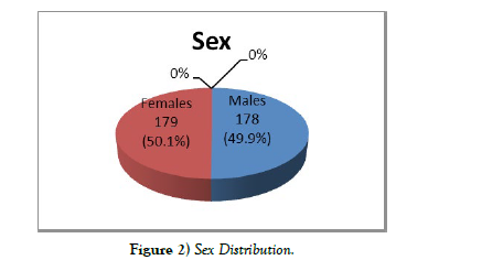
The populations of this study were adults (18 years to 70 years; 357 radiographs) who visited the radiology department of Delta State University Teaching Hospital, Oghara for pelvic radiography. The radiographs selected showed the hip joints, intact Shenton’s lines, no fracture at the femoral necks, no pathological disorders, intact cortices at femoral heads and were of known sexes.
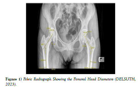
The radiographs were those taken at a routine object-film distance of 5 cm and focal film distance of 92 cm in the anterior-posterior view with the two big toes touching on their medial aspects. With the aid of a certified radiographer, the measurements were taken by locating the appropriate points using a mouse-operated cursor at the defined anatomical coordinates. The transverse diameter of the femoral shaft was measured at two sites to identify the longitudinal axis of the femoral shaft: one beneath the lesser trochanter and one under in diaphysis [21,22]. The midpoints of the two diameters was then determined and connected by a line, the longitudinal axis of the femoral shaft [23].
The obtuse angle (interior) of the femoral neck and shaft axes was gotten as Neck-Shaft angle and the measurements was performed by extending the lines joining the midpoints of the femoral neck and the femoral shaft until they intersected [24-26]. Vertical diameter of the femur head was measured at right angle to the long axis of the neck of femur [27]. This was gotten as the straight distance between the highest and deepest points of the femoral head [28] Transverse diameter of the femur head was gotten by drawing a best fit circleof the femur head [29]. And measurement taken as an extension of the femoral neck axis from the border of the circle opposite the acetabulum to the border of the circle directly underneath the rim of the acetabulum.
The data was analyzed using the Statistical Package for the Social Sciences (SPSS) software version 23 (IBM Corp., Armonk, NY, USA).The level of statistical significance was set at P < 0.05. Firstly, the general descriptive statistics for the different parameter were obtained. The independent T-tests was employed to find differences between the sexes while the paired T-test was used to determine the side difference between the right and left femoral head diameters. The sexual dimorphism ratio was calculated as: (male mean/female mean) × 100. Lastly, stepwise and direct discriminant function analyses were performed to discriminate between sexes [Figures 1 and 2].
RESULTS
Table 1 showed the means of the different parameters within age group 18-27 years. The means of males and females were seen to have variations and these variations were statistically significant for all parameters. The sex differences observed in RFHVD, RFHTD, LFHVD and LFHTD were highly statistically significant with P-Value of P<0.001.
| N | Minimum | Maximum | Mean | Std. Deviation | |
|---|---|---|---|---|---|
| Age (years) | 357 | 18 | 70 | 43.33 | 14.632 |
| Right femoral head vertical diameter(cm) | 357 | 3.81 | 5.22 | 4.7137 | 0.25936 |
| Right femoral head transverse diameter(cm) | 357 | 3.78 | 5.19 | 4.6562 | 0.25366 |
| Left femoral head vertical diameter(cm) | 357 | 3.79 | 5.2 | 4.6573 | 0.25975 |
| Left femoral head transverse diameter(cm) | 357 | 3.7 | 5.12 | 4.5975 | 0.25029 |
Table 1) Descriptive Statistics.
The means of the different parameters within age group 28-37 years were observed. There was statistically significant sex difference for all parameters. The sex differences observed in RFHVD, RFHTD, LFHVD and LFHTD were highly statistically significant with P<0.001 [Table 2].
| Parameters | Gender | N | Mean | Std. Deviation | P-Value |
|---|---|---|---|---|---|
| RFHVD (cm) | M | 35 | 4.7740 | 0.19090 | <0.001 |
| F | 32 | 4.3072 | 0.20073 | ||
| RFHTD (cm) | M | 35 | 4.7014 | 0.16714 | <0.001 |
| F | 32 | 4.2581 | 0.20628 | ||
| LFHVD (cm) | M | 35 | 4.7283 | 0.17606 | <0.001 |
| F | 32 | 4.2550 | 0.20304 | ||
| LFHTD (cm) | M | 35 | 4.6549 | 0.16008 | <0.001 |
| F | 32 | 4.2134 | 0.23076 |
Table 2) Sex Differences of Studied Parameters within Age Group 18-27 Years.
Table 3 showed the means of the different studied parameters. The table revealed that the study comprised of 178 males and 179 females whose means showed statistically significant sex differences for all parameters (P<0.001).
| Parameters | Gender | N | Mean | Std. Deviation | P-Value |
|---|---|---|---|---|---|
| RFHVD (cm) | M | 178 | 4.8985 | 0.16484 | <0.001 |
| F | 179 | 4.5299 | 0.19842 | ||
| RFHTD (cm) | M | 178 | 4.8337 | 0.16213 | <0.001 |
| F | 179 | 4.4798 | 0.19944 | ||
| LFHVD (cm) | M | 178 | 4.8452 | 0.15739 | <0.001 |
| F | 179 | 4.4706 | 0.19987 | ||
| LFHTD (cm) | M | 178 | 4.7684 | 0.16065 | <0.001 |
| F | 179 | 4.4274 | 0.20330 |
Table 3) Sex Differences of Studied Parameters.
Stepwise discriminant function analysis was developed for all variables and is presented in Table 4. The LFHVD was found to be most dimorphic followed by RFHVD, RFHTD and LFHTD. This classification was based on their Eigen, Canonical correlation, Wilk’s Lambda and Chi-square values. Those with the highest values of Eigen, Canonical correlation and Chi-square values were said to be more dimorphic while the lower the Wilk’s Lambda value the more dimorphic the variables [30,31]. The table also showed that these values were statistically significant with P < 0.001.
| Variables | Eigen | Cannonical Correlation | Wilk’s Lambda | Chi-Square | Df | Significance |
|---|---|---|---|---|---|---|
| RFHVD | 1.026 | 0.712 | 0.493 | 250.368 | 1 | <0.001 |
| RFHTD | 0.952 | 0.698 | 0.512 | 237.160 | 1 | <0.001 |
| LFHVD | 1.090 | 0.722 | 0.479 | 261.245 | 1 | <0.001 |
| LFHTD | 0.870 | 0.682 | 0.535 | 221.950 | 1 | <0.001 |
| All Variables | 1.832 | 0.804 | 0.353 | 366.425 | 6 | <0.001 |
Table 4) Stepwise Discriminant Function Analysis for Sex Determination from Femoral Head Diameters.
DISCUSSION
In forensic anthropology and bioarchaeology, sex estimation is one of the steps to characterize and/or achieve a successful identification of unknown individuals. For the Sexual Dimorphism of the human bones, the male bones are more massive and heavier than female bones. The crests, ridges, tuberosities, and lines of attachment of muscles and ligaments are more strongly marked in males. However, the femur remains by far the masterpiece of skeletal specimen in establishing sex. In this study 357 pelvic radiographs were used (males=178 and females-179) to determine sex and ascertain the percentage of accuracy.
From this study it was found that for all the parameters, the mean for the males were more than the means for the females and this was in agreement with Igbigbi and Msamati [32]. Tahir et al [33]. Abdul-Rafik et al [34]. And Kumar et al [35]. who found out that the femoral head diameters, collodiaphysial angles and all measured parameters respectively were greater in males than in females.
From this study, it was observed that generally, all the parameters have a strong positive correlation with their opposite side with the femoral head vertical diameter having the highest (0.974). It was also revealed that this correlation was highly statistically significant with P < 0.001. This was in accordance with Barnali et al [36]. But was inconsistent with Prabha et al [37].
Who reported that there was no statistically significant association between the right and left side of the femoral head diameters? Discrepancies in this finding may be due to differences in methodology and genetic variations. Identification points (IP) are levels of femoral head diameters above which values are those of male bones and below which are female bones. According to Igbigbi and Msamati [32] the identification point for the different parameters were derived from the range of their values (minimum and maximum values) and this was adopted in this study. In this study, the male identification point was derived as the maximum value of the female femoral head diameters, while the identification point for the female was derived as the minimum value of the female femoral head diameters. The identification point of the right femoral head transverse diameter had the highest identification point (5.12 cm) in femoral head diameters in males.
This suggests that any value below 5.12 cm for the right femoral head transverse diameter is considered a female. While in females, theidentification point of theright and left femoral head vertical diameter had the highest identification point (4.30 cm each) in femoral head diameters. This study revealed that the identification points for the males are generally greater than those of the females and this was in unison with Igbigbi and Msamati [32].
CONCLUSION
Focusing on the results of this study, it was concluded that the mean values of the studied parameters are significantly higher in males than females. These parameters are useful and reliable for sexual dimorphism in anthropometric and Forensic studies, especially in identifying skeletal remains.
REFERENCES
- Kranioti E, Vorniotakis N, Galiatsou C, Iscan M, Michalodimitrakis M. Sex identification and software development using digital femoral head radiographs. Forensic Science International Journal, 2009; 189: 1131-1137.
- Mitra A, Khadijeh B, Vida A, Ali R, Farzaneh M et al. Sexing based on measurements of the femoral head parameters on pelvic radiographs. Journal of Forensic Leg Medicine, 2014; 23: 70-75.
- Reynolds A. The fractured femur. Radiological Technology, 2013; 84(3): 273-294.
- Boese C, Jostmeier J, Oppermann J. The neck shaft angle CT reference values of 800 adult hips. Skeletal Radiology, 201; 45: 455-463.
- Curate F. The Estimation of Sex of Human Skeletal Remains in the Portuguese Identified Collections History and Prospects. Forensic Sciences, 2022; 2(1):272-286.
- Rissech C, Schaefer M, Malgosa A. Development of the femur implications for age and sex determination. Forensic Science International Journal, 2008; 180: 1-9.
- Pujol A, Rissech C, Ventura J, Badosa J, Turbón D. Ontogeny of the female femur: geometric morphometric analysis applied on current living individuals of a Spanish population. Journal of Anatomy, 2014; 225: 346-357.
- Chatterjee P, Krishan K, Singh R, Kanchan T. Sex estimation from the femur using discriminant function analysis in a Central Indian population. Medical Scientific Law, 2020; 60: 112-121.
- Cuzzullin M, Curate F, Freire A, Costa S, Prado F et al. Validation of anthropological measures of the human femur for sex estimation in Brazilians. Austria Journal of Forensic Science, 2020; 54(1): 1-14.
- Ernest EO, Enaohwo MT, Vincent Junior I, Nwaokoro IC, Azubike IT et al. Sciatic nerve morphometry in the gluteal region in a Nigerian population an anatomical study. 2023:7.
- Iman FG, Alaa MS, Khaled AB. Sex determination in femurs of modern Egyptians: A comparative study between metric measurements and SRY gene detection. Egyptian Journal of Forensic Sciences, 2014; 4 (4):109-115.
- Monum T, Prasitwattanseree S, Das S, Siriphimolwat P, Mahakkanukrauh P. Sex estimation by femur in modern Thai population. Clin Ter, 2017; 168: 203-207.
- Enaohwo TM, Okoro OG. Anthropometric study of the frontal sinus on plain radiographs in Delta State University Teaching Hospital. Journal of Experimental and Clinical Anatomy, 2018; 17(2): 49-49.
- Carballido Gamio J, Nicolella D. Computational anatomy in the study of bone structure. Current Osteoporosis, 2013; 1(3): 237-245.
- Li M, Cole P. Anatomical considerations in adult femoral neck fractures: how anatomy influences the treatment issues. Injury, 2015; 46(3): 453-458.
- Nwaokoro IC, Enemodia OE, Ehebha SE and Okoro OG. Anthropometric study of the canthal parameters among the Hausa and Yoruba Ethnic groups in Nigeria. International Journal of Medical Research and Health Sciences, 2023; 12(5):48-51.
- Bonneau N, Libourel P, Simonis C, Puymerail L, Baylac M et al. A three-dimensional axis for the study of femoral neck orientation. Journal of Anatomy, 2012; 221: 465-476.
- Okoro OG, Isioma CN, Owhefere GO, Akindugbagbe TF, Oladunni AE. Stages of Epiphyseal Fusion at the Distal End of Radius and Ulna in Nigeria; A Radiological study. International Research in Medical and Health Sciences, 2023; 5(5): 1-6.
- Srisaarn T, Salang K, Klawson B, Vipulakorn K, Chalayon O et al. Surgical correction of coxavara Evaluation of neck shaft angle Hilgenreiner epiphyseal angle for indication of recurrence. Journal of Clinical Orthopedic Trauma, 2019; 10:593-598.
- Enaohwo TM, Okoro OG. Morphometric study of hypoglossal canal of occipital bone in dry skulls of two states in southern nigeria. Bangladesh Journal of Medical Science, 2020; 19(4): 670-672.
- Oladunni AE, Ogheneyebrorue GO, Joyce EI. Radiological assessment of age from epiphyseal fusion at the wrist and ankle in Southern Nigeria. Forensic Science International Reports, 2021; 3:100164.
- Lakati K, Ndelev B, Mouti N, Kibet J. Proximal femur geometry in the adult Kenyan femur and its implications in orthopaedic surgery. East Africa Orthopaedic Journal, 2017; 11: 22-27.
- Siwach R. Anthropometric study of proximal femur geometry and its clinical application. Ann National Academy of Medical Sciences (India), 2018; 54:203-215.
- Igbigbi P. Collo-diaphysial angle of the femur in East African subjects. Clinical Anatomy, 2003; 16: 416-419.
- Laumonerie P, Ollivier M, Li S, Faizan A, Cavaignac E et al. Which factors influence proximal femoral asymmetry? A 3D CT analysis of 345 femoral pairs. Bone and Joint Journal, 2018; 100: 839-844.
- Mokoena P, Billings B, Gibbon V, Bidmos M, Mazengenya P. Development of discriminant functions to estimate sex in upper limb bones for mixed ancestry. South Africans Science Justice, 2019; 59: 660-666.
- Mukhia R, Poudel P. Morphometric study of proximal end of femur of Nepalese people. Nepal Journal of Medical Sciences, 2019; 4:9-14.
- Ravi G, Saheb S, Abraham RA. Morphometric study of femur and its clinical importance. International Journal of Integrated Medical Sciences, 2016; 3:341-344.
- Guodong W, Ai G, Yichao Z, Hua Q, Haomiao Y et al. Measurement of the relative position of the femoral head center greater trochanter and lesser trochanter. Annals of Palliative Medicine, 2021; 10(11):11524-11528.
- Büyüköztürk Ş, Çokluk-Bökeoğlu O. Discriminant function analysis: Concept and application. Egitim Arastirmalari Eurasian Journal of Educational Research, 2008; 33: 73-92.
- Green SB, Salkind NJ, Akey TM. Using SPSS for Windows and Macintosh: Analyzing and understanding data. New Jersey Prentice Hall, 2008.
- Igbigbi P, Msamati B. Sex determination from femoral head diameters in black Malawians. East Africa Medical Journal, 2000; 77.
- Tahir A, Abdul wahab H, Umar I. A study of the collodiaphyseal angle of the femur in the North-Eastern Sub-Region of Nigeria. Nigerian journal of medicine journal of the National Association of Resident Doctors of Nigeria, 2001; 10(1):34-36.
- Abdul Rafik A, Moses B, Yussif A. Models for estimating age and sex from variables of the proximal femur in a Ghanaian population. Forensic Science International: Reports, 2022; 5: 100270.
- Kumar K, Subhadarsini S, Mishra D, Mohapatra C. Sexual dimorphism of femur in the population of Odisha an anthropometric observational study. International Journal of Pharmaceutical Sciences and Research, 2022; 13(1): 478-483.
- Barnali M, Anirban S, Sudeshna M. Evaluation of neck shaft angle of dry femora in the gangetic region of West Bengal. National Journal of Clinical Anatomy, 2021; 10(3): 148-154.
- Prabha N, Shirol V, Rajendra DA. Morphometric study of femoral length, anterior neck length, and neck-shaft angle in dry femora: A cross-sectional study. Indian Journal of Health Science and Biomedical Research Kleu, 2017; 10(3): 331-334.
Indexed at, Google Scholar, Crossref
Indexed at, Google Scholar, Crossref
Indexed at, Google Scholar, Crossref
Indexed at, Google Scholar, Crossref
Indexed at, Google Scholar, Crossref
Indexed at, Google Scholar, Crossref
Indexed at, Google Scholar, Crossref
Indexed at, Google Scholar, Crossref
Indexed at, Google Scholar, Crossref
Indexed at, Google Scholar, Crossref
Indexed at, Google Scholar, Crossref
Indexed at, Google Scholar, Crossref
Indexed at, Google Scholar, Crossref
Indexed at, Google Scholar, Crossref
Indexed at, Google Scholar, Crossref
Indexed at, Google Scholar, Crossref
Indexed at Google Scholar, Crossref
Indexed at, Google Scholar, Crossref
Indexed at, Google Scholar, Crossref


