Significance of variations in the interconnection between flexor digitorum longus and flexor hallucis longus of the lower limb
Vaishali Kiran Yagain*, Mitesh R. Dave and Samir Anadkat
Department of Anatomy, Medical University of the Americas, Nevis, West Indies
- *Corresponding Author:
- Dr. Vaishali Kiran Yagain
Assistant Professor of Anatomy, Medical University of the Americas Potworks, Nevis St. Kitts and Nevis, West Indies
Tel: +1 869 660 8859
E-mail: vaishalikiran31@gmail.com
Date of Received: September 28th, 2011
Date of Accepted: July 6th, 2012
Published Online: November 12th, 2012
© Int J Anat Var (IJAV). 2012; 5: 90–92.
[ft_below_content] =>Keywords
flexor digitorum longus, flexor hallucis longus, tendon transfer, knot of Henry, quadratus plantae
Introduction
Flexor digitorum longus, flexor hallucis longus and tibialis posterior are the three deep muscles of the posterior compartment of the leg. Flexor digitorum longus arises from the posterior surface of tibia below the soleal line medial to tibialis posterior, and by a broad tendon from the fibula, then courses deep to the flexor retinaculum in the tarsal tunnel to enter the sole of foot. In the sole, the flexor digitorum longus muscle passes superficial to flexor hallucis longus tendon, crosses it from medial to lateral side, and divides into four tendons which insert into the plantar surface of the base of distal phalanx of the lateral four toes [1]. The tendon of flexor digitorum longus receives a strong slip from the tendon of flexor hallucis longus (and may also send a slip to it, so called knot of Henry) [2].
Flexor hallucis longus is a powerful plantar flexor of the foot, and arises from the inferior two thirds of the posterior surface of fibula and inferior part of interosseous membrane, and inserts into the base of distal phalanx of great toe. Flexor hallucis longus, as it crosses the flexor digitorum longus in the sole of foot at the knot of henry, gives strong slips to its medial two tendons (for the second and third toes). The connecting slip to flexor digitorum longus varies in size: it usually continues into the tendons for the second and third toes but sometimes restricted to the second (present case) and occasionally extends to the fourth [2].
Multiple accessory, supernumerary and variant muscles have been described in the anatomical, radiological and surgical literature. Present case reports a connecting slip from flexor digitorum longus tendon to flexor hallucis longus tendon on the left side and from flexor hallucis longus tendon to flexor digitorum longus tendon on the right side.
Case Report
During routine dissection of the lower limbs of a 62-year-old American male cadaver in the Department of Anatomy, Medical University of the Americas, Nevis, West Indies, we found a strong tendinous connecting slip between the tendons of flexor digitorum longus and flexor hallucis longus in the sole of the left foot. The slip originated from the tendon of the flexor digitorum longus, just proximal to its division into the four tendons for the lateral four toes, and also before the attachment of the quadratus plantae muscle to the tendon. The slip then coursed forward and medially to get attached to the tendon of the flexor hallucis longus on its way to the first toe. It was 7.5 mm long (Figures 1,2).
However, in the sole of the right foot, we found a connecting slip running from the tendon of the flexor hallucis longus to the first tendon of the flexor digitorum longus (Figure 3).
Discussion
Singh et al., Gumusalan et al. have reported bilateral accessory flexor digitorum longus muscles in the literature. According to Singh et al., in the left sole, the accessory muscle tendon received slips from the flexor hallucis longus tendon and the flexor digitorum longus tendon distally, with muscle insertion of the quadratus plantae, on its way to final insertion at the second toe; whereas in the right sole, the tendon got inserted to the calcaneus with fibrous insertions to the quadratus plantae, flexor hallucis tendon and flexor digitorum tendon [3]. According to Gumusalanet al., the accessory tendon terminated in the sole of the foot by merging into the tendon of the quadratus plantae muscle [4].
O’Sullivan et al. carried out sixteen cadaveric dissections at the region of the knot of Henry, based on which they proposed that identification of the tendon to be transected for tendon transfer should be decided at the time of surgery depending on the anatomical pattern. They also suggested that transection of flexor digitorum longus tendon proximal to the region of the knot of Henry for the repair of tibialis posterior dysfunction would result in retention of function of the great and lesser toes in the majority of cases [5].
Mahajan et al. have described the flexor hallucis longus tendon transfer for the reconstruction of chronically ruptured Achilles tendon to be effective, safe and easy surgery in patients with low to moderate demands [6].
A dissection study of 24 cadaveric legs by Larue et al. showed the distal anatomical relationship between flexor hallucis longus and flexor digitorum longus tendons, which revealed three different configurations. According to this, the present case is categorized as type 1 on the right side. However, no reports were found in the literature where a tendinous slip arose from the flexor digitorum longus tendon to the flexor hallucis longus tendon [7].
The present case shows rare variation with dissimilar arrangement of the tendons on both sides. Such variation has to be kept in mind during radiodiagnostic and surgical procedures.
Acknowledgements
Our sincere thanks to the faculty members of our university who helped and supported during the writing of this manuscript.
References
- Moore KL, Agur AR, Dalley AF. Essential Clinical Anatomy. 4th Ed., Philadelphia, Wolters Kluwer Company. 2011; 366.
- Standring S, Ellis H, Healy JC, Johnson D, Williams A, Collins P, Wigley C, eds. Gray’s Anatomy. 39th Ed., New York, Churchill Livingstone. 2005; 1500.
- Singh R, Shamal SN. Bilateral accessory flexor digitorum muscle in the posterior compartment of the leg. Int J Anat Variat (IJAV). 2010; 3: 176–177.
- Gumusalan Y, Kalaycioglu A. Bilateral accessory flexor digitorum longus muscle in man. Ann Anat. 2000; 182: 573–576.
- O’Sullivan E, Carare–Nnadi R, Greenslade J, Bowyer G. Clinical significance of variations in the interconnections between flexor digitorum longus and flexor hallucis longus in the region of the knot of Henry. Clin Anat. 2005; 18: 121–125.
- Mahajan RH, Dalal RB. Flexor hallucis longus tendon transfer for reconstruction of chronically ruptured Achilles tendons. J Orthop Surg (Hong Kong). 2009; 17: 194–198.
- LaRue BG, Anctil EP. Distal anatomical relationship of the flexor hallucis longus and flexor digitorum longus tendons. Foot Ankle Int. 2006; 27: 528–532.
Vaishali Kiran Yagain*, Mitesh R. Dave and Samir Anadkat
Department of Anatomy, Medical University of the Americas, Nevis, West Indies
- *Corresponding Author:
- Dr. Vaishali Kiran Yagain
Assistant Professor of Anatomy, Medical University of the Americas Potworks, Nevis St. Kitts and Nevis, West Indies
Tel: +1 869 660 8859
E-mail: vaishalikiran31@gmail.com
Date of Received: September 28th, 2011
Date of Accepted: July 6th, 2012
Published Online: November 12th, 2012
© Int J Anat Var (IJAV). 2012; 5: 90–92.
Abstract
Surgical importance of the interconnection between the tendons of flexor digitorum longus and flexor hallucis longus is applied in the tendon transfer surgery in patients with tibialis posterior dysfunction, and in reconstruction of chronically ruptured Achilles tendon. The anatomical distal connection of these tendons varies considerably and the presence of strong link in the region of Knot of Henry can preserve distal function if one of the tendons is used for transfer surgery. The present case is of a connecting tendinous slip from flexor digitorum longus to flexor hallucis longus tendons in the left foot and from flexor hallucis longus to flexor digitorum longus tendons in the right foot. The importance of such variations is to be kept in mind during radiodiagnostic procedures and surgical interventions.
-Keywords
flexor digitorum longus, flexor hallucis longus, tendon transfer, knot of Henry, quadratus plantae
Introduction
Flexor digitorum longus, flexor hallucis longus and tibialis posterior are the three deep muscles of the posterior compartment of the leg. Flexor digitorum longus arises from the posterior surface of tibia below the soleal line medial to tibialis posterior, and by a broad tendon from the fibula, then courses deep to the flexor retinaculum in the tarsal tunnel to enter the sole of foot. In the sole, the flexor digitorum longus muscle passes superficial to flexor hallucis longus tendon, crosses it from medial to lateral side, and divides into four tendons which insert into the plantar surface of the base of distal phalanx of the lateral four toes [1]. The tendon of flexor digitorum longus receives a strong slip from the tendon of flexor hallucis longus (and may also send a slip to it, so called knot of Henry) [2].
Flexor hallucis longus is a powerful plantar flexor of the foot, and arises from the inferior two thirds of the posterior surface of fibula and inferior part of interosseous membrane, and inserts into the base of distal phalanx of great toe. Flexor hallucis longus, as it crosses the flexor digitorum longus in the sole of foot at the knot of henry, gives strong slips to its medial two tendons (for the second and third toes). The connecting slip to flexor digitorum longus varies in size: it usually continues into the tendons for the second and third toes but sometimes restricted to the second (present case) and occasionally extends to the fourth [2].
Multiple accessory, supernumerary and variant muscles have been described in the anatomical, radiological and surgical literature. Present case reports a connecting slip from flexor digitorum longus tendon to flexor hallucis longus tendon on the left side and from flexor hallucis longus tendon to flexor digitorum longus tendon on the right side.
Case Report
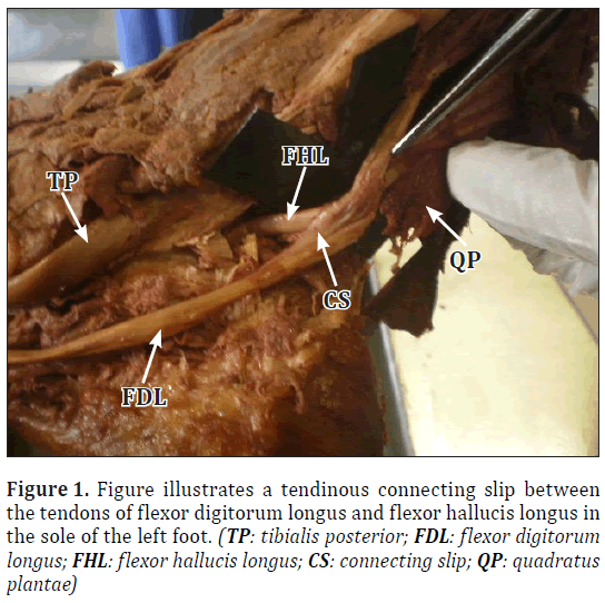
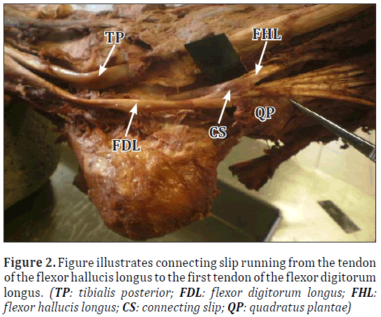
During routine dissection of the lower limbs of a 62-year-old American male cadaver in the Department of Anatomy, Medical University of the Americas, Nevis, West Indies, we found a strong tendinous connecting slip between the tendons of flexor digitorum longus and flexor hallucis longus in the sole of the left foot. The slip originated from the tendon of the flexor digitorum longus, just proximal to its division into the four tendons for the lateral four toes, and also before the attachment of the quadratus plantae muscle to the tendon. The slip then coursed forward and medially to get attached to the tendon of the flexor hallucis longus on its way to the first toe. It was 7.5 mm long (Figures 1,2).
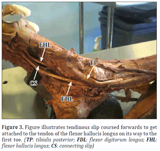
However, in the sole of the right foot, we found a connecting slip running from the tendon of the flexor hallucis longus to the first tendon of the flexor digitorum longus (Figure 3).
Discussion
Singh et al., Gumusalan et al. have reported bilateral accessory flexor digitorum longus muscles in the literature. According to Singh et al., in the left sole, the accessory muscle tendon received slips from the flexor hallucis longus tendon and the flexor digitorum longus tendon distally, with muscle insertion of the quadratus plantae, on its way to final insertion at the second toe; whereas in the right sole, the tendon got inserted to the calcaneus with fibrous insertions to the quadratus plantae, flexor hallucis tendon and flexor digitorum tendon [3]. According to Gumusalanet al., the accessory tendon terminated in the sole of the foot by merging into the tendon of the quadratus plantae muscle [4].
O’Sullivan et al. carried out sixteen cadaveric dissections at the region of the knot of Henry, based on which they proposed that identification of the tendon to be transected for tendon transfer should be decided at the time of surgery depending on the anatomical pattern. They also suggested that transection of flexor digitorum longus tendon proximal to the region of the knot of Henry for the repair of tibialis posterior dysfunction would result in retention of function of the great and lesser toes in the majority of cases [5].
Mahajan et al. have described the flexor hallucis longus tendon transfer for the reconstruction of chronically ruptured Achilles tendon to be effective, safe and easy surgery in patients with low to moderate demands [6].
A dissection study of 24 cadaveric legs by Larue et al. showed the distal anatomical relationship between flexor hallucis longus and flexor digitorum longus tendons, which revealed three different configurations. According to this, the present case is categorized as type 1 on the right side. However, no reports were found in the literature where a tendinous slip arose from the flexor digitorum longus tendon to the flexor hallucis longus tendon [7].
The present case shows rare variation with dissimilar arrangement of the tendons on both sides. Such variation has to be kept in mind during radiodiagnostic and surgical procedures.
Acknowledgements
Our sincere thanks to the faculty members of our university who helped and supported during the writing of this manuscript.
References
- Moore KL, Agur AR, Dalley AF. Essential Clinical Anatomy. 4th Ed., Philadelphia, Wolters Kluwer Company. 2011; 366.
- Standring S, Ellis H, Healy JC, Johnson D, Williams A, Collins P, Wigley C, eds. Gray’s Anatomy. 39th Ed., New York, Churchill Livingstone. 2005; 1500.
- Singh R, Shamal SN. Bilateral accessory flexor digitorum muscle in the posterior compartment of the leg. Int J Anat Variat (IJAV). 2010; 3: 176–177.
- Gumusalan Y, Kalaycioglu A. Bilateral accessory flexor digitorum longus muscle in man. Ann Anat. 2000; 182: 573–576.
- O’Sullivan E, Carare–Nnadi R, Greenslade J, Bowyer G. Clinical significance of variations in the interconnections between flexor digitorum longus and flexor hallucis longus in the region of the knot of Henry. Clin Anat. 2005; 18: 121–125.
- Mahajan RH, Dalal RB. Flexor hallucis longus tendon transfer for reconstruction of chronically ruptured Achilles tendons. J Orthop Surg (Hong Kong). 2009; 17: 194–198.
- LaRue BG, Anctil EP. Distal anatomical relationship of the flexor hallucis longus and flexor digitorum longus tendons. Foot Ankle Int. 2006; 27: 528–532.


