situs Inversus – Macroscopic work-up and analysis of Cerebral lateralization Using Manual and Imaging Techniques
Received: 01-Sep-2022, Manuscript No. ijav-22-5326; Editor assigned: 05-Sep-2022, Pre QC No. ijav-22-5326 (PQ); Accepted Date: Sep 23, 2022; Reviewed: 19-Sep-2022 QC No. ijav-22-5326; Revised: 23-Sep-2022, Manuscript No. ijav-22-5326 (R); Published: 30-Sep-2022, DOI: 10.37532/1308-4038.15(9).215
Citation: Schoen M, Leupold M, Boeckers TM, Boeckers A, et al. Situs Inversus â?? Macroscopic work-up and Analysis of Cerebral lateralization Using Manual and Imaging Techniques. Int J Anat Var. 2022;15(9):213-215.
This open-access article is distributed under the terms of the Creative Commons Attribution Non-Commercial License (CC BY-NC) (http://creativecommons.org/licenses/by-nc/4.0/), which permits reuse, distribution and reproduction of the article, provided that the original work is properly cited and the reuse is restricted to noncommercial purposes. For commercial reuse, contact reprints@pulsus.com
Abstract
Within the last decade three body donors with previously diagnosed situs inversus (SI) were admitted to the institute of anatomy. Herein, we will present one of these cases regarding its past medical history and findings after dissection of thoracic and abdominal organs. In order to obtain further information concerning its cerebral lateralization, manual measurements on skull and brain structures as well as on post mortem cranial computer tomograms (cCT) and 3T-MRI were performed. Osseous, vascular, parenchymal structures and hemispheric volumes were compared intra individually between left and right hemispheres, or with data gained from ten control donors with situs solitus (SS). Only hemispheric petalia suggested a distinct reversed lateralization. This study adds further data to the existing record on this rare anatomical variant for research and teaching purposes. Neuroimaging techniques such as MRI and cCT proved superior to manual techniques without revealing definite signs of cerebral lateralization.
Keywords
Situs inversus; Cerebral lateralization; Neuroimaging; Gross anatomy; anatomical education
INTRODUTION
Situs inversus (SI) describes an anatomical variant characterized by the mirror-imaged transposition of unpaired thoracic and abdominal organs. The physiological development and lateralization of body organs with the common left-right body asymmetry results in the predominant appearance of a Situs solitus (SS) in most individuals. Despite extensive research, the underlying regulatory processes are not yet fully understood [1]. Disorders of the lateralization process cause atypical organ positions, so-called heterotaxies. There are different types of lateralization defects: isolated lateralization defects and the phenomena of Situs ambiguus or Situs inversus [2]. Situs inversus (totalis) occurs in a frequency of approximately 1:10.000 and is associated with a chiral arrangement of all inner organs including a dextrocardia [3]. There are currently only a few studies that deal with the mirroring of macro anatomical brain structures like Petelia and Bending or volumetric measurements of dedicated cerebral areas like the peri-Sylvian regions on the base of radiological methods such as MRT. On the current state of knowledge, no structure could be clearly identified, which consistently presents itself in the mirror-image position in Situs inversus, also due to the little number of SI cases [4-9].
Three body donors previously diagnosed with SI (year of death 2010/2013/2019) were admitted to the Institute for Anatomy and Cell Biology, Ulm, Germany, for teaching and research purposes. Herein, one of these SI cases will be presented regarding its medical history followed by a description of the macroscopic findings related to organ location revealed by dissection of the thoracic and abdominal cavities. In order to obtain further information concerning cerebral lateralization, manual measurements on skull and brain structures as well as on post mortem neuro-imaging were performed. Study designs including case analysis were previously approved by the ethics committee of Ulm University, Germany (Nr. 389/16).
CASE REPORT
Medical history: In 2013, the Institute for Anatomy and Cell Biology of Ulm University accepted a female 82-year-old body donor (150 cm/80 kg). At the age of 18, she had to undergo an appendectomy and was incidentally diagnosed with Situs inversus. She was right-handed and did not show any noticeable medical problems during her lifetime related to her diagnosis of a SI. At an advanced age the patient suffered from adipositas, lower leg varcosis, generalized arthrosis, and insulin-dependent diabetes mellitus type II, agerelated pulmonary emphysema, arterial hypertension and bilateral peripheral arterial occlusive disease (PAOD) of her tibial arteries.
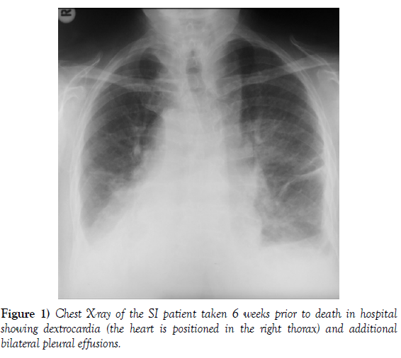
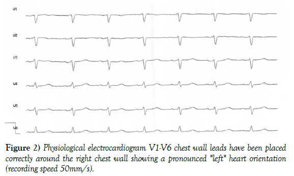
About 6 weeks prior to her death, she was admitted to hospital with dyspnea and bilateral lower limb edema. Clinically, bronchial rales and bilateral pleural effusions were found and confirmed radiologically (Figure 1). Further clinical examinations such as an electrocardiogram and an echocardiography confirmed a decompensated heart failure with tachymyopathy (ejection fraction 30%) (Figure 2). The coronaries were angiographically open. The patient could be discharged from the hospital, but had to be readmitted three weeks later after a domestic fall. On admittance no fractures or neurological abnormalities could be diagnosed, but the patient died several days later due to an acute pulmonary edema in April 2013.
Post mortem workup: Within 48 hours after death the SI body donor was fixated and preserved using a 4% formaline solution according to standard institutional procedures. Macroscopic dissection and photo documentation was conducted according to previously published data [4] about three years post-fixation. Manual measurements of cranial osseous structures and specified vessels (A. carotis interna C1, A. cerebri ant A1, A. cerbri media M1, Labbé/Trolard-Veins) were performed according to Tubbs et al. [6].
Postmortem cranial computer tomograms (cCT) taken in situ on the fixated body donor were used to measure petalia according to Vingerhoets [9] and to record cerebral hemispheres bending in the frontal and sagittal planes. Collected data were compared with data from 10 female control body donors following the same post mortem workup. Hereafter the brain was retrieved from the scull and stored in dielectric inert fluid (3MTM, IoLiTec Ionic Liquids). Remaining air bubbles in ventricles and cortical folds were removed and a 3T-MRImaging for hemispheric volumetric measurements performed (software of AG Neuroimaging, Uni Ulm, Germany).
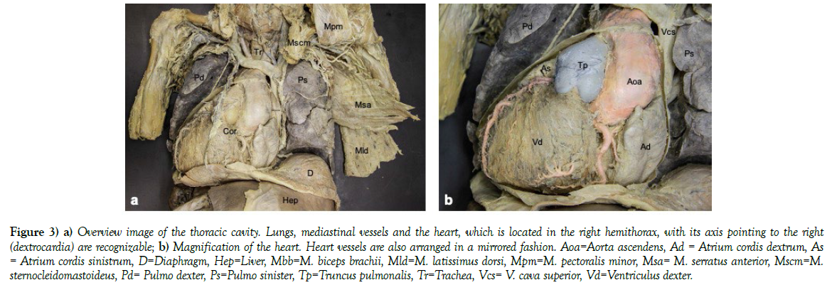
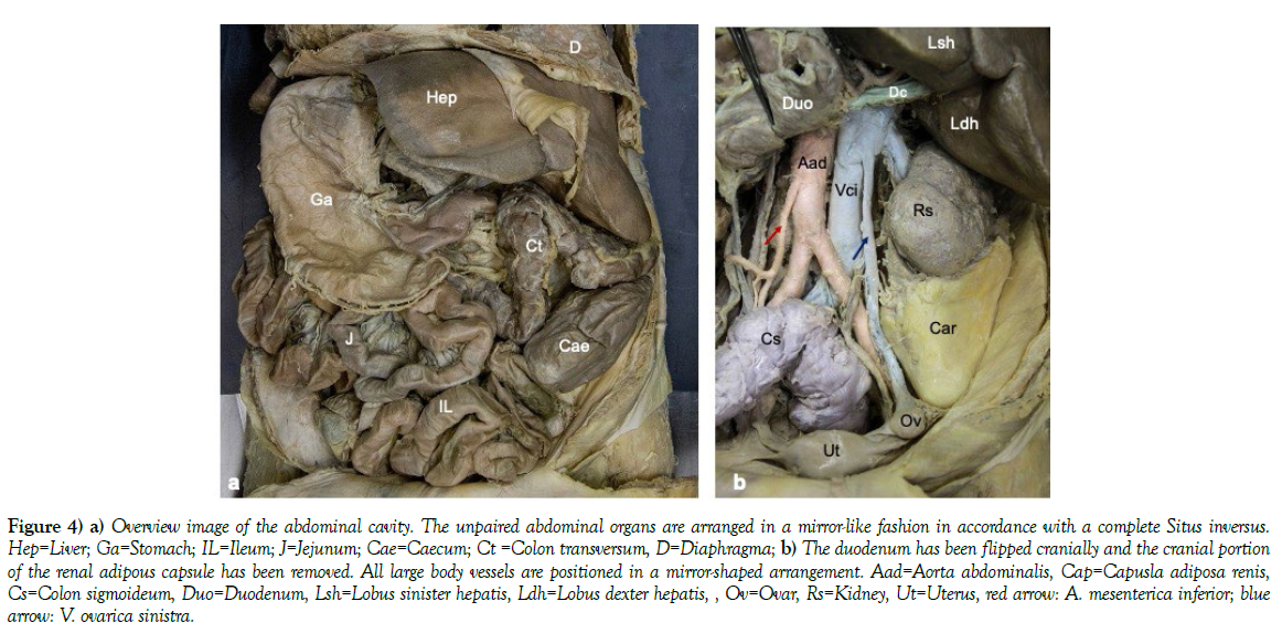
The macroscopic visualization of all body organs confirmed a complete Situs inversus totalis with dextrocardia and dextroversion (cardiac axis pointing to the right). Figures 3 and 4 illustrate the topography of thoracic and abdominal organs. Examination of cerebral structures in terms of lateralization yielded inconsistent results. The diameters of cerebral arterial vessels showed no obvious lateralization. The diameter of venous vessels was greater on the right hemispheric side. The average size of the right medial cranial fossa of SI donors including the presented case (42 vs 39.4 mm) was larger than the left. Converted results were measured for the average width of the posterior cranial fossa with this SI showing a larger right fossa (56.3 vs. 55.6 mm) and a larger left sided anterior cranial fossa on the cCT (12.6 vs. 15.2 mm). All three SI brains exhibited on CT scans a left sided frontal petalia (SI 2013: 0.5 mm) and a right sided petalia occipitally (SI 2013: 1.0 mm). Six brains of the Situs solitus control donors exhibited rightsided frontal petalia, five leftsided occipital petalia. Bending of the cerebral hemispheres yielded inhomogeneous results with the presented SI case revealing a frontal (0.6 mm) and occipital (0.7 mm) bending of the left hemisphere. Volumetric analysis of MRI images revealed a slightly larger left hemisphere in this case (404.5 vs. 390.1 cm3) without a definite side preference comparing average sizes in all three cases.
Figure 3: a) Overview image of the thoracic cavity. Lungs, mediastinal vessels and the heart, which is located in the right hemithorax, with its axis pointing to the right (dextrocardia) are recognizable; b) Magnification of the heart. Heart vessels are also arranged in a mirrored fashion. Aoa=Aorta ascendens, Ad = Atrium cordis dextrum, As = Atrium cordis sinistrum, D=Diaphragm, Hep=Liver, Mbb=M. biceps brachii, Mld=M. latissimus dorsi, Mpm=M. pectoralis minor, Msa= M. serratus anterior, Mscm=M. sternocleidomastoideus, Pd= Pulmo dexter, Ps=Pulmo sinister, Tp=Truncus pulmonalis, Tr=Trachea, Vcs= V. cava superior, Vd=Ventriculus dexter.
Figure 4: a) Overview image of the abdominal cavity. The unpaired abdominal organs are arranged in a mirror-like fashion in accordance with a complete Situs inversus. Hep=Liver; Ga=Stomach; IL=Ileum; J=Jejunum; Cae=Caecum; Ct =Colon transversum, D=Diaphragma; b) The duodenum has been flipped cranially and the cranial portion of the renal adipous capsule has been removed. All large body vessels are positioned in a mirror-shaped arrangement. Aad=Aorta abdominalis, Cap=Capusla adiposa renis, Cs=Colon sigmoideum, Duo=Duodenum, Lsh=Lobus sinister hepatis, Ldh=Lobus dexter hepatis, , Ov=Ovar, Rs=Kidney, Ut=Uterus, red arrow: A. mesenterica inferior; blue arrow: V. ovarica sinistra.
DISCUSSION
The dissection confirmed a complete Situs inversus with dextrocardia and opposed lateralization of all organs without any signs of Situs ambiguus. Examination of cerebral structures in terms of lateralization yielded in this case-and additionally in all three SI donors - only inconsistent results. The diameters of arterial and venous cerebral vessels, hemispheric bending on cCT and hemispheric volume on MRT showed no obvious lateralization.
In accordance with previous studies, the average size of the medial cranial fossae of SI donors was larger on the right side and petalia occurred with higher frequency left frontally and right occipitally [5-9].
Study data complement and confirm existing data on SI. Conclusions regarding brain functionality cannot be drawn, in particular since information on the donor’s handedness could not be provided in all cases. In addition to the scientific workup of Situs inversus, its exemplary macroscopic visualization and workup of its medical history rendered it a valuable study object in anatomy teaching for medical and dental undergraduate students.
ACKNOWLEDGEMENT
The authors would like to give their acknowledgements to all body donors of the Institute of Anatomy and Cell Biology of the Ulm University, Germany, for their willingness to donate their bodies for medical training and research. In particular, the authors would like to give their thanks to the three body donors with a previously diagnosed Situs inversus for their body donation and for the chance to get further information about their past medical history by interviewing themselves, their physicians or their relatives. Additionally, the authors would like to thank Prof. Dr. Bernd Schmitz, Prof. Dr. Meinrad Beer, and Prof. Dr. Volker Rasche for their support in CT radiological imaging and measuring methods and Prof. Dr. Hans-Peter Müller for his support in the MRI volumetric analysis of the hemispheres.
CONFLICTS OF INTEREST
None
FUNDING
None
REFERENCES
- Bisgrove B, Essner J, Yost H. Multiple pathways in the midline regulates concordant brain, heart and gut left-right asymmetry. Development. 2000; 127(16):3567-3579.
- Lee SE, Kim HY, Jung SE, Lee SC et al. Situs anomalies and gastrointestinal abnormalities. J Pediatr Surg. 2006; 41:1237-1242.
- Eitler K, Bibok A, Telkes G. Situs inversus total is: a clinical review. Int J Gen Med. 2022; 15:2437-2449.
- Sharma S, Chaitanya KK, Suseelamma D. Situs inversus (Dextroversio) - An anatomical study. Anat Physiol. 2012; 2:5.
- Kennedy DN, O’Craven KM, Ticho BS, Goldstein AM et al. ‘Structural and functional brain asymmetries in human situs inversus totalis’. Neurology. 1999; 53:1260-1265.
- Tubbs, SR, Wellons JC, Salter G, Blound JP et al. Intracranial asymmetry in situs inverus totalis. Anat Embryol. 2003; 206:199-202.
- Ihara A, Hirata M, Fujimaki N, Goto T et al. Neuroimaging study on brain asymmetries in situs inversus totalis’. J Neurol Sci. 2010; 288:72-78.
- Schuler A L, Kasprian G, Schwartz E, Seidl R et al. Mens inversus in corpore inverso? Language lateralization in a boy with situs inversus totalis. Brain Lang. 2017; 174:9-15.
- Vingerhoets G, Gerrits R, Bogaert S. A typical brain functional segregation is more frequent in situs inverus totalis. Cortex. 2018; 106:12-25.
Indexed at, Google Scholar, Crossref
Indexed at, Google Scholar, Crossref
Indexed at, Google Scholar, Crossref
Indexed at, Google Scholar, Crossref
Indexed at, Google Scholar, Crossref
Indexed at, Google Scholar, Crossref
Indexed at, Google Scholar, Crossref
Indexed at, Google Scholar, Crossref


